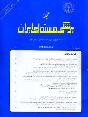
فهرست مطالب

Iranian Journal of Nuclear Medicine
Volume:11 Issue: 1, 2003
- تاریخ انتشار: 1382/01/20
- تعداد عناوین: 9
-
-
Pages 1-7Today, nuclear medicine has rapidly been developed so that in some cases, plays a unique role in diagnosis but unfortunalty in spite of diagnostic and therapeutic advantages, the term “NUCLEAR” can induce worries in patients and society. In this article, base on new documents, we intend to show that this worries has no scientific basis. To produce a realistic view, regarding to radiation protection we used several ways such as natural origin of radiation, high natural background radiation areas’ data, non-linear dose-effect model, risk versus benefit, use of arbitrary unit for measurement of radiation, radioadaptive response and radiation hormesis. Harmful effects of radiation on biologic systems has obviously been shown, but most of related documents are based on receiving high doses in nuclear and atomic accidents and explosions and radiation protection regulations are based on this observation. So, it sometimes causes patients are afraid of low doses of radiation in medical diagnostic procedures so that some of them even resist against performing this procedures. Thus, being aware of scientific realities and new related findings can significantly reduce fear from radiation in patients and all society.
-
Pages 9-16Contamination with radiopharmaceuticals in nuclear medicine centers in addition to being a health concern requires time-consuming decontamination efforts. According to NRC (Nuclear Regulatory Commission) contamination should be monitored in nuclear medicine centers where radiopharmaceuticals are prepared and administrated at the end of each working session; otherwise, contamination spread to other areas not only equipment but also personnel and other people will be expected. The wipe test for the presence of radioactivity is accomplished by wiping the surface over an area approximately 100cm² with an absorbent paper, then counting it in an appropriate radiation detector. In this study, contamination monitoring of patient’s rooms (4 rooms), entrance corridor, patient’s corridor, waiting room, control room (Nursing station), radiopharmaceutical storage room in therapy unit of Research Institute for Nuclear Medicine, Shariati hospital was performed by indirect method. Based on the results, some areas including storage room were contaminated. There was also a direct relationship between dose administrated and levels of contamination in patient’s rooms. Regarding high uptake of iodine by thyroid gland and damaging effects of Na¹³¹I, weekly wipe tests are required to determine the level of contamination. Patient’s rooms after discharging the patients and before rehospitalization specially should be checked. If these tests reveal contamination over standard levels, appropriate decontamination procedures should be carried out immediately.
-
Pages 17-21Salivary gland involvement is one of the radioiodine therapy complications. Salivary gland scintigraphy in quantitative mode can accurately evaluate salivary gland function.MethodsSalivary gland scintigraphy was performed with Tc-99m Pertechnetate, at the time of iodine therapy as a basic study and then 3 weeks and 3 months afterwards. Ejection Fraction (EF) of parotid and submandibular glands was obtained at each stage of the study.Results36 patients (10 male, 16 female) were studied. Mean of EF 3 weeks and 3 months following radioiodine therapy was reduced. There was no significant involvement in 12 patients (33.3%). With increase in radioiodine dose, more salivary gland involvement was noted in 3 weeks (P=0.07), but not after 3 months (P=0.5). No difference was noted between two sexes (P=0.6). Parotid gland involvement was more than submandibular gland (P<0.05), confirming more radiosensitivity of parotid gland. No meaningful relation was noted between salivary gland involvements with age (P=0.1). Parotid gland dysfunction was not related to radioiodine dose, but in submandibular gland, with dosage increase, more involvement was noted (P=0.02). Clinical symptoms were not reliable in evaluating salivary gland dysfunction.
-
Pages 23-28Somatostatin analoges labeled with different radionuclides are able used for imaging and treatment of somatostatin receptor positive tumors. In this study Tyr³-Octreotide protected at lysine as an analoge of somatostatin was conjugated with bifunctional chelating agent 6-BOC-hydrazinopyridine-3-carboxylic acid (BOC-HYNIC) and after deprotecting of conjugate, purification was performed with preparative-RP-HPLC. The conjugated peptide was labeled with 99mTc and tricine as coligand. Radiolabelling efficiency, radiochemical purity, stability in human serum, internalization of receptor bound 99mTc-labeled peptide and biodistribution in normal mice were studied.
-
Pages 29-32It is of value to determine the amount of viable myocardial tissue in patients suffering from chronic coronary artery disease and ventricular dysfunction. Having the capability of evaluating both myocardial perfusion and function, simultaneously, myocardial scanning by ECG-Gated is an appropriate method for this purpose. The aim of this study was to compare the results of myocardial perfusion and wall motion before and after Coronary Artery Bypass Grafting (CABG), as well as assessment of the efficiency of these parameters for detection of myocardial viability. Forty patients with positive history of previous myocardial infarction and candidate for CABG underwent ECG-Gated SPECT scanning 1 month before and 2-3 months after surgery. Findings of myocardial perfusion and wall motion, obtained from the two phases of the study, were compared. The results showed that functional status of some preoperatively severely hypoperfused segments, recovered significantly after CABG, which proved existence of viable tissue in these regions. Also, septal wall motion presented no statistically significant changes after CABG. These results suggest that septal perfusion and motion are not reliable parameters for the assessment of surgical outcome in this region. Most of the myocardial walls demonstrated considerable wall motion improvement after operation. Concerning the results of this research, as a marker of viable against nonviable myocardial tissue, we recommend revision of applying the severity of perfusion defect, alone, as well as taking the wall motion parameter into consideration, to improve diagnostic accuracy of the SPECT method.
-
Pages 33-39The aim of this study was to assess the value of 99mTc-hexakis-2-methoxyisobutylisonitrile (MIBI) scintigraphy in patients with clinical and radiologic features of primary or metastatic musculoskeletal tumors.MethodsThe scintigraphic findings for 84 patients were compared with the surgical and histologic findings. Each patient underwent three-phase bone scan with 99mTc-methylene diphosphonate (MDP) and dynamic and static MIBI scintigraphy. The MIBI scans were evaluated by visual and quantitative analysis. The count ratio of the lesion to the adjacent or contralateral normal area (Lesion/Normal) was calculated from the region of interest drawn on the MIBI scan. The Mann-Whitney test was used to determine the differences between the uptake ratios of malignant and benign lesions.ResultsAlthough increased MDP uptake was not specific for bone malignancy, a significant difference was found between benign tumors (Lesion/Normal=1.22±0.43) and malignant tumors (L/N=2.25±1.03) on MIBI scans. Sensitivity and specificity were 81% and 87%, respectively. 43 of 53 proven benign lesions did not show significant MIBI uptake. The negative predictive value was 88%. In all seven sites of pathologic fracture, significant uptake was seen. However, three malignant lesions were not detected by MIBI scintigraphy, whereas seven benign lesions showed false positive results.ConclusionThe major diagnostic worth of MIBI scintigraphy is its high negative predictive value. Although not capable of replacing tissue biopsy as a definitive diagnostic modality for musculoskeletal neoplasm, MIBI scintigraphy does appear to have a role in better preoperative assessment, and in distinguishing between pathologic and simple fractures.
-
Pages 41-50Myocardial perfusion SPECT imaging with Tc99m-sestamibi is the most accurate non-invasive means of detecting coronary artery disease and assessing the severity of perfusion abnormalities in the patients with coronary stenosis. Though simple and straight forward the results produced by the technique is very much affected by the details being used. One of the main problems with the test, which has created a lot of discrepancies, is the selection of proper filter and adjustment of filter parameters to individual cases. In this study we have analyzed and compared for widely used filters for myocardial Tc-99m sestamibi SPECT study i.e. Hanning, Butterworth, Metz and Wiener. The aim of the study was to find the most suitable filter for this type of study and to verify the theoretical peculiarity of the filters in practice. Patients with proven coronary artery abnormality were selected. We have investigated four conventional filters commonly used in nuclear medicine using quantitative and qualitative method. Filters were compared to each other and to the results of angiography. Our results show that the best filter in terms of contrast; smoothing and spatial resolution is the classical Metz filter with the FWHM of 4, order of 0.5. The best filter in the myocardial viability was Hanning filter with the cutoff of 0.5
-
Pages 51-55Fixed myocardial defects in both stress and rest images, could be artifactual as a result of soft tissue attenuation. To increase specificity and identify the false positive results, we used gated technique to evaluate the wall motion and wall thickening as an index to differentiate real ischemic lesions from artifactual defects. 93 patients were studied. In 46 patients (48.8%) fixed perfusion defects were identified. Of these patients 10 (21.7%) had definite evidence of previous MI, 6 of whom (60%) showed abnormal gated SPECT findings. Among 36 patients without history of MI, 11 cases (30%) showed abnormal ventricular function (Probably due to silent MI) and 25 patients (70%) had normal function. The latter group was comprised of 13 females (52%) with fixed perfusion defects seen in the anterior, septal and apical segments of the myocardium. He results of the investigation indicate that using gated SPECT method with 99mTc-MIBI myocardial perfusion studies increases specificity of the fixed myocardial defects and is highly recommended.
-
Pages 57-65IntroductionThe lymphatic system provides one of the main paths for the spread (Metastasis) of cancer from one part of body to another. Hodgkin’s diseases, lymphocytic leukemia, various metastatic disease and many lymph node disorders can be assessed by lymphoscintigraphy. Radionuclide lymphoscintigraphy has been used for many years to define lymphatic drainage of melanoma. The most common radiopharmaceuticals used for lymphoscintigraphy are 99mTc-SC, 99mTc-antimony sulfide colloid and 99mTc-HSA-nanocolloid. Preparation of 99mTc-antimony sulfide colloid has been chosen among other colloids.Materials And MethodsFor antimony colloid preparation, hydrogen sulfide gas was passed through D.W. until saturation. Antimony potassium tartrate is then added to the solution to form Sb2S3 colloid. The colloid was stabilized with P.V.P. Excess H2S was removed by bubbling with nitrogen. The preparation was then filtered through a 0.22μm membrane filter and aliquots containing 1.017mg Sb2S3 were dispensed into kit vials. Labeling was accomplished by adding 99mTcO-4 and HCl to the vial then heating it at 100°C in boiling water bath for 10 min. The pH was adjusted by adding a phosphate buffer.ResultsThe radiochemical purity of 99mTc-antimony trisulfide colloid by ITLC-SG/normal saline was more than 95 percent. The amount of Sb in reaction vial was 0.729mg.ConclusionThe study demonstrated that our formulation of antimony sulfide, which has 0.0486mg (Sb) in 0.2ml for injection per patient (Total volume after labeling with 99mTc is 3ml).

