فهرست مطالب

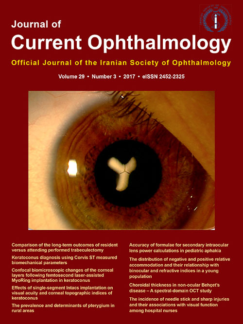
Journal of Current Ophthalmology
Volume:29 Issue: 3, Sep 2017
- تاریخ انتشار: 1396/07/01
- تعداد عناوین: 16
-
-
Pages 143-144Over the past few years, a number of studies have provided new information about the proper dosage, relative risk, fundus distribution, and screening guidelines for the use of hydroxychloroquine (HCQ) and chloroquine (CQ). These are excellent drugs for systemic lupus erythematosis (SLE) and rheumatoid diseases, but excessive and prolonged intake can cause irreversible retinopathy. However, the ocular safety profile is very good if the drugs are used wisely. Current standards of care are presented in the 2016 revision of the American Academy of Ophthalmology recommendations for screening, which illustrates findings and is available Open Access.1 This editorial summarizes the key information.
Dose and duration of use
The most important new study was an analysis of almost 2500 patients using HCQ long-term.2 The results showed that risk is a balance of daily dose and duration of use, and risk rises markedly with over-dosage (>5 mg/kg real weight) or durations beyond 10 years. For daily dose below 5 mg/kg real weight, prevalence of toxicity was Dose measurement and adjustment
Dose should now be measured in real rather than ideal weight. The evidence for ideal weight (used in past years) was actually rather weak, and human demographics show that real weight gives more accurate assessment of risk over all body types. Bottom line: stay below 5 mg/kg real weight for HCQ, and an estimated 2.3 mg/kg for CQ. These drugs do not come in small tablets, and even a single CQ tablet is too big for most women. However, blood levels stabilize slowly, and one can vary the dose on different days to achieve the desired intake for a week.
Fundus distribution
Another important new finding was that people of Asian descent (including Filipinos) often show a different pattern of early fundus damage from those of Caucasian descent (including the Middle East and India).3 Instead of a parafoveal bull's eye, Asians may show damage out near the vascular arcades.
Screening schedule
Toxicity cannot be prevented, but it can be detected before most patients will notice scotomas and before any loss of central vision. In the first year of use, patients should have a good fundus exam to rule significant macular degeneration, retinal dystrophy, or field loss (such as glaucoma) that might interfere with diagnosis. If that exam is unremarkable (e.g. a few hard drusen are not a contraindication), and there are no major risk factors (see above), then the risk of toxicity is so low in the first 5 years that annual screening can be deferred until that time.
Screeningvisual fields
Probably the most sensitive screening tests are central automated fields, and I suggest using the SITA protocol that provides statistical pattern deviation plots. A 10-2 field is critical for Caucasians, as 24-2 patterns do not have sufficient parafoveal test points. A 24-2 field can be added for Asians, and with the SITA Fast protocol one can do both a 10-2 and 24-2 field in the time for one standard 10-2. Learn to recognize the pattern of early losses 25 deg from center, usually superotemporal or superonasal.
ScreeningSD-OCT
Not all patients are good field takers, and the most specific screening test is the SD-OCT. Ideally, annual screening should do both fields and SD-OCT (or at least add the SD-OCT at occasional intervals). The goal of SD-OCT exams is to recognize in the parafovea (or arcade region in many Asian eyes) early thinning of the outer nuclear layer, and early break up of the ellipsoid zone (EZ) line. ETDRS thickness plots often show abnormal thinning in the parafovea, and the OCT cross-sections may have a sombrero or flying saucer appearance with normal appearance in the fovea, and again beyond the ring of damage.
Toxicity and progression
These changes are all recognizable before damage to the retinal pigment epithelium (RPE). Fundus examination is not sensitive enough for screening HCQ and should never be relied upon. If pathology is detected at an early stage, before RPE damage, there is little progression after stopping the drug.4 However, once RPE changes are visible, retinal damage may progress and expand for many years after the drug is stopped, and eventually destroy the fovea. Early detection is critical.
Stoppage of drugs
HCQ or CQ should be stopped once toxicity is clearly identified. But keep in mind that these drugs are very useful medically, and should not be stopped for questionable or borderline findings. Retinopathy does not develop that fast, and there is always time to bring the patient back in a few months for re-testing to see whether ambiguous findings are consistent, or for the addition of confirmatory studies such as multifocal electroretinography (mfERG) or fundus autofluorescence (FAF). The risk to vision remains low as long as the foveal anatomy is good, and the RPE is not involved.
Proper dose ( -
Pages 145-153PurposeThe aim of this study was to review the safety and stability of cornea cross-linking (CXL) for the treatment of keratectasia after Excimer Laser Refractive Surgery.
MethodsEligible studies were identified by systematically searching PubMed, Embase, Web of Science and reference lists. Meta-analysis was performed using Stata 12.1 software. The primary outcome parameters included the changes of corrected distant visual acuity (CDVA), uncorrected visual acuity (UCVA), the maximum keratometry value (Kmax) and minimum keratometry value (Kmin), the surface regularity index (SRI), the surface asymmetry index (SAI), the keratoconus prediction index (KPI), corneal thickness, and endothelial cell count. Efficacy estimates were evaluated by weighted mean difference (WMD) and 95% confidence interval (CI) for absolute changes of the interested outcomes.
ResultsSeven studies involving 118 patients treated with CXL for progressive ectasia after laser-assisted in situ keratomileusis (LASIK) or photorefractive keratectomy (PRK) (140 eyes; the follow-up time range from 12 to 62 months) were included in the meta-analysis. The pooled results showed that there were no significant differences in Kmax and Kmin values after CXL (WMD = 0.584; 95% CI: −0.289 to 1.458; P = 0.19; WMD = 0.466; 95% CI: −0.625 to 1.556; P = 0.403, respectively). The CDVA improved significantly after CXL (WMD = 0.045; 95% CI: 0.010 to 0.079; P = 0.011), whereas UCVA did not differ statistically (WMD = 0.011; 95% CI: −0.055 to 0.077; P = 0.746). The changes were not statistically significant in SRI, SAI, and KPI (WMD = 0.116; 95% CI: −0.090 to 0.322; P = 0.269; WMD = 0.240; 95% CI: −0.200 to 0.681; P = 0.285; WMD = 0.045; 95% CI: −0.001 to 0.090; P = 0.056, respectively). Endothelial cell count and corneal thickness did not deteriorate (WMD = 12.634; 95% CI: −29.460 to 54.729; P = 0.556; WMD = 0.657; 95% CI: −9.402 to 10.717; P = 0.898, respectively).
ConclusionThe study showed that CXL is a promising treatment to stabilize the keratectasia after Excimer Laser Refractive Surgery. Further long-term follow-up studies are necessary to assess the persistence of the effect of the CXL.Keywords: Cross-linking, Keratectasia, Refractive surgery, Meta-analysis -
Pages 154-168PurposeSince different subspecialties are currently performing a variety of upper facial rejuvenation procedures, and the level of knowledge on the ocular and periocular anatomy and physiology is different, this review aims to highlight the most important preoperative examinations and tests with special attention to the eye and periocular adnexal structures for general ophthalmologist and specialties other than oculo-facial surgeons in order to inform them about the fine and important points that should be considered before surgery to have both cosmetic and functional improvement.
MethodsEnglish literature review was performed using PubMed with the different keywords of periorbital rejuvenation, blepharoptosis, eyebrow ptosis, blepharoplasty, eyelid examination, facial assessment, and lifting. Initial screening was performed by the senior author to include the most pertinent articles. The full text of the selected articles was reviewed, and some articles were added based upon the references of the initial articles. Included articles were then reviewed with special attention to the preoperative assessment of the periorbital facial rejuvenation procedures.
ResultsThere were 254 articles in the initial screening from which 84 articles were found to be mostly related to the topic of this review. The number finally increased to 112 articles after adding the pertinent references of the initial articles.
ConclusionStatic and dynamic aging changes of the periorbital area should be assessed as an eyelid-eyebrow unit paying more attention to the anthropometric landmarks. Assessing the facial asymmetry, performing comprehensive and detailed ocular examination, and asking about patient's expectation are three key elements in this regard. Furthermore, taking standard facial pictures, obtaining special consent form, and finally getting feedback are also indispensable tools toward a better outcome.Keywords: Blepharoplasty, Cheek, Eyebrow, Eyelid, Lifting, Rejuvenation -
Pages 169-174PurposeTo compare the long-term outcomes obtained by residents and attending surgeons performing trabeculectomy.
MethodsAfter reviewing medical records of the patients, 41 residents performing trabeculectomy under supervision of attendings were compared to 41 attendings performing trabeculectomy. The primary outcome measure was the surgical success defined in terms of intraocular pressure (IOP) ≤ 21 mmHg (criterion A) and IOP ≤ 16 mmHg (criterion B), with at least 20% reduction in IOP, either with no medication (complete success) or with no more than 2 medications (qualified success). IOP, number of glaucoma medications, surgical complications, and visual acuity were analyzed as secondary outcome measures.
ResultsMean age of the patients was 59.5 ± 8.6 years in the resident group and 59.6 ± 12.31 years in the attending group (P = 0.96). Furthermore, mean duration of the follow-up was 62.34 ± 5.51 months in the resident group and 64.80 ± 7.80 months in the attending group (P = 0.10). The cumulative success according to criterion A was 87.8% in the resident group and 85.3% in the attending group (P = 0.50). Moreover, according to criterion B, it was 87.8% and 83% in the resident and attending groups, respectively (P = 0.62). Repeated glaucoma surgery was required in 12.2% and 2.4% of the patients in the resident and attending groups, respectively (P = 0.09). Rate of complications was 12.2% and 4.8% in the resident and attending groups, respectively (P = 0.23).
ConclusionThere were comparable results with respect to success rates and complications between residents and attending surgeons performing trabeculectomy in the long-term follow-up.Keywords: Glaucoma, Trabeculectomy, Intraocular pressure -
Pages 175-181PurposeTo assess the diagnostic power of the Corneal Visualization Scheimpflug Technology (Corvis ST) provided corneal biomechanical parameters in keratoconic corneas.
MethodsThe following biomechanical parameters of 48 keratoconic eyes were compared with the corresponding ones in 50 normal eyes: time of the first applanation and time from start to the second applanation [applanation-1 time (A1T) and applanation-2 time (A2T)], time of the highest corneal displacement [highest concavity time (HCT)], magnitude of the displacement [highest concavity deformation amplitude (HCDA)], the length of the flattened segment in the applanations [first applanation length (A1L) and second applanation length (A2L)], velocity of corneal movement during applanations [applanation-1 velocity (A1V) and applanation-2 velocity (A2V)], distance between bending points of the cornea at the highest concavity [highest concavity peak distance (HCPD)], central concave curvature at the highest concavity [highest concavity radius (HCR)]. To assess the change of parameters by disease severity, the keratoconus group was divided into two subgroups, and their biomechanical parameters were compared with each other and with normal group. The parameter's predictive ability was assessed by receiver operating characteristic (ROC) curves. To control the effect of central corneal thickness (CCT) difference between the two groups, two subgroups with similar CCT were selected, and the analyses were repeated.
ResultsOf the 10 parameters compared, the means of the 8 were significantly different between groups (P 0.05). ROC curve analyses showed excellent distinguishing ability for A1T and HCR [area under the curve (AUC) > 0.9], and good distinguishing ability for A2T, A2V, and HCDA (0.9 > AUC > 0.7). A1T reading was able to correctly identify at least 93% of eyes with keratoconus (cut-off point 7.03). In two CCT matched subgroups, A1T showed an excellent distinguishing ability again.
ConclusionsThe A1T seems a valuable parameter in the diagnosis of keratoconic eyes. It showed excellent diagnostic ability even when controlled for CCT. None of the parameters were reliable index for keratoconus staging.Keywords: Keratoconus, Cornea, Corvis ST, Biomechanics -
Pages 182-188PurposeTo evaluate the effect of the femtosecond laser-assisted MyoRing implantation on the confocal biomicroscopic findings in different corneal layers of the patients with keratoconus.
MethodsTwelve eyes of 12 patients with mild to moderate keratoconus (keratometry between 48 and 52 diopters) and intolerance to hard contact lens entered the study. All the included patients underwent femtosecond laser-assisted MyoRing (Dioptex GmBH, Linz, Austria) implantation. The confocal biomicroscopy of the cornea was performed for all corneal layers in the center and periphery preoperatively and 3 and 6 months postoperatively. The cell counts and the qualitative findings in each layer of the cornea were compared between preoperative and 3 and 6 months postoperative images.
ResultsCompared with preoperative values, the central epithelial and the central and peripheral midstromal cell counts were significantly decreased 6 months after MyoRing implantation (P = 0.015, P = 0.010 and 0.005, respectively). Furthermore, compared with preoperative values, the peripheral posterior stromal cell count was significantly decreased 3 months after MyoRing implantation (P = 0.033). In the qualitative analysis, highly reflective nuclei in the basal epithelium, transient disruption in the subepithelial nerve plexus, increase in the reflectivity of the stromal keratocyte, and normal endothelial cell morphology were seen.
ConclusionsOur study demonstrated some findings similar to that reported in intrastromal corneal ring segments (ICRS): decreased central epithelial cell counts, highly reflective nuclei in the basal epithelium, transient disruption in the subepithelial nerve plexus, and normal endothelial cell count and morphology. In addition, a decrease in the central and peripheral midstromal, transient decrease in posterior stromal cell counts, and absence of amorphous depositions were in contrast with the findings reported in ICRS.Keywords: Keratoconus, Confocal biomicroscopy, MyoRing -
Pages 189-193PurposeTo assess the changes in visual acuity and topographic indices after implantation of single-segment Intacs.
MethodsForty-two keratoconic eyes received Femtosecond-assisted single-segment Intacs. Uncorrected distance visual acuity (UDVA) and best spectacle corrected visual acuity (BSCVA), refractive error, keratometry (K1, K2, Km, and KMax.), and seven Pentacam measured topographical indices; index of surface variance (ISV), index of vertical asymmetry (IVA), keratoconus index (KI), central keratoconus index (CKI), index of height asymmetry (IHA), index of height decentration (IHD), and minimum radius of curvature (R Min) were assessed 4 months after surgery. Correlations between changes of visual acuity and topographical indices changes were evaluated.
ResultsUDVA increased from 0.92 ± 0.35 to 0.49 ± 0.31 logMAR (P ConclusionIntacs implantation in keratoconic eyes increased visual acuity and made corneal shape less irregular. However, the improvements of visual acuity and corneal shape were not strongly correlated.Keywords: Intracorneal ring segment, Pentacam, Corneal topographic indices, Keratoconus -
Pages 194-198PurposeTo evaluate the prevalence of pterygium and its determinants in the underserved, rural population of Iran.
MethodsIn this cross-sectional study of 3851 selected individuals, 86.5% participated in the study, and the prevalence of pterygium was evaluated in 3312 participants. A number of villages were selected from the north and south of Iran using multistage cluster sampling. Pterygium was diagnosed by the ophthalmologist using slit-lamp examination.
ResultsThe mean age of the study participants was 37.3 ± 21.4 years (293 years), and 56.3% (n = 1865) of them were women. The prevalence of pterygium was 13.11% [95%confidence interval (CI):11.7514.47]. The prevalence of pterygium was 14.99 (95%CI:12.7917.19) in men and 12.07 (95%CI:10.313.84) in women. Pterygium was not seen in children below the age of 5 years. The prevalence of pterygium increased linearly with age; the lowest and highest prevalence of pterygium was observed in the age group 520 years (0.19%) and 6170 years (28.57%). Evaluation of the relationship between pterygium with age, sex, educational level, and place of living using a multiple model showed that age, living in the south of Iran, and low educational level were correlated with pterygium.
ConclusionThe prevalence of pterygium was significantly higher in Iranian villages when compared with the results of previous studies. This finding may represent the effect of a rural lifestyle and its risk factors.Keywords: Pterygium, Prevalence, Rural population, Middle East -
Pages 199-203PurposeTo compare the accuracy of axial length vergence formulas versus refractive vergence formulas for secondary intraocular lens (IOL) implantation in pediatric aphakia.
MethodsThis retrospective comparative study, evaluated 31 eyes of 31 patients aged ≤3.5 years, who had undergone secondary IOL implantation. The median absolute error (MedAE) was compared between axial length vergence formulas (Hoffer Q, Holladay I, SRK II, and SRK/T) and refractive vergence formulas (Lanchulev, Holladay R, Mackool, and Khan) as well as between formulas within the same vergence.
ResultsThere was a significant difference (P = 0.010) between MedAE for axial length vergence formulas [1.19 Diopter(D)] and MedAE for refractive vergence formulas (2.48 D). The MedAE of axial length vergence formulas were comparable as to Hoffer (1.59 D), Holladay (1.27 D), SRK/T (1.23 D), and SRK II (1.30 D). Among refractive vergence formulas, Lanchulev (5.00 D) and Holladay R (2.51 D) had significantly larger MedAE as compared to Khan (2.06 D) and Mackool (2.15 D).
ConclusionAxial length vergence formulas performed significantly better than refractive vergence formulas; however, axial length vergence formulas were comparable within the same vergence.Keywords: Axial length, Refractive, Intraocular lens, Pediatric, Aphakia, Intraocular lens power calculation -
Pages 204-209PurposeTo determine the distribution of negative relative accommodation (NRA) and positive relative accommodation (PRA) and its relationship with binocular vision indices in a young population.
MethodsIn this cross-sectional study conducted in a student population, samples were selected through multistage cluster sampling. All the samples underwent the measurement of uncorrected and corrected visual acuity and refraction. Then far and near cover tests were performed. The near point of convergence (NPC) and accommodation, accommodation facility, PRA and NRA were evaluated in all participants.
ResultsThe mean age of the 382 participants was 22.5 ± 4.4 years (1835 years). Mean NRA and PRA in the total sample was .08 ± 0.33 D and −2.92 ± 0.76 D, respectively. Mean NRA was highest in hyperopic (P = 0.002) and mean PRA was highest in myopic (P = 0.003) participants.
The multiple model showed that NRA had a direct relationship with accommodation facility and spherical refractive error, while PRA had a direct relationship with amplitude of accommodation (AA).
ConclusionThis study provides the normal range of the NRA and PRA and their relationship with accommodation facility, spherical refractive error, and AA in a sample of the Iranian population.Keywords: Negative relative accommodation, Positive relative accommodation, Population based study, Accommodation facility -
Pages 210-213PurposeTo evaluate choroidal thickness in patients with non-ocular Behçet's disease (BD) using spectral domain optical coherence tomography (SD-OCT) and to compare the results to normal eyes.
MethodsIn this retrospective observational comparative study, we collected OCT and clinical data from the charts of 4 patients (7 eyes) with BD who had been referred for a screening eye exam and had a normal ocular examination. Data from 9 healthy volunteers (17 eyes) were collected as age-matched controls. The choroid was manually segmented from volume OCT scans using custom Doheny Image Reading Center OCT grading software (3D-OCTOR). Main outcome measures were choroidal thickness and intensity were compared between eyes of patients with BD and those of healthy controls.
ResultsEyes of patients with non-ocular BD had significantly thinner mean central subfield choroidal thickness (227.5 ± 56.93 versus 306.85 ± 17.85, P = 0.04) and central subfield choroidal volume (0.18 ± 0.04 vs 0.24 ± 0.02, P = 0.005). There was no significant difference in mean choroidal thickness in the whole ETDRS grid or in mean choroidal intensity in the central subfield and the whole ETDRS grid between eyes of patients with non-ocular BD and those of controls.
ConclusionThis study demonstrates that BD may have subclinical manifestations in the choroid, resulting in thinning of the choroid relative to normal eyes, even without overt signs of ocular involvement.Keywords: Choroidal thickness, Behçet's disease, Spectral domain optical coherence tomography, Choroidal intensity, Choroidal reflectivity -
Pages 214-220PurposeTo determine the one-year incidence of needle stick and sharp injuries (NSIs and SIs) and their associations with visual function among Iranian nurses.
MethodsIn this cross-sectional study, 278 nurses working at one hospital were selected through stratified random sampling. After applying the exclusion criteria, the final analysis was performed on the data of 267 nurses. The data of occupational injuries were collected through a researcher-administered questionnaire. Visual function indices including distance and near best corrected visual acuities (BCVAs), color vision, stereoacuity, distance and near heterophorias, accommodative amplitude and facility, contrast sensitivity (CS) for high and low spatial frequencies (SFs), near point of convergence (NPC), saccadic and pursuit eye movements, distance and near convergence and divergence fusional reserves and peripheral vision were evaluated through optometric examinations using standard protocols.
ResultsThe one-year incidence of NSIs and SIs was 41.2% [95% Confidence interval (CI): 35.347.1] and 19.1% (95% CI: 14.423.8), respectively. Color vision deficiency, pursuit deficiency, abnormal near heterophoria, and decreased CS for high SF had a significant association with the increased incidence of NSIs with odds ratios of 3.26, 2.32, and 1.35, respectively. Moreover, saccadic deficiency, abnormal near heterophoria, and decreased near fusional divergence reserve were significantly associated with the increased incidence of SIs with odds ratios of 2.42, 2.40, and 1.27, respectively.
ConclusionsOur findings showed a relatively high incidence of NSIs and SIs in Iranian nurses and their associations with some visual function indices. Therefore, pre-employment and periodic visual examinations are recommended to detect and remove the corresponding visual risk factors. Moreover, preventive strategies should be adopted to decrease the occurrence of the aforementioned injuries.Keywords: Needle stick injuries, Sharp injuries, Visual function, Nurses -
Pages 221-223PurposeWe report a patient with abnormal head posture following ocular blunt trauma.
MethodsThis is report of a case that despite findings compatible with diagnosis of left superior oblique (SO) palsy, the patient acquired an ipsilateral (left) head tilt. The interesting observation in our patient was reduction of left hypertropia and consequent less diplopia with ipsilateral head tilt.
ResultsAfter blunt trauma, our patient adopted paradoxical left head tilt and consequently less diplopia despite acquired left SO palsy. Left inferior oblique myectomy resulted in significant improvement of patient's strabismus and abnormal head position.
ConclusionTraumatic SO palsy may present with paradoxical head tilt.Keywords: SO palsy, Head tilt, Traumatic strabismus -
Pages 224-227PurposeTo present the clinical, histological, and radiographic findings of a case of orbital myofibroma in an unusual location. The literature is reviewed and the clinical relevance discussed.
MethodsA 5-year-old boy was examined with a 1.5-month history of progressive swelling in the left supraorbital region.
ResultsExamination revealed a firm, painless mass in the supralateral region of the left orbit with slight reddish discoloration of the overlying skin. Computerized tomography (CT) scan images showed a well demarcated, homogenous, solid mass with extension to the lacrimal gland region and adjacent to frontal bone erosion. The mass was surgically excised and was confirmed to be myofibroma in diagnostic histological studies. There has been no evidence of recurrence in the first year after surgery.
ConclusionsClinical appearance and imaging findings are unspecific for this tumor, and histological examination still remains the definite method of diagnosis. Therefore, it is important to be able to differentiate myofibromas from other malignant tumors with a similar presentation in pediatric patients to avoid mismanagement.Keywords: Orbital tumor, Lacrimal gland, Children, Histopathology -
Pages 228-231PurposeTo investigate the short-term outcomes after intravitreal injection of ziv-aflibercept in the treatment of choroidal and retinal vascular diseases.
MethodsThirty-four eyes of 29 patients with age-related macular degeneration (AMD), diabetic retinopathy, and retinal vein occlusion (RVO) received a single dose intravitreal injection of 0.05 ml ziv-aflibercept (1.25 mg). Visual acuity, spectral domain optical coherence tomography (SD-OCT) activity, and possible side effects were assessed before and at 1 week and 1 month after the intervention.
ResultsAt 1 month after treatment, mean central macular thickness (CMT) significantly decreased from 531.09 μm to 339.5 μm (P ConclusionOur findings suggest that a single dose intravitreal injection of ziv-aflibercept may have acceptable relative safety and efficacy in the treatment of patients with intraocular vascular disease.
The trial was registered in the Iranian Registry of Clinical Trials (IRCT2015081723651N1).Keywords: Ziv-aflibercept, Age-related macular degeneration, Diabetic retinopathy, Anti-vascular endothelial growth factor, Retinal vein occlusion -
Page 232As it was correctly mentioned in the text, in the results, The mean of the best corrected visual acuity (BCVA) with MSD lenses, was 0.05 logMAR (range from 0.4 to −0.04 logMAR). and the visual acuity of the cases improved by MSD lenses and it reached 20/20 or logMAR = 0 in half of the cases. Unfortunately, the final visual acuity (last column) in the Table 2 and 3 of the manuscript has been typed incorrectly and should be substituted by 0 in cases number 2, 3, 7 and 8. The authors would like to apologize for any inconvenience.


