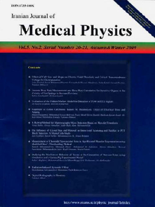
فهرست مطالب

Iranian Journal of Medical Physics
Volume:13 Issue: 1, Winter 2016
- تاریخ انتشار: 1395/02/06
- تعداد عناوین: 8
-
-
Pages 1-7IntroductionThe Gamma Knife system is designed solely for non-invasive treatment of brain disorders, and it benefits from stereotactic surgical techniques. Dose calculations required in the system are performed by GammaPlan code; in this code, brain tissue is considered uniform. In the present study, we evaluated the effect of Gamma Knife system on the obtained dose through simulating a real human brain phantom.Materials And MethodsIn this study, a Monte Carlo simulation code (MCNPX2.7) was employed to simulate Gamma Knife system. Brain tissue equivalent Snyder phantom and combinations were considered according to International Commission on Radiological Units (ICRU)-44 report.ResultsTo ensure accuracy of the simulations, patients head was modeled by a spherical water phantom. At this point, the dosimetry parameters were compared with those obtained by the Monte Carlo code EGS4 and good consistency was observed (less than 7% difference). At the next stage, the above dosimetry parameters were compared with those obtained experimentally by polystyrene phantom and EDR2 dosimetry film and improved consistency was detected (less than 0.5% difference). Finally, the Snyder phantom, as the human brain, was simulated. The Full Width at Half Maximum (FWHM) and penumbra decreased by 4.7% and 18%, respectively. Moreover, an isocenter dose reduction of 30-40%, compared to the water phantom, was noted.ConclusionThe calculation of the real phantom showed that water and polystyrene could function similarly, while evaluating dosimetry parameters in the Gamma Knife system; thus, water and polystyrene are not appropriate phantom matters for this purpose.Keywords: Film Dosimetry, Gamma Knife radiosurgery, Monte Carlo method, Snyder Phantom
-
Pages 8-16IntroductionThe negative health effects of electromagnetic radiation and psychological dependence are among the major consequences of widespread cell phone use in the general population, especially among adolescents. In this study, the relationship between cell phone use and sleep quality parameters was evaluated.Materials And MethodsThe study sample consisted of 820 students (305 males and 515 females), recruited from Arak University of Medical Sciences, Arak, Iran. The participants completed Pittsburgh Sleep Quality Index (PSQI) and Cell-phone Overuse Scale (COS); the validity of these questionnaires had been previously confirmed in the Iranian population. Information on demographic characteristics and variables associated with cell phone exposure, such as the frequency and duration of phone calls and number of messages was collected in a separate questionnaire.ResultsData analysis showed that cell phone overuse was significantly correlated with sleep quality and its components. Moreover, the results indicated that the global PSQI score and some sleep components were significantly correlated with several variables related to cell phone use. Based on the findings, the mean PSQI score was significantly different among heavy and light cell phone users (PConclusionAccording to the literature and the present study, services provided by modern cell phones, along with the extensive use of these devices, could be considered as potential threats to public health, especially adolescents sleep quality. In order to reach a definitive conclusion, further systematic and laboratory studies are required.Keywords: Cell Phone, Sleep quality, Medical students
-
Pages 17-24IntroductionBlue light is a part of the spectrum with the highest energy content, which can reach the retina. The damage that it can cause to the retina is called photochemical or blue-light retinal injury. For the retinal injury assessment of the photochemical and aphakic retinal hazards in the wavelength range of 300-700 nm, use of effective spectral radiance limits (W.m-2.sr-1) seems to be slightly perplexing for ophthalmologists. However, in this study, the temperature (OC) that can emit the same effective spectral radiance limit was detected using a computer code; this method could help prevent blue-light retinal injury.Materials And MethodsThe limits proposed by International Commission on Non-Ionizing Radiation Protection for blue-light induced photochemical and aphakic eye hazards were expressed in terms of temperature by a computer code for 13 Planckian sources that produce the same radiance. The calculated temperature by the computer code, here known as threshold temperature, is the maximum source temperature that for a specified viewing distance and source diameter does not cause the exposure at the receptor position to exceed the exposure limit.ResultsIn terms of threshold temperature, the exposure limits for aphakia or infant retinal injury are much lower than retinal photochemical damage. For light sources with more effective radiances, these differences reach 800 K.ConclusionThis method allows evaluation of photochemical and aphakic retinal hazard only by comparing the calculated threshold temperature by a computer code with the temperature of the radiant source, which may be beneficial for hygienist and ophthalmic clinicians.Keywords: Chest, Film, Image Quality, Pediatric, Radiation dose, X-ray
-
Pages 25-35IntroductionApplication of quality control (QC) programs at diagnostic radiology departments is of great significance for optimization of image quality and reduction of patient dose. The main objective of this study was to perform QC tests on stationary radiographic X-ray machines, installed in 14 hospitals of Kerman province, Iran.Materials And MethodsIn this cross-sectional study, QC tests were performed on 28 conventional radiographic X-ray units in Kerman governmental hospitals, based on the protocols and criteria recommended by the Atomic Energy Organization of Iran (AEOI), using a calibrated Gammex QC kit. Each section of the QC kit incorporated different models.ResultsBased on the findings, kVp accuracy, kVp reproducibility, timer accuracy, timer reproducibility, exposure reproducibility, mA/timer linearity, and half-value layer were not within the acceptable limits in 25%, 4%, 29%, 18%, 11%, 12%, and 7% of the evaluated units (n=28), respectively.ConclusionAs radiographic X-ray equipments in Kerman province are relatively old with a high workload, it is recommended that AEOI modify the current policies by changing the frequency of QC test implementation to at least once a year.Keywords: Diagnostic X-ray, Quality control, Radiology Device
-
Pages 36-42IntroductionIn order to discharge the patients receiving treatment with large radiation doses of 131I for thyroid cancer, it is necessary to measure and evaluate the external dose rates of these patients. The aim of the study was to assess a new method of external dose rate measurement, and to analyze the obtained results as a function of time.Materials And MethodsIn this study, a telescopic radiation survey meter was utilized to measure the external dose rates of a sample population of 192 patients receiving treatment with high-dose 131I at one, 24, and 48 hours after dose administration.ResultsThe proposed technique could reduce the occupational radiation exposure of the physicist by a factor of 1/16. Moreover, the external dose rates of both genders rapidly decreased with time according to bi-exponential equations, which could be attributed to the additional factors associated with iodine excretion, as well as the physiology of the body in terms of 131I uptake.ConclusionAccording to the results of this study, telescopic radiation survey meter could be used to measure the external dose rates of patients receiving treatment with 131I. Furthermore, the average difference in the radiation exposure between female and male patients was calculated to be less than 17%.Keywords: Radiation protection, Radiation dose, Iodine, Radiation Monitoring
-
Pages 43-48IntroductionBlue light is a part of the spectrum with the highest energy content, which can reach the retina. The damage that it can cause to the retina is called photochemical or blue-light retinal injury. For the retinal injury assessment of the photochemical and aphakic retinal hazards in the wavelength range of 300-700 nm, use of effective spectral radiance limits (W.m-2.sr-1) seems to be slightly perplexing for ophthalmologists. However, in this study, the temperature (OC) that can emit the same effective spectral radiance limit was detected using a computer code; this method could help prevent blue-light retinal injury.Materials And MethodsThe limits proposed by International Commission on Non-Ionizing Radiation Protection for blue-light induced photochemical and aphakic eye hazards were expressed in terms of temperature by a computer code for 13 Planckian sources that produce the same radiance. The calculated temperature by the computer code, here known as threshold temperature, is the maximum source temperature that for a specified viewing distance and source diameter does not cause the exposure at the receptor position to exceed the exposure limit.ResultsIn terms of threshold temperature, the exposure limits for aphakia or infant retinal injury are much lower than retinal photochemical damage. For light sources with more effective radiances, these differences reach 800 K.ConclusionThis method allows evaluation of photochemical and aphakic retinal hazard only by comparing the calculated threshold temperature by a computer code with the temperature of the radiant source, which may be beneficial for hygienist and ophthalmic clinicians.Keywords: Aphakic eye, Blue, light photochemical, Exposure limit, Effective spectral radiance, Threshold temperature
-
Pages 49-57IntroductionNatural and artificial radionuclides are the main sources of human radiation exposure. These radionuclides, which are present in the environment, can be dissolved into water. Evidence suggests that radionuclides being entered the human body through drinking or hot spring water can be harmful for human health.Materials And MethodsIn this study,10 samples were collected from ground water resources of Arak, one sample from the surface water of Kamal-Saleh Dam, and four samples from the hot springs of Mahallat region. The specific activities of 226Ra, 232Th, 40K, and 137Cs were determined in the samples, using gamma ray spectrometry and a high-purity germanium (HPGe) detector.ResultsSpecific activities of 226Ra, 232Th, 40K, and 137Cs were determined in the water samples. The mean 226Ra activity concentrations in drinking water samples from Aman Abad, Mobarak Abad, and Taramazd wells were 7.65±1.64, 1.56±1.04, and 1.45±1.39 Bq/l, while the corresponding values for 232Th were 2.70±0.18, 0.41±0.16, and 1.27±0.44 Bq/l, respectively. The annual effective dose due to drinking water varied from 0.01 to 0.78 mSv/y. Moreover, the specific activity of 226Ra in the water samples from the orifice of Donbe, Shafa, Soleymani, and Souda hot springs varied from 0.47±0.16 to 1.90±0.21.ConclusionThe calculated annual effective dose due to water consumption by Iranians was within the average annual global range. Therefore, based on the present results, radionuclide intake due to water consumption had no consequences for public health; however, it is recommended that hot spring baths use air conditioning devices.Keywords: Effective dose, Hot spring, Radionuclide, Water resource
-
Pages 58-64IntroductionThe continuous and rapid growth of telecommunication industries, along with the common application of cell phones, hasraised debates on the associated risks for humanhealth due to exposure to radiofrequency fields, caused bycell phonesorother sources, and industrial and medical applications. Accordingly, in this study, we aimed to investigate the effects of cell phone radiation on BALB/c mice blood factors.Materials And MethodsIn this study, 48 BALB/c mice were divided into six groups, each consisting of eight animals. Fourexposure groups were exposed to radiation waves twice a day for 30 min, 1 h, 2 h, and 4 h, respectivelyover one month. On the other hand,one exposure group wasexposed to 900 and 1800 MHz radiations (2 W)for 4 h once a day. Afterwards, the blood samples were taken from the heart, and blood factors including white blood cell count, red blood cell count, platelet count, hemoglobin level, hematocrit level, MCH, MCHC,neutrophil count, and lymphocyte count weremeasured and analyzed by SPSS version 15.ResultsBased on the findings, MCHC in the exposure groupreceiving 0.5 h of radiation and MCH in the exposure groupsreceiving 0.5, 2, and 4 h of radiationwere significantlydifferentfromthe control group (PConclusionAccording to the results of the present study, it can be concluded that microwaves have significant effects on the blood of mice. Therefore, it is suggested that further studies be conducted in this area to confirm the findings.Keywords: Electromagnetic Waves, BALB, C Mice, Cell Phone, Hematological Factors

