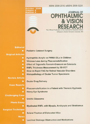
فهرست مطالب

Journal of Ophthalmic and Vision Research
Volume:4 Issue: 4, Oct-Dec 2009
- تاریخ انتشار: 1388/08/11
- تعداد عناوین: 14
-
-
Page 199
-
Page 201PurposeTo compare primary implantation of foldable hydrophilic acrylic with polymethylmethacrylate (PMMA) intraocular lenses (IOLs) in pediatric cataract surgery in terms of short-term complications and visual outcomes.MethodsThis randomized clinical trial included 40 eyes of 31 consecutive pediatric patients aged 1 to 6 years with unilateral or bilateral congenital cataracts undergoing cataract surgery with primary IOL implantation. Two types of IOLs including foldable hydrophilic acrylic and rigid PMMA were randomly implanted in the capsular bag during surgery. Primary posterior capsulotomy and anterior vitrectomy were performed in all eyes. Patients were followed for at least 1 year. Intra- and postoperative complications, visual outcomes and refractive errors were compared between the study groups.ResultsMean age was 3.2±1.8 years in the hydrophilic acrylic group and 3.7±1.3 years in the PMMA group. Mean follow-up period was 19.6±5 (12-29) months. No intraoperative complication occurred in any group. Postoperative uveitis was seen in 2 (10%) eyes in the acrylic group versus 5 (25%) eyes in the PMMA group (P=0.40). Other postoperative complications including pigment deposition (30%), iridocorneal adhesions (10%) and posterior synechiae formation (10%), were seen only in the PMMA group. The visual axis remained completely clear and visual outcomes were generally favorable and comparable in the study groups.ConclusionIn pediatric eyes undergoing lensectomy with primary posterior capsulotomy and anterior vitrectomy, hydrophilic acrylic IOLs are comparable to PMMA IOLs in terms of biocompatibility and visual axis clarity, and seem to entail less frequent postoperative complications.
-
Page 208PurposeTo determine the rate and risk factors of vitreous loss during phacoemulsification in patients with cataracts operated by ophthalmology residents and fellows at Labbafinejad Medical Center.MethodsThis prospective descriptive study included consecutive patients with cataracts undergoing phacoemulsification over a one year period. All patients were operated under local or general anesthesia using the divide and conquer technique. Preoperatively, all patients underwent a complete ocular examination including measurement of visual acuity, slitlamp biomicroscopy, intraocular pressure measurement, and dilated funduscopy. Main outcome measures included the rate of posterior capsular rupture and vitreous loss as well as associated risk factors such as surgical experience, ocular and systemic conditions, and type and severity of the cataract.ResultsOverall, 767 eyes of 767 patients with mean age of 63.7±10.3 (range, 25-91) years were operated. The overall rate of vitreous loss was 7.9% which was 5-fold greater in the hands of residents as compared to fellows. Among different factors, older age, female sex, small pupil, small capsulorrhexis, presence of pseudoexfoliation, and high myopia were significantly associated with vitreous loss. The highest rate of vitreous loss occurred in patients with dense nuclear cataracts.ConclusionConsidering the higher rate of vitreous loss in patients operated by ophthalmology residents; patients with known risk factors for vitreous loss should better be operated by more experienced surgeons.
-
Page 213PurposeTo evaluate the in vitro and in vivo anti-cataract potential of Trigonella foenum-graecum (TF) on galactose induced cataracts in an animal model.MethodsIn the in vitro group, enucleated rat lenses were maintained in organ culture containing Dulbecco''s Modified Eagles Medium alone (normal group), or with the addition of 30 mM galactose (control group). The medium in the test group was supplemented with both galactose and TF. All lenses were incubated at 37°C for 24 hours and then processed for determination of levels of reduced glutathione and malondialdehyde. In the in vivo group, cataracts were induced in rats by a 30% galactose diet alone (control) or with the addition of TF (treated group).ResultsReduction (26%) in glutathione level and elevation (31%) in malondialdehyde content were observed in controls as compared to normal lenses. TF significantly (P < 0.01) restored glutathione and reduced malondialdehyde levels as compared to controls. A significant delay in the onset and progression of cataract was observed with 2.5% TF diet; after 30 days none of the treated eyes developed mature cataracts as compared to 100% of control eyes.ConclusionsTF can delay the onset and progression of cataracts in an experimental rat model of galactose induced cataracts both in vitro and in vivo.
-
Page 220PurposeTo determine peripapillary retinal nerve fiber layer (RNFL) thickness values by three-dimensional optical coherence tomography (3D-OCT) in a normal Iranian population and to evaluate the concordance of these measurements with those obtained by the second generation of optical coherence tomography (OCT II).MethodsIn a cross-sectional observational study, 96 normal Iranian subjects 20-53 years old were enrolled. Peripapillary RNFL thickness in one randomly selected eye of each subject was measured by 3D-OCT and also by OCT II. Standard achromatic perimetry, corneal pachymetry and A-scan ultrasonographic biometry were also performed. Other study variables included age, gender, laterality (right versus left eye), refractive error, corneal diameter and disc area.ResultsMean peripapillary RNFL thickness measured by 3D-OCT (75.50±8.38) µm was significantly less than that measured by OCT II (144.10±33.32 µm) (P < 0.001). Using 3D-OCT, no significant difference in peripapillary RNFL thickness was observed by gender (P=0.90) or laterality (P=0.17); RNFL thickness had no correlation with age (P=0.95), axial length (P=0.32), spherical equivalent refractive error (P=0.21), central corneal thickness (P=0.66) and disc area (P=0.31). However, a positive correlation was found between peripapillary RNFL thickness and corneal diameter (P=0.03).Conclusion3D-OCT seems to yield lower RNFL thickness values as compared to OCT II. It seems advisable to obtain separate baseline measurements when using different generations of OCT machines.
-
Page 228PurposeTo compare the incidence of adverse reactions following rapid versus slow fluorescein injection for fundus angiography.MethodsThis randomized controlled trial was performed on 500 patients with retinal vascular disorders. Subjects with central serous retinopathy, age-related macular degeneration and retinal pigment epithelial changes were excluded. Pregnancy, asthma, allergic diseases and previous history of reactions to fluorescein were other exclusion criteria. Patients were randomly divided into two equal groups who received slow infusion of dye (over 15-25 seconds) versus the usual rapid injection (in 5-8 seconds), and were compared for adverse effects.ResultsOverall, 47 (9.4%) patients including 34 (13.6%) subjects in the rapid group and 13 (5.2%) cases in the slow group developed adverse reactions (P=0.001, relative risk=2.6). All adverse reactions were categorized as mild; no instance of moderate or severe reactions was observed. There was a lower incidence of nausea and vomiting with slow infusion of fluorescein (P=0.02), however no statistically significant difference was observed in the frequency of vertigo and vasovagal reactions between the study groups.ConclusionSlow fluorescein injection during fundus angiography, instead of the usual rapid application, can be an effective way to reduce the incidence of nausea and vomiting in patients whose first phase of angiography is of little diagnostic importance.
-
Page 232PurposeTo determine the histopathological diagnosis of ocular tumor specimens and to assess their correlation with preoperative clinical diagnosis.MethodsSurgical records of all patients who had undergone ocular surgery yielding a tissue specimen at the ophthalmology department of the University of Benin Teaching Hospital, Benin City, Nigeria, from March 1999 to February 2007 were extracted. Parameters included age, sex, preoperative clinical diagnosis, type of surgery, and histopathological diagnosis.ResultsOverall, 148 patients including 88 male (59.5%) and 60 female (40.5%) subjects were operated during the study period. The most prevalent histopathological diagnoses included squamous cell carcinoma (SCC) of the conjunctiva and eyelids (16.9%), pterygium (12.2%) and retinoblastoma (10.8%). Excisional conjunctival biopsies were performed in 30 cases to rule out SCC which was confirmed in 16 cases (53.3%). Enucleation was performed in 19 children with suspicion of intraocular malignancy of whom 16 had retinoblastoma and one had teratoid medulloblastoma; yielding a correct clinical diagnosis in 89.5% of cases. Of 24 cases of enucleation in adults, the preoperative diagnosis was confirmed by histology in 21 cases (87.5%). The preoperative diagnosis was confirmed histologically in 8 cases (53.3%) of 15 orbital specimens and 11 cases (50%) of 22 eyelid samples.ConclusionThe most common ophthalmic malignancies were SCC of the conjunctiva and eyelids, and retinoblastoma. Clinicopathological correlation was lowest in eyelid lesions and highest in enucleation specimens.
-
Page 238Normal vision depends on the optimal function of ocular barriers and intact membranes that selectively regulate the environment of ocular tissues. Novel pharmacotherapeutic modalities have aimed to overcome such biological barriers which impede efficient ocular drug delivery. To determine the impact of ocular barriers on research related to ophthalmic drug delivery and targeting, herein we provide a review of the literature on isolated primary or immortalized cell culture models which can be used for evaluation of ocular barriers. In vitro cell cultures are valuable tools which serve investigations on ocular barriers such as corneal and conjunctival epithelium, retinal pigment epithelium and retinal capillary endothelium, and can provide platforms for further investigations. Ocular barrier-based cell culture systems can be simply set up and used for drug delivery and targeting purposes as well as for pathological and toxicological research.
-
Page 253PurposeTo introduce a simple way for achieving the routine position for phacoemulsification in a patient with a marked thoracic kyphosis. CASE REPORT: A 74-year-old man with marked thoracic kyphosis and visually significant cataracts presented for surgery; he was unable to lie flat due to the severe deformity. The best possible surgical position was achieved by placing a chair with an adjustable top between a standard operating table and another small table. The wheels of the table and the chair were securely immobilized by adhesive tape. The space between the operating table and the small table was filled with rolled towels and covered with a blanket. The patient lay down with his head placed on the small table while the kyphotic portion of his thorax fitted into the free space between the small table and the operating table. The variable top of the chair allowed adjusting the space in order to accommodate his kyphotic thorax. Successful temporal approach phacoemulsification was performed comfortably while the patient lay in the standard position required for cataract surgery.ConclusionIt is possible to position patients with thoracic problems on a standard operating table using readily available equipment in the operating theater.
-
Page 256PurposeTo report the clinical features and surgical outcomes of two patients with heavy eye syndrome who underwent partial Jensen''s procedure. CASE REPORT: A 21-year-old man and a 24-year-old woman with high myopia (-18 and -8 diopters, respectively), high axial length (27.5 and 24.6 mm), progressive esotropia (40 and 50 prism diopters), hypotropia (5 and 2 prism diopters), abduction limitation, and inferior displacement of the lateral rectus on computed tomography were diagnosed with heavy eye syndrome and underwent partial Jensen''s procedure. The technique consisted of splitting the lateral and superior recti from their insertion up to the equator and uniting their superior and temporal halves respectively, with non-absorbable sutures without scleral fixation. Two months postoperatively, esotropia was reduced to 10 prism diopters in case #1 and to 25 prism diopters in case #2; limitation of abduction was also considerably diminished.ConclusionPatients with heavy eye syndrome, large angle esotropia and limitation of abduction, may benefit from partial Jensen''s procedure which is a simple and safe surgical option.
-
Page 260
-
Page 266Several techniques have been employed for repositioning dislocated intraocular lenses (IOLs). Herein, we describe a simplified and modified technique in which scleral fixation is performed together with temporary externalization of IOL haptics through a small, superior clear corneal incision. The sutures are tied to the externalized haptics; the haptics are then repositioned into the anterior chamber followed by IOL reimplantation into the ciliary sulcus. Using this technique, the dislocated IOL is repositioned under direct visualization without need for IOL extraction or extensive intraocular manipulations.

