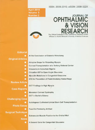
فهرست مطالب

Journal of Ophthalmic and Vision Research
Volume:5 Issue: 2, Apr-Jun 2010
- تاریخ انتشار: 1389/02/11
- تعداد عناوین: 14
-
-
Pages 75-81To investigate whether seasonal modification in the concentration of atropine drops is effective in retarding the progression of myopia.MethodsTwo hundred and forty eyes of 120 healthy preschool- and school-age children in Chiayi region, Taiwan were recruited. The treatment group consisted of 126 eyes of 63 children who received atropine eye drops daily for one year and the control group included 114 eyes of 57 children who received nothing. The concentration of atropine eye drops was modified by seasonal variation as follows: 0.1% for summer, 0.25% for spring and fall, and 0.5% for winter. Refractive error, visual acuity, intraocular pressure (IOP), and axial length were evaluated before and after intervention.ResultsMean age was 9.1±2.8 years in the atropine group versus 9.3±2.8 years in controls (P=0.88). Mean spherical equivalent, refractive error and astigmatism were -1.90±1.66 diopters (D) and -0.50±0.59 D in the atropine group; corresponding values in the control group were -2.09±1.67 D (P=0.97) and -0.55±0.60 D (P=0.85), respectively. After one year, mean progression of myopia was 0.28±0.75 D in the atropine group vs 1.23±0.44 D in controls (P < 0.001). Myopic progression was significantly correlated with an increase in axial length in both atropine (r=0.297, P=0.001) and control (r=0.348, P < 0.001) groups. No correlation was observed between myopic progression and IOP in either study group.ConclusionModifying the concentration of atropine drops based on seasonal variation, seems to be effective and tolerable for retarding myopic progression in preschool- to school-age children.
-
Pages 82-86To report the indications and techniques of corneal transplantation at a tertiary referral center in Tehran over a 3-year period.MethodsRecords of patients who had undergone any kind of corneal transplantation at Labbafinejad Medical Center, Tehran, Iran from March 2004 to March 2007 were reviewed to determine the indications and types of corneal transplantation.ResultsDuring this period, 776 eyes of 756 patients (including 504 male subjects) with mean age of 41.3±21.3 years underwent corneal transplantation. The most common indication was keratoconus (n=317, 40.8%) followed by bullous keratopathy (n=90, 11.6%), non-herpetic corneal scars (n=62, 8.0%), infectious corneal ulcers (n=61, 7.9%) previously failed grafts (n=61, 7.9%), endothelial and stromal corneal dystrophies (n=28, 3.6%), and trachoma keratopathy (n=26, 3.3%). Other indications including Terrien''s marginal degeneration, post-LASIK keratectasia, trauma, chemical burns, and peripheral ulcerative keratitis constituted the rest of cases. Techniques of corneal transplantation included penetrating keratoplasty (n=607, 78.2%), deep anterior lamellar keratoplasty (n=108, 13.9%), conventional lamellar keratoplasty (n=44, 5.7%), automated lamellar therapeutic keratoplasty (n=8, 1.0%), and Descemet stripping endothelial keratoplasty (n=6, 0.8%) in descending order. The remaining cases were endothelial keratoplasty and sclerokeratoplasty.ConclusionIn this study, keratoconus was the most common indication for penetrating keratoplasty which was the most prevalent technique of corneal transplantation. However, deep anterior lamellar keratoplasty is emerging as a growing alternative for corneal pathologies not involving the endothelium.
-
Pages 87-91To determine the incidence and contribution of different types of glaucoma to blindness at Irrua Specialist Teaching Hospital, a suburban tertiary care hospital in Edo State, Nigeria.MethodsMedical records of all new patients with glaucoma who presented to the eye clinic of the hospital from June 2007 to May 2009 were reviewed.ResultsOut of a total of 2,742 new patients seen over the study period, 177 (6.5%) subjects had glaucoma which included primary open angle glaucoma (130 cases, 73.4%), juvenile glaucoma (31 patients, 17.5%), secondary glaucoma (10 subjects, 5.6%), congenital glaucoma (3 cases, 1.7%) and primary angle closure glaucoma (3 persons, 1.7%). Of patients with primary open angle glaucoma, 23 (17.7%) were blind based on visual acuity criteria and 67 (51.5%) were blind based on visual field criteria.ConclusionGlaucoma remains a blinding scourge; late presentation, especially in rural areas, is an important factor predisposing to blindness. In this Nigerian population primary open angle glaucoma was the most prevalent subtype of the disease but primary angle closure glaucoma was rare.
-
Pages 92-100To evaluate circadian intraocular pressure (IOP) profiles in eyes with different types of chronic open-angle glaucoma (COAG) and normal eyes.MethodsThis study included 3,561 circadian IOP profiles obtained from 1,408 eyes of 720 Caucasian individuals including glaucoma patients under topical treatment (1,072 eyes) and normal subjects (336 eyes). IOP profiles were obtained by Goldmann applanation tonometry and included measurements at 7 am, noon, 5 pm, 9 pm, and midnight.ResultsFluctuations of circadian IOP in the secondary open-angle glaucoma (SOAG) group (6.96±3.69 mmHg) was significantly (P < 0.001) higher than that of the normal pressure glaucoma group (4.89±1.99 mmHg) and normal eyes (4.69±1.95 mmHg); but the difference between the two latter groups was not significant (P=0.47). Expressed as percentages, IOP fluctuations did not vary significantly among any of the study groups. Inter-ocular IOP difference for any measurement was significantly (P < 0.001) smaller than the profile fluctuations. In all study groups except the SOAG group, IOP was highest at 7 am, followed by noon, 5 pm, and finally 9 pm or midnight. In the SOAG group, mean IOP measurements did not vary significantly during day and night.ConclusionsIn contrast to normal eyes and eyes with primary open-angle glaucoma under topical antiglaucoma treatment, eyes with SOAG under topical treatment do not show the usual circadian IOP profile in which the highest IOP values occur in the morning, and the lowest in the evening or at midnight. These findings may have implications for timing of tonometry. Fluctuation of circadian IOP was highest in
-
Pages 101-104To assess the frequency of mutations in the Myocilin (MYOC) gene in Iranian patients affected with primary congenital glaucoma (PCG).MethodsThe individuals evaluated herein are among a larger cohort of 100 patients who had previously been screened for CYP1B1 mutations. Eighty subjects carried mutations in CYP1B1, but the remaining 20 patients who did not, underwent screening for MYOC mutations for the purpose of the study. MYOC exons in the DNA were polymerase chain reaction (PCR) amplified and sequenced. Sequencing was performed using PCR primers, the ABI big dye chemistry and an ABI3730XL instrument. Sequences were analyzed by comparing them to reference MYOC sequences using the Sequencher software.ResultsFour MYOC sequence variations were observed among the patients, but none of them were considered to be associated with disease status. Three of these variations were single nucleotide polymorphisms already reported not to be disease causing, the fourth variation created a synonymous codon and did not affect any amino acid change.ConclusionIn this cohort, MYOC mutations were not observed in any Iranian subject with PCG. It is possible that in a larger sample, a few subjects carrying disease causing MYOC mutations could have been observed. But our results show that the contribution of MYOC to PCG status in Iran is small if any.
-
Pages 105-109To compare the hemostatic effect of sulfur hexafluoride 20% (SF6 20%) with lactated Ringer''s solution for prevention of early postoperative vitreous hemorrhage following diabetic vitrectomy.MethodsIn a prospective randomized clinical trial, 50 eyes undergoing diabetic vitrectomy were divided into two groups. At the conclusion of surgery, in one group the vitreous cavity was filled with SF6 20% while in the other group lactated Ringer''s solution was retained in the vitreous cavity. The two groups were compared for the rate of early postoperative vitreous hemorrhage.ResultsThe incidence of vitreous hemorrhage was lower in the SF6 group than the Ringer''s group 4 days (20% vs 68%, P=0.001), 7 days (24% vs 60%, P=0.01) and 4 weeks (16% vs 40%, P=0.059) after vitrectomy.ConclusionIn comparison with lactated Ringer''s solution, SF6 20% had a significant hemostatic effect especially in the early postoperative period after diabetic vitrectomy and reduced the incidence of vitreous hemorrhage.
-
Pages 110-121Optical coherence tomography (OCT) has enhanced our understanding of changes in different ocular layers when axial myopia progresses and the globe is stretched. These findings consist of dehiscence of retinal layers known as retinoschisis, paravascular inner retinal cleavage, cysts and lamellar holes, peripapillary intrachoroidal cavitation, tractional internal limiting membrane detachment, macular holes (lamellar and full thickness), posterior retinal detachment, and choroidal neovascular membranes. In this review, recent observations regarding retinal changes in highly myopic eyes explored by OCT are described to highlight structural findings that cannot be diagnosed by simple ophthalmoscopy.
-
Pages 122-126To report the microstructural features of Meesmann corneal dystrophy (MCD) in two patients. CASE REPORT: The first patient was a 10-year-old boy who presented with bilateral visual loss, diffuse corneal epithelial microcystic changes, high myopia and amblyopia. With a clinical impression of MCD, automated lamellar therapeutic keratoplasty was performed in his left eye. Histopathologic examination of the corneal button disclosed epithelial cell swelling and cyst-like intracytoplasmic inclusions. The cells contained moderate amounts of periodic acid-Schiff-positive and diastase-sensitive material (glycogen). Transmission electron microscopy revealed numerous vacuoles and moderate numbers of electron-dense membrane-bound bodies in the cytoplasm, similar to lysosomes, some engulfed by the vacuoles. The second patient was a 17-year-old female with a clinical diagnosis of MCD and episodes of recurrent corneal erosion. On confocal scan examination of both corneas, hyporeflective round-shaped areas measuring 6.8 to 41.4 µm were seen within the superficial epithelium together with irregular and ill-defined high-contrast areas in the sub-basal epithelial region. The subepithelial nervous plexus was not visible due to regional hyperreflectivity.ConclusionThis case report further adds to the microstructural features of Meesmann corneal dystrophy and suggests confocal scan as a non-invasive method for delineating the microstructural appearance of this rare dystrophy.
-
Pages 127-129To describe optical coherence tomography (OCT) findings in a patient with Berlin''s edema following blunt ocular trauma. CASE REPORT: A 26-year-old man presented with acute loss of vision in his left eye following blunt trauma. He underwent a complete ophthalmologic examination and OCT. Fundus examination revealed abnormal yellow discoloration in the macula. OCT disclosed thickening of outer retinal structures and increased reflectivity in the area of photoreceptor outer segments with preservation of inner retinal architecture. Re-examination was conducted one month later at the time which OCT changes resolved leading to a surprisingly normal appearance.ConclusionOCT can be a useful tool in the diagnosis and follow-up of eyes with Berlin''s edema and may reveal ultrastructural macular changes.
-
Pages 136-137
-
Pages 138-141The surgical results of severe or complex deviations such as those due to complete third nerve palsy, aberrant innervation of extraocular muscles (EOMs) and Duane syndrome are usually not completely successful. Herein, we describe the surgical technique of EOM fixation to the orbital wall. After a limbal or fornix based conjunctival incision, the related EOM is identified and dissected; the muscle insertion is sutured with non-absorbable sutures and detached from the sclera. The adjacent periosteum is exposed approximately 5 mm posterior to the orbital rim. The sutured muscle is then fixed to the orbital wall with two periosteal bites. The cut edges of the intermuscular membrane are closed over the sclera to avoid adherence of the muscle to the sclera. Finally the conjunctiva is reapproximated or recessed if necessary. This method of EOM inactivation completely eliminates all muscle forces from the globe and can provide better alignment in the above mentioned types of strabismus. The procedure is reversible and can be converted to other types of weakening operations if necessary

