فهرست مطالب

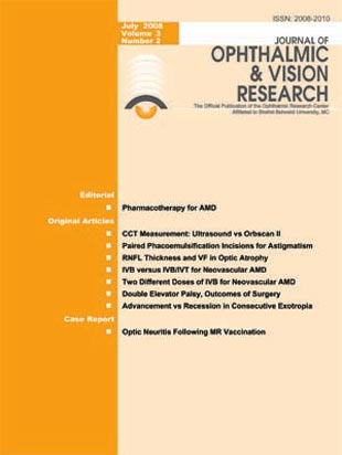
Journal of Ophthalmic and Vision Research
Volume:3 Issue: 2, Automn and Winter 2008
- تاریخ انتشار: 1386/10/11
- تعداد عناوین: 9
-
Page 83PurposeTo compare Orbscan II and ultrasonic pachymetry for measurement of central corneal thickness (CCT) in eyes scheduled for keratorefractive surgery.MethodsCCT was measured using Orbscan II (Bausch & Lomb, USA) and then by ultrasonic pachymetry (Tomey SP-3000, Tomey Ltd, Japan) in 100 eyes of 100 patients with no history of ocular surgery scheduled for excimer laser refractive surgery.ResultsMean CCT was 544.7±35.5 (range 453-637) µm by ultrasonic pachymetry versus 546.9±41.6 (range 435-648) µm measured by Orbscan II applying an acoustic factor of 0.92 (P=0.14). The standard deviation of measurements was greater with Orbscan pachymetry but the difference was not statistically significant.ConclusionCCT measurements by Orbscan II (applying an acoustic factor) and by ultrasonic pachymetry are not significantly different; however, when CCT readings by Orbscan II are in the lower range, it is advisable to recheck the measurements using ultrasonic pachymetry.
-
Page 87PurposeTo compare the efficacy of adding an opposite clear corneal incision (OCCI) on the steep meridian versus performing surgery on the steep meridian alone during phacoemulsification in reducing pre-existing corneal astigmatism.MethodsThis randomized clinical trial was performed on 120 eyes with corneal astigmatism of > 1D undergoing phacoemulsification. Incisions were made based on the type of astigmatism as follows: superior or superior + OCCI for with-the-rule and temporal or temporal + OCCI for against-the-rule astigmatism. Patients were followed with refraction, keratometry and topography. Statistical analysis was performed using one- and two-way ANOVA and Tukey-a test.ResultsMean corneal astigmatism was 1.82±0.86 D in the superior + OCCI group and 1.74±0.86 D in the temporal + OCCI group preoperatively which decreased to 1.31±0.59 (P=0.013) and 1.19±0.64 (P=0.009) postoperatively respectively. No significant change occurred in the amount of astigmatism in any of the two single incision groups.ConclusionPaired OCCI on the steep axis is a useful technique to correct mild to moderate pre-existing astigmatism with no need for particular skill or additional instruments.
-
Page 91PurposeTo investigate the correlation between retinal nerve fiber layer (RNFL) thickness determined by optical coherence tomography (OCT) and visual field (VF) parameters in patients with optic atrophy.MethodsThis study was performed on 35 eyes of 28 patients with optic atrophy. RNFL thickness was measured by OCT (Carl Zeiss, Jena, Germany) and automated perimetry was performed using the Humphrey Field Analyzer (Carl Zeiss, Jena, Germany). The correlation between RNFL thickness and VF parameters was evaluated.ResultsMean global RNFL thickness was 44.9±27.5 µm which was significantly correlated with mean deviation score on automated perimetry (r=0.493, P=0.003); however, no significant correlation was observed between visual field pattern standard deviation and the corresponding quadrantic RNFL thickness. In a similar manner, no significant association was found between visual acuity and RNLF thickness.ConclusionMean global RNFL thickness as determined by OCT seems to be correlated with VF defect depth as represented by the mean deviation score on Humphrey VF testing. OCT may be used as an objective diagnostic tool in the evaluation of patients with optic atrophy.
-
Page 95PurposeTo compare the short-term outcomes of intravitreal bevacizumab (IVB) with the combination of IVB and intravitreal triamcinolone acetonide (IVB/IVT) for treatment of neovascular age-related macular degeneration (AMD).MethodsThis randomized clinical trial was performed on 92 eyes of 90 patients with subfoveal and juxtafoveal choroidal neovascularization (CNV) secondary to AMD. The eyes were randomly assigned to receive IVB 1.25 mg alone (53 eyes) or in combination with IVT 2 mg (39 eyes). Best-corrected visual acuity (BCVA) and fundus autofluorescence were assessed, and fluorescein angiography (FA) and optical coherence tomography (OCT) were performed at baseline and repeated 6 weeks after treatment.ResultsMean age was 70.6±8.7 (range 50-89) years and 57.7% of the patients were male. BCVA improved from 1.03±0.40 to 0.93±0.38 logMAR (P=0.001) in the IVB group and from 1.08±0.33 to 0.91±0.38 logMAR (P=0.008) in the IVB/IVT group. There was a trend toward greater visual improvement with combined therapy (P=0.06). BCVA improvement was greater in eyes with +1 versus those with +2 (P=0.049) and +3 (P < 0.001) fundus autofluorescence at baseline. Mean decrease in central macular thickness was 113±115 µm (P < 0.001) in the IVB group versus 53.96±125 µm (P=0.008) in the IVB/IVT group with no intergroup difference (P=0.38). FA showed decreased leakage in 57.4%, increased leakage in 12.8% and no change in 29.8% of patients in the IVB group. Corresponding figures were 60.0%, 5.7% and 34.3% in the IVB/IVT group (P=0.556).ConclusionAddition of triamcinolone acetonide to bevacizumab for treatment of neovascular AMD does not seem to significantly increase its short-term efficacy. More severe fundus autofluorescence appears to be predictive of poorer response to treatment.
-
Page 102PurposeTo compare the efficacy and safety of 1.25 mg versus 2.5 mg intravitreal bevacizumab (IVB) for treatment of choroidal neovascularization (CNV) associated with age-related macular degeneration (AMD).MethodsIn this randomized clinical trial, consecutive patients with active CNV associated with AMD received 1.25 mg or 2.5 mg IVB. Best corrected visual acuity (BCVA), foveal thickness and side effects of therapy were evaluated one and three months after intervention.ResultsOverall 86 subjects were enrolled and completed the scheduled follow-up. Forty seven and 39 patients received 1.25 and 2.5 mg IVB respectively. The study groups were balanced in terms of baseline characteristics such as age, BCVA and foveal thickness. Mean improvement in BCVA was 0.06±0.3 logMAR in the 1.25 mg group and 0.07±0.34 logMAR in the 2.5 mg group (P=0.9). Mean decrease in foveal thickness was 49±36 µm in the 1.25 mg group and 65±31µm in the 2.5 mg group (P=0.6). Three cases of vitreous reaction and one case of massive subretinal hemorrhage were observed in the 2.5 mg group.ConclusionDouble dose (2.5 mg) IVB does not seem to be more effective than regular dose (1.25 mg) injections for treatment of CNV due to AMD and may lead to more complications.
-
Page 108PurposeTo describe the clinical manifestations of subtypes of double elevator palsy and to report the outcomes of surgery in these patients.MethodsThis retrospective study was conducted on hospital records of patients with double elevator palsy at Labbafinejad Medical Center over a ten-year period from 1994 to 2004. Patients were classified into three subgroups of primary elevator muscle palsy (9 subjects), primary supranuclear palsy with secondary inferior rectus restriction (4 subjects) and pure inferior rectus restriction (7 subjects) according to forced duction test (FDT), force generation test (FGT) and Bell''s reflex. Patients in the first group underwent Knapp procedure, the second group received Knapp procedure and inferior rectus recession simultaneously and in the third group vertical recess-resect or mere inferior rectus recess operation was performed. Success was defined as final residual deviation of 5 PD or less and 25% improvement or more in restriction after all operations.ResultsOverall 20 subjects including 10 male and 10 female patients with mean age of 12.6±9.3 (range 1.5-32) years were operated during the mentioned period which included 9 cases of primary elevator muscle palsy, 4 patients with primary supranuclear palsy and secondary inferior rectus restriction, and 7 subjects with pure inferior rectus restriction. Mean follow-up was 22.0±20.0 (range 3-63.5) months. Mean pre and post-operative deviation was 32.0±8.0 PD and 3.8±8.0 PD (P < 0.001) respectively, and mean restriction before and after the operation(s) was -3.5±0.7 and -2.3±1.2 (P < 0.001), respectively. Success rate was 77% for correction of deviation and 80% for improvement in muscle restriction.ConclusionSurgery for double elevator palsy must be individualized according to FDT, FGT and Bell''s reflex. The outcomes are favorable with appropriate surgical planning.
-
Page 114PurposeTo compare bilateral medial rectus advancement (BMRA) and bilateral lateral rectus recession (BLRR) for the treatment of consecutive exotropia.MethodThis randomized clinical trial was performed on 14 patients with consecutive exotropia. Inclusion criteria were history of bilateral medial rectus recession, exotropia of 20 PD or more with far-near discrepancy < 10 PD. Exclusion criteria consisted of more than once medial rectus recession, restricted adduction, history of operation on the lateral rectus, positive forced duction test of the lateral rectus, concomitant neurologic disorders and follow-up less than 6 months'' duration.ResultsSeven patients underwent BMRA and 7 patients underwent BLRR. Mean age was 11.4±6.9 (range 5 to 21) years in the BMRA group and 13.7±7.1 (range 5-22) years in the BLRR group (P=0.44). Two patients in the BMRA group and 3 subjects in the BLRR group were amblyopic. Mean preoperative exotropia was 27.8±6.3 PD and 39.2±14.8 PD (P=0.09) which was reduced to 4.2±2.3 PD and 3.4±2.2 PD (P=0.94) in the BMRA and BLRR groups respectively. Successful alignment was achieved in 71.4% and 85.7% of cases in the BMRA and BLRR groups respectively (P=0.94). All amblyopic patients achieved successful alignment postoperatively.ConclusionBilateral medial rectus advancement and bilateral lateral rectus recession are comparable in efficacy for treatment of consecutive exotropia.
-
Page 118PurposeTo report two cases of optic neuritis with onset less than 24 hours following measles-rubella (MR) vaccination. CASE REPORT: Two teenage patients developed acute optic neuritis 6 to 7 hours after MR booster vaccination. The first patient demonstrated bilateral papillitis and severe visual loss but improved significantly with pulse intravenous steroid therapy with methylprednisolone 500 mg/day. The second patient had unilateral retrobulbar optic neuritis and demonstrated excellent visual recovery without intervention.ConclusionAcute optic neuritis is a rare complication of MR vaccination and may occur early after immunization.

