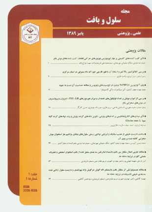
فهرست مطالب

مجله سلول و بافت
سال یکم شماره 1 (پاییز 1389)
- 88 صفحه،
- تاریخ انتشار: 1390/03/20
- تعداد عناوین: 8
-
- مقالات پژوهشی
-
صفحه 1هدفهدف از این تحقیق بررسی نقش کلیت کننده های کلسیم بر مهار اپوپتوزیس نورون های حرکتی قطعات کشت شده نخاع موش بالغ بود.مواد و روش هاناحیه سینه ای نخاع موش بالغ توسط دستگاه قطعه کننده بافت به قطعات 400 میکرونی بریده و به چهار گروه تقسیم شدند: 1- قطعات لحظه زمانی صفر 2- قطعات کنترل 3- قطعات تیمار با اتیلن دی آمین تترا استیک اسید (EDTA) 4- قطعات تیمار با اتیلن گلیکول تترا استیک اسید (EGTA). قطعات کنترل و تیمار به مدت 6 ساعت در محیط کشت انکوبه شدند. سنجش MTT [3-(4،5-dimethylthiazol-2-yl)-2،5-diphenyltetrazolium bromide] جهت ارزیابی قابلیت حیات قطعات نخاع مورد استفاده قرار گرفت. جهت مطالعه مورفولوژیکی نورون های حرکتی از رنگ آمیزی پروپیدیوم آیوداید و هوخست 33342 استفاده شد. داده ها با روش آنالیز واریانس یکطرفه و تست Tukey مورد تجزیه و تحلیل آماری قرار گرفت و تفاوت میانگین ها در سطح p<0.05 معنی دار لحاظ شد.نتایجنورون های حرکتی قطعات کشت شده نخاع پس از 6 ساعت نشانه های مورفولوژیک اپوپتوزیس را نشان دادند. کاربرد جداگانه کلیت کننده های کلسیمی (EDTA و EGTA) نه تنها توانست قابلیت حیات قطعات کشت شده به مدت 6 ساعت را افزایش دهد بلکه توانست مرگ سلولی اپوپتوزیس را در نورون های حرکتی این قطعات مهار و همچنین درصد تعداد نورون های حرکتی زنده را افزایش دهد.نتیجه گیریاز آنجا که کاربرد کلیت کننده های کلسیمی درمحیط کشت قطعات نخاع توانست قابلیت حیات این قطعات را افزایش، نشانه های اپوپتوزیس را در نورون های حرکتی مهار و درصد تعداد نورون های حرکتی زنده را در این قطعات افزایش دهند، بیانگر آن است که افزایش کلسیم داخل سلولی احتمالا یکی از دلایل اپوپتوزیس نورون های حرکتی در قطعات کشت شده نخاع می باشد.
کلیدواژگان: اپوپتوزیس، نخاع، نورون حرکتی، EDTA و EGTA -
صفحه 9هدفمصرف خوراک دام آلوده به Aspergilus سبب تولید آفلاتوکسین و ایجاد اختلال در چرخه سلامت دام، شیر و افراد مصرف کننده می گردد. در این پژوهش بررسی آفلاتوکسین M1 شیر و ارتباط آن با فلور قارچی خوراک دام مصرفی در استان مرکزی انجام گردید.مواد و روش هاخوراک دام سالانه مصرفی ده دامداری استان مرکزی در 1388 و 1389 بررسی گردید. جداسازی، کشت و تشخیص قارچ های موجود در آنها انجام و آفلاتوکسین موجود درشیر تولیدی به روش الایزا اندازه گیری شد و ارتباط بین ترکیب خوراک دام، کپک و آفلاتوکسین M1 در شیر دام سنجش گردید.نتایجنتایج نشان داد بیشترین مواد تشکیل دهنده خوراک دام شامل ذرت، کنجاله پنبه دانه و کلزا، مکمل های غذایی، جو، سبوس گندم، نان خشک، پودر چربی و یونجه می باشند. بیشترین عامل آلودگی وجود کپک های Aspergilus clavatus، A. flavus وRhizopus stolonifer بودند. نتایج مطالعه سالانه آفلاتوکسین در شیر دامداری ها وجود آفلاتوکسین M1 را در همه آنها نشان داد. آنالیز آماری داده های سالیانه خوراک دام و آفلاتوکسین وجود ضریب همبستگی قوی بین کنجاله کلزا و سویا با کپک های آسپرژیلوس در خوراک دام و آلودگی شیر به آفلاتوکسین را تایید نمود.نتیجه گیریپس کنترل آلودگی خوراک دام به کفک ها، بهترین روش برای جلوگیری از آلودگی شیر و فرآورده های آن به آفلاتوکسین هاست که به بهبود سلامت جامعه کمک می کند.
کلیدواژگان: آسپرژیلوس، آفلاتوکسین M1، خوراک دام، شیر -
صفحه 19هدفحرکت آب از عرض غشاءهای سلولی در اثر حضور کانال های آبی به نام آکواپورین ها (AQPs) تسهیل می شود. ترکیبات جیوه یکی از بازدارنده های قوی آکواپورین های گیاهی و جانوری می باشند. به هر حال چندین فرم مشابه از آکواپورین ها مانند NtAQP1 در گیاه توتون توسط جیوه ممانعت نمی شوند. هدف از این تحقیق بررسی حساسیت آکواپورین NtPIP2;1 جداسازی شده از توتون نسبت به جیوه است.مواد و روش هادر این مطالعه تجربی پس از سنتز cRNA مربوط به NtPIP2;1، تزریق آن به داخل اووسیت های زنوپوس توسط میکرواینجکشن صورت گرفت. پس از دو روز انکوبه کردن اووسیت ها، آنها به مدت 10 دقیقه در معرض 1 میلی مولار کلرور جیوه قرار گرفته، سپس آزمون انبساط حجم اووسیت در محیط هیپوتونیک انجام و ضریب نفوذپذیری غشاء (Pf) اووسیت تعیین شد.نتایجPf محاسبه شده برای اووسیت های گروه کنترل و تیمار شده با کلرور جیوه که هر دو آکواپورین NtPIP2;1 را بیان می کنند به ترتیب برابر با 2-10×99/0 و2-10×98/0 سانتیمتر بر ثانیه بود که تفاوت معنی داری با یکدیگر نشان ندادند(p>0.05). مقایسه توالی اسید آمینه آکواپورین NtPIP2;1 با NtAQP1 (غیرحساس به جیوه) نشان داد که NtPIP2;1 همانند NtAQP1 در نزدیک منفذ آبی دارای اسید آمینه ترئونین به جای سیستئین می باشد.نتیجه گیریNtPIP2;1 یک آکواپورین غیر حساس به جیوه را در گیاه توتون کدگذاری می کند و این به دلیل جایگزینی اسیدآمینه سیستئین در نزدیک منفذ آبی با ترئونین می باشد.
کلیدواژگان: آکواپورین، توتون، جیوه، اووسیت، زنوپوس -
صفحه 27هدفاثر امواج تلفن های همراه بر بافت تخمدان موش های صحرایی بررسی شد.مواد و روش ها28 سر موش صحرایی نژاد ویستار با وزن 20 200 گرم و سن90-80 روزه انتخاب و به 4 گروه (کنترل، شاهد، تجربی1و تجربی2) تقسیم شدند. گروه تجربی 1 دو هفته و تجربی 2 یک ماه روزانه 10 دقیقه در مجاورت تلفن همراه در حال مکالمه قرار گرفتند. گروه شاهد همین مدت در مجاورت تلفن همراه روشن بدون مکالمه قرار گرفتند. غلظت هورمون ها به روش الیزا اندازه گیری شد. تعداد فولیکول های تخمدان با تکنیک دیسکتور فیزیکی شمارش شد.نتایجتفاوت معنی داری در وزن تخمدان، تعداد فولیکول های اولیه و جسم زرد در گروه های مختلف مشاهده نشد. تعداد فولیکول های ثانویه در گروه های تجربی 1و2 و شاهد نسبت به گروه کنترل کاهش معنی داری و تعداد فولیکول های گراف در گروه تجربی 1 و شاهد نسبت به کنترل کاهش معنی دار ولی تعداد فولیکول آترتیک در گروه های تجربی 1و2 نسبت به کنترل و شاهد افزایش معنی داری نشان داد. میزان هورمون LH در گروه تجربی1 نسبت به کنترل و هورمون FSH در گروه تجربی 2 نسبت به گروه های کنترل و شاهد افزایش معنی دار نشان دادند. هورمون های استروژن و پروژسترون در گروه های تجربی 1و2 نسبت به کنترل و شاهد افزایش معنی داری نشان دادند.نتیجه گیریامواج موبایل آترزی فولیکول های تخمدانی را افزایش و با اختلال در ترشح هورمون ها باروری را تحت تاثیر قرار می دهد.
کلیدواژگان: امواج مایکروویو، تلفن همراه، تخمدان، تولید مثل، موش صحرایی -
صفحه 35هدفمیدان های الکترو مغناطیسی عامل محیطی اجتناب ناپذیری برای جانداران هستند که اخیرا تحقیقات زیادی برای بررسی اثر آن انجام شده است. دراین پژوهش تاثیر میدان الکترومغناطیسی بر اندامهای رویشی، تکوین دانه های گرده، رویش و رشد لوله گرده در گیاه سویا بررسی شده است.مواد و روش هامیدان الکترومغناطیسی توسط منبع تغذیه ای با ولتاژ 220 ولت و شدت جریان 1/0 آمپر در سیم پیچ مسی با 300 دور دراستوانه ای از پلی وینیل کلرایدP.VC)) به قطر و ارتفاع 20 سانتی متر ایجاد شد و سپس بذرهای سترون شده 24 ساعت با شدت 20 گوس تیمار شدند. ساختار تشریحی اندام های رویشی و زایشی به روش های متداول سلول– بافت شناختی بررسی شد.نتایجدر ساقه نمونه های تحت تیمار، افزایش لایه های کلانشیمی، تسریع تشکیل بافتهای چوبی و در برگ بی نظمی سلولهای پارانشیم اسفنجی، افزایش تعداد کرکها و نیز تاخیر در تکوین برگ و ساقه دیده شد. بساک ها کوچک تر و دیواره ٱنها نامنظم بود. تعداد تتراسپورها و گرده ها کاهش داشت و گرده ها شکل غیر طبیعی داشتند. رویش گرده ها تحت تاثیر میدان تا چهار برابر کمتر و لوله های گرده پیچیده و کوتاه تر بودند.نتیجه گیریمیدان های الکترومغناطیسی با شدتهای کم بر ساختار و تکوین اندامها در گیاه سویا اثردارند.
کلیدواژگان: سویا، دانه گرده، میدان الکترومغناطیس -
صفحه 43هدفاخیرا استفاده از بافت بدون سلول به عنوان داربست طبیعی در مهندسی بافت مورد توجه پژوهشگران قرار گرفته است. هدف تهیه داربست طبیعی از عصب سیاتیک بدون سلول و ارزیابی توانایی زیستی سلول های بنیادی مزانشیم مغز استخوان موش صحرایی کاشته شده برروی آن بود.مواد و روش هادر این مطالعه تجربی سلول های عصب سیاتیک با استفاده از تریتونX-100، سدیم دودسیل سولفات و سدیم دزوکسی کولات حذف و داربست طبیعی تهیه شد. سلول های بنیادی مزانشیم مغز استخوان در محیط کشت DMEM (Dulbecco Modified Eagle Medium) حاوی 15% FBS (Fetal Bovine Serum) با روش سانتریفیوژ بر روی داربست کاشته شد. توانایی زیستی سلول ها در 1، 5، 10، 15 و 20 روز پس از کاشت با آزمون MTT (3-(4،5-Dimethylthiazol-2-yl)-2،5-diphenyltetrazolium-bromide) ارزیابی گردید. مورفولوژی داربست و سلول های مزانشیم کاشته شده بر روی آن توسط رنگ آمیزی های هماتوکسیلین-ائوزین و آکریدین اورنج بررسی شد. داده ها با روش آنالیز واریانس یک طرفه و تست Tukey در سطح P<0.05 بررسی شد.نتایجمطالعات بافت شناسی حذف کامل سلول ها از عصب سیاتیک تیمار شده با دترجنت های فوق را نشان داد. تعداد سلول های زنده بر روی داربست 20 روز بعد از کاشت نسبت به روزهای 1 و 5 افزایش معنی داری (P<0.05) را نشان داد اما افزایش معنی داری بین تعداد سلول ها بر روی داربست طی روزهای 10، 15 و 20 مشاهده نشد (P>0.05).نتیجه گیرینتایج نشان داد که داربست طبیعی تهیه شده از عصب سیاتیک فاقد سلول دارای شرایط لازم برای کاشت، رشد و تکثیر سلول های بنیادی مزانشیم مغز استخوان می باشد.
کلیدواژگان: ماتریکس فاقد سلول، داربست، سلول بنیادی مزانشیم، عصب سیاتیک -
صفحه 53هدفماتریکس خارج سلولی علاوه بر نقش فیزیکی، می تواند کنترل کننده رفتارهای سلولی از قبیل تکثیر، تمایز و مهاجرت سلولی نیز باشد. مطالعه رفتار سلولی در ماتریکس های سه بعدی می تواند از نظر بعد، معماری و قطبیت سلولی ریز محیطی مشابه با شرایط in vivo فراهم کند. در اینجا از ماتریکس خارج سلولی مشتق شده از استخوان اسفنجی و غضروف مفصلی گاو به عنوان بستری سه بعدی برای مطالعه مهاجرت و قطبیت سلول های بافت بلاستمایی استفاده گردید.مواد و روش هابرای حذف سلول ها از ماتریکس خارج سلولی، از روش فیزیکی و شیمیایی سلول زدایی شامل فریز- ذوب سریع و شوینده یونی سدیم دودسیل سولفات (SDS) استفاده گردید. سپس ماتریکس های سه بعدی تهیه شده با حلقه بافت بلاستمایی حاصل از پانچ لاله گوش خرگوش نر نژاد نیوزلندی در شرایط in vitro، در روز های مختلف کشت داده شد.نتایجبا استفاده از رنگ آمیزی های بافتی، حذف سلول ها از بافت تائید شد. آنچه که بعد از کشت بافت بلاستما در کنار ماتریکس خارج سلولی استخوان اسفنجی و غضروف مفصلی اتفاق افتاد، چسبندگی، قطبیت و مهاجرت سلول های بافت بلاستما در این ماتریکس بود.نتیجه گیرینتایج این تحقیق نشان می دهد کشت بافت بلاستما که دارای سلول های پویایی است، در کنار داربست مشتق شده از ماتریکس خارج سلولی که ویژگی سه بعدی دارد، می تواند مدل مناسبی جهت بررسی رفتارهایی همچون قطبیت و حرکت سلولی در شرایط in vitro فراهم نماید.
کلیدواژگان: ماتریکس خارج سلولی، غضروف مفصلی، سدیم دودسیل سولفات، قطبیت سلولی -
صفحه 63هدفهدف از این پژوهش بررسی بر هم کنش و رفتارهای سلولی بافت بلاستما در مجاورت داربست آئورتی بود.مواد و روش هادر این تحقیق آئورت گاو به عنوان داربست استفاده شد. ابتدا سلول ها و کلاژن بافت آئورت با استفاده از محلول برمید سیانوژن و فرمیک اسید حذف گردید تا داربستی بسیار متخلخل به دست آید. سپس داربست های تهیه شده درون حلقه هایی از بافت بلاستمایی که تجمعی از سلول های تمایز نیافته با قابلیت تقسیم و تمایز سلولی مشابه با سلول های بنیادی جنینی می باشند، قرار داده شده و در محیط کشت به مدت 40 روز نگهداری شدند. سپس ارتباط بین بافت بلاستما و داربست الاستیک به فاصله هر 10 روز مورد بررسی قرار گرفت.نتایجمطالعات میکروسکوپی در مورد بلاستما و داربست همراه آن در روزهای مختلف کشت، علاوه بر تایید حذف سلول ها و رشته های کلاژن، نفوذ تدریجی سلول های بلاستمایی به داخل داربست الاستیک و تغییراتی از قبیل رگزایی، شکل گیری بافت همبند و تمایز احتمالی سلول های بلاستمایی به فیبروبلاست و میوسیت در اثر القاء داربست الاستیک را نشان داد.نتیجه گیرینتایج نشان دادند که امکان تهیه یک داربست طبیعی الاستیک از آئورت به وسیله تیمار با برمید سیانوژن وجود دارد. از طرف دیگر، این داربست می تواند دارای اثر القایی بر رفتارهای سلولی از قبیل مهاجرت، چسبندگی، تقسیم و احتمالا تمایز باشد. هرچند مطالعات بیشتری برای اثبات هویت سلول ها و سایر ویژگی های این داربست و همچنین امکان استفاده از آن در روش های مهندسی بافت عروقی مورد نیاز است.
کلیدواژگان: بافت بلاستمایی، تمایز، داربست سه بعدی، سلول زدایی
-
Page 1AimThe aim of this study was to investigate the inhibitory effect of calcium chelators on apoptosis of motor neurons in adult spinal cord slices.Materials And MethodsThe thoracic region of adult mouse spinal cord was sliced by a tissue chopper into 400 µm sections and divided into four groups: 1- freshly dissected slices (0 hour), 2- control slices, 3- slices treated by ethylene deamin tetra acetic acid (EDTA) and 4- slices treated by ethylene glycol tetra acetic acid (EGTA). The control and treated slices were incubated in a culture medium for 6 hours. MTT [3-(4, 5-dimethylthiazol-2-yl)-2, 5-diphenyltetrazolium bromide] assay was used to evaluate the viability of the slices. To study motor neurons morphology, propidium iodide and Hoechst 33342 were used. Data were analyzed using one- way ANOVA and Tukey test, and means difference was considered significant at p<0.05.ResultsMotor neurons from slices cultured for 6 hours displayed morphological features of apoptosis. The application of calcium chelators (EDTA and EGTA) not only increased the viability of the cultured slices, but also inhibited apoptosis in the motor neurons and increased the percentage of viable motor neurons.ConclusionIt could be concluded that apoptosis in motor neurons of cultured spinal cord slices might be due to increased level of intracellular calcium.
-
Page 9AimUtilizing Aspergillus polluted feed causes aflatoxin production, disturbing disordered domestic health, milk and consumers cycle. In this research the relationship of milk aflatoxin M1 and feed fungi flora was studied in Markazi Province.Materials And MethodsIn this study the feed composition used in ten grazieries in Markazi Province in years 2009 and 2010 were examined. Isolation, cultivation and identification of feed fungi were done. Also the resulting produced milk aflatoxin M1 was measured using ELISA method.Then the relationship between feed composition, molds and milk aflatoxin were calculated.ResultsResults showed that the most feed composition were zea, cotton and kolza cakes, feed complementary, barley, wheat bran, dried bread, fat powder and alfalfa. The most comon feed pollutants were Aspergillus flavus, A. clavatus and Rhizopus stolonifere. Anuall studies on collected milk from grazieries showed existing aflatoxin M1 in all of them. Statistical analysis of data confirmed a strong correlation between soya and kolsa cakes with Aspergillus molds in feed and milk pollution to aflatoxin.ConclusionControl al feed mold pollution is the best method for the prevention of milk and its products bing polluted to aflatoxins that helps to improve community health.
-
Page 19AimMovement of water across cellular membranes is facilitated by the presence of water channels named aquaporins (AQPs). Mercurial compounds are potent blockers of plant and animal aquaporins. However some aquaporin isoforms such as NtAQP1 in tobacco are mercury-insensitive. The aim of this research was to determine the sensitivity of tobacco NtPIP2;1 to mercury.Materials And MethodsIn this experimental study, after invitro transcription of NtPIP2;1 and cRNA synthesis, cRNA was injected to Xenopus oocytes by microinjection. Two days after incubation, oocytes were subjected to 1 mM mercury chloride for 10 min. Then, oocyte volume swelling assay in hypotonic medium was conducted and the membrane osmotic water permeability coefficient (Pf) was determined.ResultsPf for control and mercury chloride-treated oocytes expressing NtPIP2;1 was respectively calculated 0.99×10⁻² and 0.98×10⁻² cm s-1, which was not significant (P>0.05). Comparing the amino acid sequence of NtPIP2;1 with NtAQP1, a mercury-insensitive aquaporin, revealed the similar to NtAQP1 replacement of cys with Thr in 233 position near the aquaporin pore in NtPIP2;1.ConclusionNtPIP2;1 encodes a mercury-insensitive aquaporin in tobacco which could be due to cysteine replacement with threonin near the aquaporin pore.
-
Page 27AimThe effects of mobile phones waves investigated on rat ovary.Materials And Methods28 rats with weight of 200±20g and age of 80-90 days selected and divided into four groups including control, sham, exposed number 1 and 2. For two weeks and one month, experimental 1 and 2 groups were exposed to mobile calls 12 times a day and each time for 10 minutes. The sham group was exposed to non-calling switched-on mobile phones. The concentration of hormones measured using ELISA method. The number of follicles calculated with physical dissector technique.ResultsThe results showed no significant difference in the ovarian weight and number of primary follicles and corpus luteum. In different groups the number of secondary follicles decreased significantly in the exposed and sham groups compared to the control group. The number of Graffian follicles decreased significantly in the exposed group number1and sham in comparison to the control group while the number of atretic follicles increased significantly in both exposed groups in compared to the control and sham groups. The level of LH increased significantly in exposed group number1 in compared to the control group while the level of FSH increased significantly in the exposed group number 2 when compared to the control and sham groups. The level of estrogen and progesterone increased significantly in the exposed groups in compared to the control and sham groups.ConclusionThe mobile phone waves may increase ovarian follicles atresia, disrupt the regulation of hormonal secretion and affect the fertility rate.
-
Page 35AimElectromagnetic field (EMF) is an unavoidable environmental factor for living beings which many investigations have been conducted to evaluate its effect. In this research the effects of EMF on vegetative organs, pollen development, pollen germination and pollen tube growth of Glycine max L. were studied.Materials And MethodsExposure to EMF was performed by a locally designed generator which its electrical power was provided by a 220 V and 0.1 A, AC power supply. This system consist of a PVC cylinder with 20 × 20 cm (diameter and length) and 300 turn coil of copper wire. The structure of vegetative parts and reproductive organs was studied using common methods of cell – histology.ResultsIn the stem of treated samples collenchymas layers were increased and formation of xylem tissues was more rapid. In the leaves, spongy parenchyma tissue was deformed and numbers of trichomes were increased, in addition leaves and shoot development were delayed and the size of anthers was also decreased with the deformation of their cell wall. The numbers of pollen and tetraspore, decreased and they were also abnormal in shape. Under EMF treatment, the germination percentage of pollen was decreased and pollen tubes were helicoidal and short.ConclusionsLow intensity electromagnetic field may have effects on the structure of some organs and developmental characteristic of them in Glycine max L.
-
Page 43AimRecently, the use of decellularized tissue as a natural scaffold has been considered in tissue engineering. The purpose of this study was to prepare a natural scaffold by decellularization of sciatic nerve and investigate the viability of seeded rat bone marrow Mesenchymal stem cells on that.Material And MethodsIn this experimental study, the rat sciatic nerve was decellularized using three detergents namely Triton X-100, Sodium dodecyl sulfate and sodium desoxy cholate, then rat bone marrow mesenchymal stem cells were seeded on the scaffold by centrifugation method in DMEM medium (Dulbecco Modified Eagle Medium) containing 15% FBS (Fetal Bovine Serum). The viability of cells was evaluated using MTT (3-(4,5-Dimethylthiazol-2-yl)-2,5-diphenyltetrazolium-bromide) test on days 1, 5, 10, 15 and 20. The morphology of the decellulized sciatic nerve and the cells seeded on that were studied using hematoxilin and eosin (H&E) and acridin orange. The data was analyzed using one-way ANOVA and Tukey test and the means difference was considered significant at P<0.05.ResultsHistological studies showed complete removal of the cells from detergent-treated sciatic nerve. The number of viable cells on the scaffold showed a significant increase (P<0.05) 20 days after seeding compared to day 1 and 5, however, the number of viable cells did not increase significantly during days 10, 15 and 20.ConclusionThe results of this study showed that the prepared scaffold provided a suitable environment for the seeding, growth and proliferation of rat bone marrow mesenchymal stem cells.
-
Page 53AimExtracellular matrix (ECM), in addition to physical role can control the cellular behavior such as proliferation, differentiation and migration. With respect to dimension, architecture, cell polarity and microenvironment similar to in vivo, it is important to study cellular behavior in the three dimensional culture compare to two dimensions. In this study, ECM derived from bovine articular cartilage and cancellous bone were used as a three dimensional environments to study the movement and polarity of cells from blastema tissues.Material And MethodsIn order to remove cells from the cancellous bone and articular cartilage, physicochemical methods including snap freeze–thaw and sodium dodecyl sulfate (SDS) as an ionic detergent were used. Then the prepared decellulized matrix were assembled with the rings of the blastema tissues originated from pinnas of male New Zealand white rabbits and cultured in different days in vitro.ResultsThe removals of the cells have been confirmed by histotechniques. In addition adhesion, polarity and migration of the blastema cells around the trabeculae of bone and articular cartilage ECM took place.ConclusionThis study showed that the co-culture of blastema tissue with dynamic cells and 3D scaffolds might be a suitable model to study cell behavior such as migration and polarity in vitro.
-
Page 63AimThe aim was to investigate the interactions and cellular behavior of the blastema tissue and natural 3D elastic scaffold in vitro.Material And MethodsIn this study cows aorta was used as a scaffold. To prepare a very porous elastic scaffold, the cells and collagen were removed by treating that with 50 mg/ml cyanogen bromide in 70% formic acid. The prepared scaffold were then placed inside the blastmea rings and kept in culture media for 40 days. The interaction between blastema tissues and elastic scaffolds were studied in 10 days intervals.ResultsMicroscopic studies in different day on blastema tissue and scaffold revealed that the cells and collagen fibers were omitted successfully from the elastic scaffold. Moreover, histological studies indicated that the cells had penetrated into the scaffold. In addition cell devision, probable differentiation of blastema cells to fibroblast and myocyte and also angiogenesis due to inductve effect of elastic scaffold were abserved.ConclusionOur results indicated that, it is possible to prepare a natural elastic scaffold from aorta by treatment with cyanogen bromide. On the other hand, this scaffold may have inductive effects on cell behaviors such as migration, adhesion, cell division and probably differentiation. Further studies are required to confirm the indentity of cells and other properties of the scaffold and also its possible use in engineering of vascular tissue.


