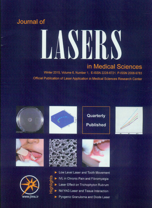
فهرست مطالب

Journal of Lasers in Medical Sciences
Volume:6 Issue: 1, Winter 2015
- تاریخ انتشار: 1393/10/04
- تعداد عناوین: 8
-
-
Pages 1-5Low-Level Laser Therapy (LLLT) provides several benefits for patients receiving orthodontic treatment. According to some literatures, Orthodontic Tooth Movement (OTM) can be enhanced but some investigators have reported contradictory results. This article reviews the literature regarding the different aspects of the use of LLLT on OTM and its alterations. The general data regarding the study design, sample size, wavelength (nm), power (mW), and duration were extracted and recorded independently. Electronic databases of PubMed and ScienceDirect from January 2009 to August 2014 were searched. Also Google Scholar and grey literature was searched for relevant references. Some investigators found that the amount of tooth movement in the Low-Energy Laser Irradiation (LELI) group was significantly greater than in the non-irradiation group by the end of the experimental period. Low-level laser irradiation accelerates the bone remodeling process by stimulating osteoblastic and osteoclastic cell proliferation and function during orthodontic tooth movement. But some researchers have reported that no statistical differences in the mean rate of tooth movement were noted between low energy and high energy experimental sides and their controls. Some evidence shows that low-level laser irradiation accelerates the bone remodeling process and some evidence shows that LLLT has not effect on OTM. In some investigations no statistical differences in the mean rate of tooth movement can be seen between low energy and high energy experimental sides and their controls. It has been shown by authors that laser irradiation can reduce the amount of OTM and a clinical usage for the inhibitory role of low level laser irradiation is enforcing the anchorage unit.Keywords: laser therapies, low, level, movement, tooth, orthodontics
-
Pages 6-9Intravenous laser blood irradiation was first introduced into therapy by the Soviet scientists EN.Meschalkin and VS.Sergiewski in 1981. Originally this method was developed for the treatment of cardiovascular diseases. Improvement of rheologic properties of the blood as well as improvement of microcirculation and reduction of the area of infarction has been proved. Further, reduction of dysrhythmia and sudden cardiac death was achieved. At first, only the Helium-Neon laser (632.8 nm) was used in this therapy. For that, a power of 1-3mW and a period of exposure of 20-60 minutes were applied. The treatments were carried out once or twice a day up to ten appointments in all1. In the years after, many, and for the most part Russian studies showed that helium-neon laser had various effects on many organs and on the hematologic and immunologic system. The studies were published mainly in Russian which were little known in the West because of decades of political separation, and were regarded with disapproval. Besides clinical research and application for patients, the cell biological basis was developed by the Estonian cell biologist Tiina Karuat the same time. An abstract is to be found in her work “The Science of Low-Power Laser-Therapy”Keywords: laser, pain, irradiation
-
Pages 10-16IntroductionTrichophyton rubrum is one of the most common species of dermatophytes which affects superficial keratinous tissue. It is not especially virulent but it can be responsible for considerable morbidity. Although there are different therapeutic modalities to treat fungal infections, clinicians are searching for alternative treatment because of the various side effects of the present therapeutic methods. As a new procedure, Laser therapy has brought on many advantages in clinical management of dermatophytes. Possible inhibitory potential of laser irradiation on fungal colonies was investigated invitro in this study.MethodsA total of 240 fungal plates of standard size of trichophyton rubrum colonies that had been cultured from the lesions of different patients at the mycology laboratory, were selected. Each fungal plate was assigned as control or experimental group. Experimental plates were irradiated by a laser system (low power laser or different wavelength of high power laser). The effects of different laser wavelengths and energies on isolated colonies were assessed. After laser irradiation, final size of colonies was measured on the first, the 7th and the 14th day after laser irradiation.ResultsAlthough low power laser irradiation did not have any inhibitory effect on fungal growth, the Q-Switched Neodymium-Doped Yttrium Aluminium Garnet (Nd:YAG) laser 532nm at 8j/cm2, Q-Switched Nd:YAG laser 1064nm at 4j/cm2 to 8j/cm2 and Pulsed dye laser 595nm at 8j/cm2 to 14j/cm2 significantly inhibited growth of trichophyton rubrum in vitro.ConclusionQ-Switched Nd:YAG 532nm at 8j/cm2, Q-Switched Nd:YAG laser 1064nm at 4j/cm2 to 8j/cm2 and pulsed dye laser (PDL) 595nm at 8j/cm2 to 14j/cm2 can be effective to suppress trichophyton rubrum growth.Keywords: laser, dermatophyte, Q, Switched, Nd:YAG lasers, PDL
-
Pages 17-21IntroductionHeat shock proteins (HSPs) are molecular chaperones involved in protein folding, stability and turnover, and due to their role in cancer progression, the effect of low power laser irradiation (LPLI) on the expression of HSP70 and HSP90 in Jurkat E6.1 T-lymphocyte leukemia (JELT) cell line was investigated in vitro.MethodsJETL cells were irradiated with LPLI at 635nm and 780m wavelengths (energy density 9.174 J/cm2), and assessed for the expression of HSP70 and HSP90 by flow cytometry after 24, 48 and 72 incubation time periods (ITPs).ResultsAt 24 hours ITP post-irradiation, control cultures showed that 10.7% of cells expressed HSP70, while LPLI cultures at 635nm and 780nm manifested a higher expression (32.1and 21.3%, respectively), and the difference was significant (P ≤ 0.05). However, at 48 hours ITP, the three means were decreased but approximated (5.6, 4.9 and 6.2%, respectively), while at 72 hours ITP, they were markedly increased (45.2, 76.5 and 66.7%, respectively). In contrast, HSP90 responded differently to LPLI. At 24 hours ITP, control cultures and 780nm cultures showed a similar expression (55.9 and 55.9%, respectively), but both means were significantly higher than that of 635nm cultures (24.0%). No such difference was observed at 48 hours ITP, and at 72 hours ITP, control cultures and 635nm cultures shared approximated means (31.7 and 35.6%, respectively); but both means were significantly higher than the observed mean in 780nm cultures (15.2%).ConclusionThe results highlighted that HSP70 and HSP90 expression responded differently to LPLI in JETL cells; an observation that may pave the way for further investigations in malignant cells.Keywords: laser Irradiation, low, Power, Jurkat cell, leukemia cell, Heat shock proteins
-
Pages 22-27IntroductionLiposuction using laser is now one of the most common cosmetic surgery. This new method has minimized the disadvantages of the conventional liposuction including blood loss, skin laxity and long recovery time. Benefits of the new liposuction methods which include less trauma, bleeding and skin tightening prove the superiority of these methods over the traditional mechanical methods. Interaction of laser with fat tissue has the vital role in the development of these new procedures because this interaction simultaneously results in retraction of skin layers and coagulation of small blood vessels so skin tightening and less bleeding is achieved.MethodLaser lipolysis uses a laser fiber inserted inside a metal cannula of 1 mm delivering the laser radiation directly to the target tissue. Laser lipolysis has a wavelength dependent mechanism, tissue heating and therefor thermal effects are achieved through absorption of radiation by the target tissue cells, causing their temperature to rise and their volumes to expand. We used Monte Carlo (MC) method to simulate the photons propagation within the tissue. This method simulates physical variables by random sampling of their probability distribution. We also simulated temperature rise and tissue heating using Comsol Multiphysics software.ConclusionBecause optimum and safe laser lipolysis operation highly depends on optical characteristics of both tissue and laser radiation such as laser fluence, laser power and etc. having physical understanding of these procedures is of vital importance. In this study we aim to evaluate the effects of these important parameters.ResultsFindings of our simulation prove that 1064 nm Neodymium-Doped Yttrium Aluminium Garnet (Nd:YAG) has good penetration depth into fat tissue and can reach inside the deeper layers of fat tissue. We see that this wavelength also resulted in good temperature rise; after irradiation of fat tissue with this wavelength we observed that tissue heated in permitted values (50-65°C), this is why this wavelength is widely used in laser lipolysis operations.Keywords: laser, lipolysis, absorption, radiation
-
Pages 28-31IntroductionCO2 (Carbon Dioxide) laser application in circumcision, for cutting and coagulation, has been reported to have excellent results. Also, tissue glue has been reported to have advantages over sutures for approximation of wound edges. Most previous studies focused on comparisons between CO2 laser and scalpel, or between tissue glue and sutures. This study prospectively compared the results and complications CO2 laser and tissue glue, with standard surgical techniques in circumcision.MethodsThirty boys were prospectively divided into two groups. Group 1 (n = 17) underwent circumcision by scalpel with approximation of the wound edges using chromic catgut sutures. Group 2 (n = 13) underwent circumcision with CO2 laser and approximation of the wound edges using tissue glue. Patient age, indications for surgery, operative time, wound swelling, bleeding, wound infection, local irritation, pain score, and cosmetic appearance were recorded.ResultsGroup 1 had a significantly longer operative time (P= 0.011), higher rate of local irritation (P= 0.016), and poorer cosmetic appearance (P< 0.001) than group 2. Bleeding only occurred in one patient in group 1. There were no significant differences in pain score, wound infection rate, or cost of surgery between the two groups.ConclusionsCO2 laser and tissue glue have advantages over standard surgical techniques in circumcision, with a significantly shorter operative time, lower rate of local irritation, and better cosmetic appearance. The cost of surgery is similar between the two groups.Keywords: circumcision, CO2 laser, tissue glue, children
-
Pages 32-39IntroductionDentin hypersensitivity (DH) is characterized by tooth pain arising from exposure of dental roots. In this study the efficiency of neodymium yttrium-aluminum-garnet (Nd:YAG) laser in association with graphite on dentinal surface changes as the alternative to the treatment of DH was evaluated.MethodsSixteen noncarious human third molars were collected and sectioned into 5 parts from cementoenamel junction (CEJ) to the furcation area. The prepared samples were randomly assigned into five groups (Gs) of each 16: Control (G1), treated by Nd:YAG laser at 0.5 W (G2), irradiation of Nd:YAG with a 0.25 W output power(G3), smeared with graphite and then using Nd:YAG laser at output powers of 0.5 W (G4) and 0.25 W (G5). For all groups the parameters were 15 Hz, 60 s, at two stages and with a right angle irradiation. The number and diameter of dentinal tubules (DT) were compared and analyzed by SPSS software, One way ANOVA and Post hoc LSD tests.ResultsThe number of open dentinal tubules varied significantly between all groups except among G1 with G3 and G2 with G5. Multiple comparison tests also exhibited significant differences regarding the diameter of tubules between the groups two by two except among G2 with G5.ConclusionNd:YAG laser used at 0.25 W and 0.5 W with application of graphite smear was able to reduce the number and diameter of dentinal tubules.Keywords: dentin hypersensitivity, Nd:YAG lasers, scanning electron microscopy
-
Pages 40-44IntroductionPyogenic granuloma (PG) is one of the inflammatory hyperplasia seen in the oral cavity. It is a reactional response to minor trauma or chronic irritation. The most common treatment of PG is surgical excision but alternative approaches such as laser excision have also been proposed especially for pediatric patients.Case report: Herein, we present a case of gingival pyogenic granuloma in a 6-year-old patient. The lesion was excised successfully with diode laser as a conservative and non-stressful method in a pediatric patient.Results andConclusionThe use of laser as modern medicine offers a new tool for treatment of oral lesions as comfortable as possible in pediatric patients, which results in less stress and fear in children.Keywords: pyogenic granuloma, hyperplasia, diode laser, pediatric

