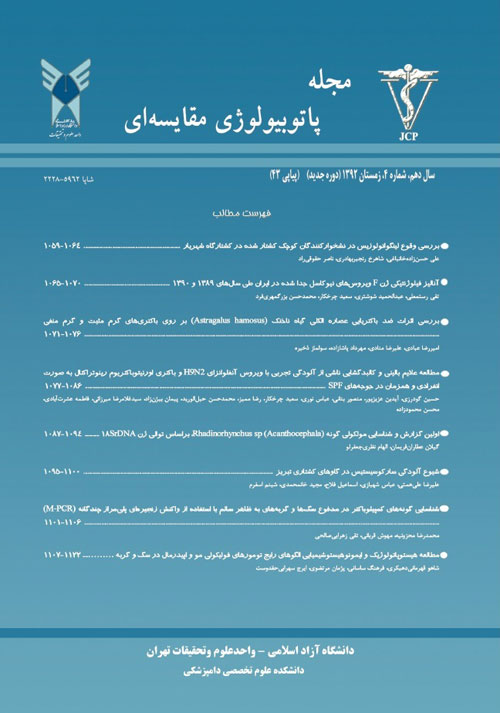
فهرست مطالب

فصلنامه پاتوبیولوژی مقایسه ای
سال دهم شماره 24 (زمستان 1392)
- تاریخ انتشار: 1392/12/12
- تعداد عناوین: 8
-
-
صفحات 1095-1100
-
Pages 1059-1064Linguatula serrata is a worldwide distribiution parasite. Dog and other carnivores are as final hosts and ruminans, equins and rodents are as intermediate hosts. Human is also as both definitive and intermediate hosts. In this study, the infection rate to Linguatula serrata and its relationship with some factors were investigated in slaughtered sheep and goats in Shahriar aboittor. To this end, mesenteric lymphatic nodes of 579 sheep and 195 goats were collected during 5 months randomely. Nymphs were followed through maceration of lymph nodes and digestion of them in digestive solution. The results showed that the infection rate was 16.23% in sheep and 53.33% in goats. Statistical analysis showed more infection rate in goats than sheep. Moreover, there was a significant relationship between sex and rate of the infection and Linguatulosis in female animals was more than males. Meanwhile, statistical analyses showed that the infection rat increased with age of animals. Therefore, regarding to presence of the infection in studied animals and its probable transmission to human, suitable meat inspections, and full-cooking of meat and animal based products can control and prevent of the infection.Keywords: Linguatula Serrata, Linguatulosis, Small Ruminants, Shahriar
-
Pages 1065-1070Newcastle disease is one of the most important diseases in poultry with huge economical damages. It is possible to detect virulence of virus base on molecular techniques and sequence of F gene. In this study, Sequence of 1674 nucleotide acid in cleavage site of F gene of Newcastle disease virus¡ was analyzed for 10 isolates in industrial poultry. These isolates were obtained from endemic of very virulent Newcastle disease in Iran during 2009 and 2010 which have caused high mortality in broiler, breeder and layers along with high egg production drop in breeders and layers. Amino acid sequences were compared phylogenetically with those of previously reported in Iran and also with the gene bank. These isolates show phylogenitically distinction from previous isolates reported in Iran. These isolates show similarity with Chinese and Israeli isolates in gene bank.Keywords: Fusion Gene, Newcastle Disease Virus, Phylogenetic Analysis
-
Pages 1071-1076The space are aware of dangerous side effects of synthetie antibiotics, the more demand will be appeared on natural alternatives of this drug. Natural materials lessen the danger of these side effects; even they would left suitable and useful side effects. Plant pterygium is the one which has most applications in traditional medicine. The aim of this study is to investigate the anti bacterial effects of plant pterygium alchol essence on pathogenic bacteria. In this experiment, pterygium plant was used with scientific name of Astragalus hamosus. After providing alcholic essence of the plant the influence of mg/m1400, mg/m1200, mg/ml100, mg/m150 densities was investigated on staphylococcus aureus, Bacillus cereus, Escherichia coli and pseudomonas aeruginosa in loop dispersion method. The least controlling density determination test of bacteria growth and minimum bacteria fatality was carried out using halo in tube method. Findings of this study indicated that Astragalus hamosus plant alcholic essence prevents staphylococcus aureus, Bacillus cereus, Escherichia coli and pseudomonas aeruginosa bacteria growth. Measuring bacteria not growing was determined in loop dispersion method. Controlling effects on bacteria growth in loop dispersion method was better and more effective than disk dispersion method in similar densities. Astragalus hamosus plant alcholic essence has considerable controlling effects on pathogenic bacteria. Clinical studies are needed to consider these essences.Keywords: Astragalus Hamosus, Loop Dispersion Method, MIC, MBC, Anti Bacterial
-
Pages 1077-1086In this study, clinical signs and gross lesions of (A/Chicken/Iran/ m.1/2010) H9N2 virus and (ORT -R87-7/1387) Ornithobacterium rhinotracheale bacteria alone and a co-infected group in SPF broiler chickens were investigated. Eighty 1-day-old specific pathogen-free White Leghorn chickens were randomly divided into four equal groups. At the age of three weeks, the chicks in the experimental groups were inoculated by virus and bacteria, individually or concurrent and in control group allantoic fluid was inoculated. We used PCR for detection of the bacteria and virus in various organs of experimentally infected broiler. Chickens of AIV and ORT coinfected group showed clinical signs such as ruffled feathers, depression, reduced appetite, cyanosis of wattles and combs, and respiratory distress. The gross lesions such as congestion in the tracheal, airsaculitis, fibrinous cast formation in tracheal and swollen kidneys were observed in birds of the AIV ORT co-infected group. While ORT and AIV groups alone had minor clinical signs and gross lesions. The results of this study indicated that concurrent infectious with H9N2 virus and ORT bacteria could exacerbates clinical signs and gross lesions in infected chickens.Keywords: H9N2 Influenza Virus, Ornithobacterium Rhinotracheale, Co, Infection, Clinical Signs, SPF Chicken
-
Pages 1087-1094Acanthocephala are a group of invertebrate and are arthropod and vertebrate parasites with cosmopolitans distribution. These parasites cause of disease in the vertebrates like fishes. The invistigastion of these parasites is essential for the identification and preventation of infection. One method of organism taxonomy and identification is molecular identification along with morphology. In this study gastrointestinal parasites of 54 tuna fishes(Thunnus albacares ) were studied in 2012. Acanthocephala which was a dominant parasits in these fishes, were separated. Acantocephalan rDNA was extracted according to the modified CTAB method and nucleotide sequence studied in SSU_rRNA region. Iranian species sequences were compared with 2 species of Acanthocephala from GenBank. Phylogenetic relationship among species assessed by ML analysis. Result showed monophyly within Acanthocephaa classes. Iranian species was sister group with Rhadinorhynchus sp and with 99% Bootstrap supported. Iranian species was belong toorder of Echinorhynchida. The results of species morphology analysis were agreed with molecular results. This is the first report of Acanthocephalan parasite of tuna fish from Oman sea.Keywords: Acanthocephala, Phylogeny, Gastrointestinal Parasite, Tuna Fish, SSU, rRNA
-
Pages 1095-1100Sarcocystis is one of the most prevalent parasites of the livestock. It is economically important and pathogenic to livestock. In this study Esophagus, heart and diaphragm muscles of 30 cattle collected from Tabriz abattoir. In corpses and samples were observed no macroscopic cysts. Microscopic cysts were identified by histopathological method and staining them by hematoxylin and eosine stain method and microscopically examined for presence of bradyzoite rate of infestation was in heart (Mean and Std.Error 8.1 ± 0.52), in esophagus (Mean SEM 6.2 ± 0.44) and in diaphragm (Mean SEM 1.2 ± 0.35). The most infective tissue was heart. A kit carried out DNA extraction. PCR conditions optimized for 18S rRNA amplification. We observed the microscopic cysts in Esophagus, heart, and diaphragm muscles. PCR analysis showed that microscopic cysts belonged to Sarcocystis cruzi.Keywords: Sarcocystis, Histopathology, PCR
-
Pages 1101-1106Campylobacteriosis is considered as a zoonotic disease and in some cases carrier are the sources of infection in humans. Dogs and Cats might have no Clinical signs but shed the bacteria in their feces and sometimes show samples of dogs and cats were examined. Primers were used related to 16S rRNA, gly A, mapA, and lpxA genes. First, positive samples which contained nucleotide sequence related to Campylobacter spp. were identified using universal Clinical sign (such as enterits). Therefore the rapid and accurate diagnosis of infected animals is important. The objective of the present study was to detection of Campylobacter species in stool samples of dogs and cats referred to small animal hospitals in Tehran, Iran, using Multiplex PCR technique. In the present study 100 stool primers. Then identification of Campylobacter spp. was performed using multiplex PCR. PCR products were analyzed and scanned using gel electrophoresis and UV illuminator, respectively. Thirty nine samples were positive for Campylobacter spp. Of these, 2 and 11 samples were related to Campylobacter jejuni and Campylobacter upsaliensis, respectively. Statistical analysis was showed that Campylobacter upsaliensis infection was more common has a higher prevalence in cats compared to dogs. It was concluded that PCR technique can be used as a useful test for rapid diagnosis of Campylobacter spp. in stool samples of diseased or carrier animals.Keywords: Campylobacter Jejuni, Campylobacter Upsaliensis, Multiplex PCR
-
Pages 1107-1122The aim of this study is to identify common patterns of epidermal and hair follicular tumors in pet animals (dogs and cats). In review of Literature, a considerable scientific background is not found in our country. 50 samples were collected from skin lesions that the 15 samples of them were skin tumors (7 epidermal tumor samples and 8 sample hair follicle tumor) and histopathological and immunohistochemical studies were performed on these tumors. The hair follicle tumor samples included: Infundibular Keratinizing Acanthoma (IKA), Tricholemmoma- Bulb type (TLB), Trichoblastoma-Trabecular type (TBT), Trichoblastoma-Ribbon type (TBR), Trichoblastoma-Granular Cell type (TBG), Trichoblastoma-Medusoid type (TBM), Trichoepithelioma (TE), Malignant Trichoepithelioma (MTE) and the epidermal tumor samples were Basal Cell Carcinoma (BCC) and Cutaneous Lymphosarcoma (CL). In addition, nonneoplastic lesions so diagnosed such as hematomas, organized abscesses and tumor-like lesions. Hair follicular tumors were examined as routine histology methods (H&E), but the malignant epidermal tumors were performed by immunohistochemical studies. According to the immunohistochemical staining results obtained from this study - regardless of the type of animal - P53 expression levels in samples of basal cell carcinoma was assessed (1) in one of the samples and (2) in three of them and all cutaneous lymphosarcoma samples were (2). Also Ck8 expression levels in samples of basal cell carcinoma was assessed (1) in three samples and was (3) in one of them, respectively. Ki67 expression levels in cutaneous lymphosarcoma samples was (1) in two samples and (2) in one of them. Finally, CD99 expression levels in all cutaneous lymphosarcoma samples were (2). Combined diagnostic panels as Ck8/ P53 markers for basal cell carcinoma and P53/Ki67/CD99 markers for cutaneous lymphosarcoma diagnosis in dogs and cats are a useful diagnostic method.Keywords: Dogs, Cats, Epidermal Tumors, Hair Follicular Tumors, Immunohistochemistry

