فهرست مطالب

Cell Journal (Yakhteh)
Volume:24 Issue: 1, Jan 2022
- تاریخ انتشار: 1400/11/20
- تعداد عناوین: 8
-
-
Page 1
Gastric cancer (GC) is one of the leading causes of cancer-related deaths worldwide. The major problems of patients with GC are the lack of proper response to the treatment, drug resistance and metastasis attributed to the presence of a subpopulation of cells inside the tumour that are called cancer stem cells (CSCs). In addition, deregulation of microRNAs (miRNAs) has been reported in different stages of GC. The aim of the present study is to determine and introduce miRNAs that contribute to regulation of stemness, metastasis and drug resistance in GC. A systematic review, we conducted data mining of available datasets and a review of previous studies to select miRNAs that target stemness, epithelial-mesenchymal transition (EMT) and drug resistance. All selected miRNAs were analysed by R software to find a common miRNA target for all three processes. Then, the target prediction of miRNAs and their related signalling pathways were obtained by using bioinformatics tools, ONCO.IO and KEGG databases, respectively. We identified seven miRNAs (miR-34a, miR-23a, miR-27a, miR-30a, miR-19b, miR-107, miR-100) from our searching approach. These miRNAs regulate pathways that contribute to stemness, EMT and drug resistance in GC. Four (miR- 34a, miR-23a, miR-30a, and miR-100) had significant interactions with each other and 52 target genes among them, from which MYC, CDK6, NOTCH1, NOTCH2, SIRT1, CD44, CD24, and AXL were involved in the regulation of several biological processes. These data suggest that the three significant properties can be regulated by common miRNAs (hsa-miR-34a, hsa-miR-23a, hsa-miR-30a and hsa-miR-100). Hence, targeting selected miRNAs or their targets might be helpful to stop tumour growth and metastasis development, and increase tumour sensitivity to chemotherapy agents. This signature can also be assumed for early detection of metastasis or drug resistance. However, there should be additional experimentation to validate these results.
Keywords: Drug Resistance, Gastric Cancer, Metastasis, MicroRNA, Stem Cells -
Page 7Objective
It is necessary to evaluate fertility effective agents to predict assisted reproduction outcomes. This study was designed to examine sperm vacuole characteristics, and its association with sperm chromatin status and protamine-1 (PRM1) to protamine-2 (PRM2) ratio, to predict assisted pregnancy outcomes.
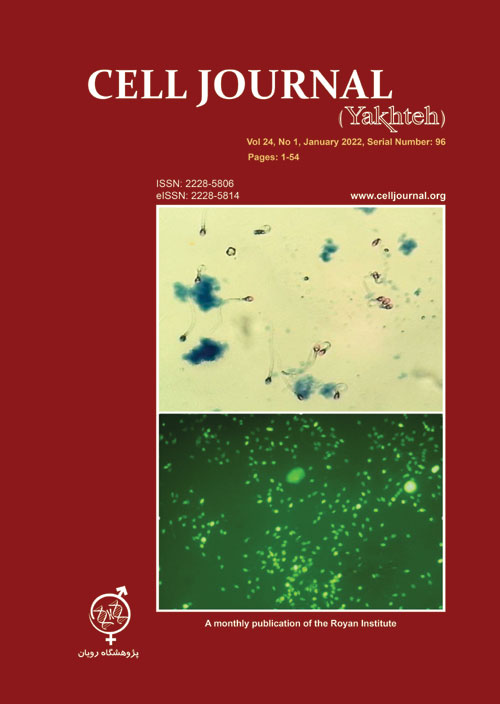
Materials and MethodsIn this experimental study, ninety eight semen samples from infertile men were classified based on Vanderzwalmen’s criteria as follows: grade I: no vacuoles; grade II: ≤2 small vacuoles; grade III: ≥1 large vacuole and grade IV: large vacuole with other abnormalities. The location, frequency and size of vacuoles were assessed using high magnification, a deep learning algorithm, and scanning electron microscopy (SEM). The chromatin integrity, condensation, viability and acrosome integrity, and protamination status were evaluated for vacuolated samples by toluidine blue (TB) staining, aniline blue, triple staining, and CMA3 staining, respectively. Also, Protamine-1 and protamine-2 genes expression was analysed by reverse transcription-quantitative polymerase chain reaction (PCR). The assisted reproduction outcomes were also followed for each cycle.
ResultsThe results show a significant correlation between the vacuole size (III and IV) and abnormal sperm chromatin condensation (P=0.03 and P=0.02, respectively), and also, protamine-deficient (P=0.04 and P=0.03, respectively). The percentage of reacting acrosomes was significantly higher in the grades III and IV spermatozoa in comparison with normal group. The vacuolated spermatozoa with grade IV showed a high protamine mRNA ratio (PRM-2 was underexpressed, P=0.01). In the IVF cycles, we observed a negative association between sperm head vacuole and fertilization rate (P=0.01). This negative association was also significantly observed in pregnancy and live birth rate in the groups with grade III and IV (P=0.04 and P=0.03, respectively).
ConclusionThe results of our study highlight the importance sperm parameters such as sperm head vacuole characteristics, particularly those parameters with the potency of reflecting protamine-deficiency and in vitro fertilization/ intracytoplasmic sperm injection (IVF/ICSI) outcomes predicting.
Keywords: Algorithm, Human Sperm, Pregnancy, Protamines, Vacuole -
Page 15Objective
The present work was aimed at uncovering the effect of circRNA-011235 (circ-011235) on irradiation-induced bone mesenchymal stem cells (BMSCs) injury and its regulatory mechanism, with a view to establish a scientific basis for its possible medical applications.
Materials and MethodsIn this experimental study, after irradiation with different doses (0, 2, 4, 6 GY), the relative expression levels of circ-011235, miR-741-3p, and cyclin-dependent kinases 6 (CDK6) were detected in the BMSCs, using the real time-quantitative polymerase chain reaction (RT-qPCR). The overexpression effects of circ-011235 and CDK6 on the cell proliferation in irradiation-treated BMSCs were measured by the Cell Counting Kit-8 (CCK8) assay. And also, their effects on the cell cycle were evaluated by flow cytometry. RT-qPCR and immunoblotting were performed to detect the effects of pcDNA-circ-011235 and pcDNA-CDK6 on the expression of cyclin D1 and cyclindependent kinases 4 (CDK4) at the gene and protein levels, respectively.
ResultsIrradiation treatment elevated the expression of circ-011235 and CDK6, but reduced miR-741-3p expression in the BMSCs with a dose-dependent effect. The proliferation of BMSCs was significantly inhibited in the irradiation treatment group, while the overexpression of circ-011235 and CDK6 effectively attenuated this inhibition. Also, overexpression of circ-011235 and CDK6 elevated the expression of cyclin D1 in irradiation-treated BMSCs, but had no significant effect on the CDK4 expression.
ConclusionOur results demonstrated that circ-011235 up-regulated the expression of cyclin D1 via miR-741-3p/ CDK6 signal pathway, thereby promoting cell cycle progression and proliferation of irradiation-treated BMSCs. This finding suggested circ-011235/ miR-741-3p/CDK6 pathway exerted a protective role in the response to irradiation and will be a potential new target for future research on the mechanism involved in the resistance of BMSCs to radiation.
Keywords: Bone Mesenchymal Stem Cell, CDK6, Cell Cycle, Irradiation -
Page 22Objective
Given the prevalence of fertility problems in couples and the defect in embryo implantation as well as the low success rate of assisted reproductive techniques, it is necessary to investigate the causes of this phenomenon. Type 2 diabetes mellitus (T2DM) is a metabolic disease with multiple effects on various organs as well as the endometrium. In this study, the effects of endometrial cell culture on the expression of α3 and β1 integrin genes and protein in type 2 diabetic rats were investigated.
Materials and MethodsIn this experimental study, 35 female rats were divided into five groups: control, sham, diabetic, Pioglitazone-treated and Metformin-treated groups. First, rats were maintained in diabetic condition for 4 weeks. Then, treatment was performed for the next four weeks. Four weeks after induction of diabetes, rats were sacrificed at the time of embryo implantation. The uterus was removed. Endometrial cells were isolated and cultured for 7 days. Immunocytochemistry staining was used to confirm endometrial cells. Expression of α3 and β1 integrin genes was determined by real-time polymerase chain reaction (PCR) technique and the α3β1 protein content measured using Western blot both before and after endometrial cell culture.
ResultThe expression level of α3 integrin gene in the Pioglitazone-treated group compared with metformin-treated group was significantly decreased (P<0.001). The same result was observed in β1 integrin gene expression (P=0.004). Also, the α3β1 protein level increased in all diabetic groups, but its reduction was significantly greater in pioglitazonetreated group (P=0.004)
ConclusionT2DM altered the expression of α3 and β1 integrin genes and related proteins, which endometrial cell culture regulated this disorder. According to these results, may be the endometrial cell culture can reduce the adverse effects of diabetes on α3 and β1 integrin expression at the level of gene and protein, in endometrial cells.
Keywords: Cell Culture, Diabetes Mellitus, Implantation, Integrin Alpha1, Integrin Beta3 -
Page 28Objective
One of the severe complications and well-known sources of end stage renal disease (ESRD) from diabetes mellitus is diabetic nephropathy (DN). Exosomes secreted from diverse cells are one of the novel encouraging therapies for chronic renal injuries. In this study, we assess whether extracted exosomes from kidney tubular cells (KTCs) could prevent early stage DN in vivo.
Materials and MethodsIn this experimental, exosomes from conditioned medium of rabbit KTCs (RK13) were purified by ultracentrifuge procedures. The exosomes were assessed in terms of morphology and size, and particular biomarkers were evaluated by transmission electron microscopy (TEM), scanning electron microscopy (SEM), Western blot, atomic force microscopy (AFM) and Zetasizer Nano analysis. The rats were divided into four groups: DN, control, DN treated with exosomes and sham. First, diabetes was induced in the rats by intraperitoneial (i.p.) administration of streptozotocin (STZ, 50 mg/kg body weight). Then, the exosomes were injected each week into their tail vein for six weeks. We measured 24-hour urine protein, blood urea nitrogen (BUN), and serum creatinine (Scr) levels with detection kits. The histopathological effects of the exosomes on kidneys were evaluated by periodic acid-Schiff (PAS) staining and expressions of miRNA-29a and miRNA-377 by quantitative real-time polymerase chain reaction (qRT-PCR).
ResultsThe KTC-Exos were approximately 50-150 nm and had a spherical morphology. They expressed the CD9 and CD63 specific markers. Intravenous injections of KTC-Exos potentially reduced urine volume (P<0.0001), and 24- hour protein (P<0.01), BUN (P<0.001) and Scr (P<0.0001) levels. There was a decrease in miRNA-377 (P<0.01) and increase in miRNA-29a (P<0.001) in the diabetic rats. KTC-Exos ameliorated the renal histopathology with regulatory changes in microRNAs (miRNA) expressions.
ConclusionKTC-Exos plays a role in attenuation of kidney injury from diabetes by regulating the miRNAs associated with DN.
Keywords: Diabetic Nephropathy, Exosomes, Kidney, miRNAs -
Page 36Objective
Poly(ε-caprolactone) (PCL) has considerable mechanical and biological properties that make it a good candidate for tissue engineering applications. PCL alongside proteins and polysaccharides, like gelatin (GEL) and chondroitin sulphate (CS), can be used to fabricate composite scaffolds that provide mechanical and biological requirements for skin tissue engineering scaffolds. The aim of this study was fabricating novel composite nanofibrous scaffold containing various ratios of GEL/CS and PCL using co-electrospinning process.
Materials and MethodsIn this experimental study, PCL mixed with a GEL/CS blend has limitations in miscibility and the lack of a common solvent. Here, we electrospun PCL and GEL/CS coincide separately on the same drum by using different nozzles to create composite nanofibrous scaffolds with different ratios (2:1, 1:1 and 1:2) of GEL to CSPCL, and we mixed them at the micro/nanoscale. Morphology, porosity, phosphate-buffered saline (PBS) absorption, chemical structure, mechanical property and in vitro bioactivity of the prepared composite scaffolds were analysed.
ResultsScanning electron microscopy (SEM) images showed beadless nanofibres at all ratios of GEL to CS-PCL. The composite scaffolds (2:1, 1:1 and 1:2) had increased porosity compared to the PCL nanofibrous scaffolds, in addition to a significant increase in PBS absorption. The mechanical properties of the composite scaffolds were investigated under different conditions. The results demonstrated that all of the composite specimens had better strength when compared with the GEL/CS nanofibres. The increase in PCL ratio led to an increase in tensile strength of the nanofibres. Human dermal fibroblasts (HDF) were cultured on the fabricated composite scaffolds and evaluated by 3-(4,5-dimethylthiazol- 2-yl)-5-(3 carboxymethoxyphenyl)-2-(4-sulfophenyl)-2H-tetrazolium (MTS) analysis and SEM. The results showed the bioactivity of these nanofibres and the potential for these scaffolds to be used for skin tissue engineering applications.
ConclusionThe fabricated co-electrospun composite scaffolds had higher porosity and PBS absorption in comparison with the PCL nanofibrous scaffolds, in addition to significant improvements in mechanical properties under wet and dry conditions compared to the GEL/CS scaffold.
Keywords: Chondroitin Sulphate, Co-electrospinning, Nanofibres, Polycaprolactone, Tissue Engineering -
Page 44Objective
The present study investigated the role of miR-181a as a small non-coding RNA molecule in acute myeloid leukemia (AML) pathogenesis and reflected on the effects of Sulforaphane (SFN) on AML progression.
Materials and MethodsThis experimental study had two parts. In vivo study, the miR-181a levels was measured in patients with symptoms of AML and compared to healthy controls (HCs) to investigate its role in AML pathogenesis. Afterward, an in vitro study was performed to examine the effects of SFN on the growth, apoptosis and proliferation rate of AML cell lines. Finally, the effect of SFN on miR-181a was evaluated as a major miRNA involved in hematopoiesis.
ResultsThe results of this study showed an increasing trend (2.9-fold, P=0.0019) in miR-181a expression levels in AML patients as compared with HCs. The data associated with MTT assay and flow cytometry (FCM) additionally demonstrated the anti-proliferative effects of SFN against AML cell lines, with a reduction in miR-181a levels. As well, no significant difference was noted between 24 hours and 48 hours treatments by SFN. It was deduced that modulation of miR-181a expression levels could be one of the mechanisms associated with the anti-proliferative effects of SFN against AML.
ConclusionMiR-181a levels contribute to AML pathogenesis and thus they can be considered as a strategy in controlling AML progression in patients. Accordingly, SFN can arrest cell proliferation and induce apoptosis in AML cell lines through retardation expression of miR-181a and affecting miR-181a pathway, which already clarified its role in the differentiation of hematopoietic stem cells and indicates another mode of anti-cancer action of sulforaphane.
Keywords: Acute Myeloid Leukemia, miR-181a, Sulforaphane -
Page 51
General control non-derepressible 5 (Gcn5) is a member of histone acetyltransferase (HAT) that plays key roles during embryogenesis as well as in the development of various human cancers. Gcn5, an epigenetic regulator of Hoxc11, has been reported to be negatively regulated by Akt1 in the mouse embryonic fibroblasts (MEFs). However, the exact mechanism by which Akt1 regulates Gcn5 is not well understood. Using protein stability chase assay, we observed that Gcn5 is negatively regulated by Akt1 at the post-translational level in MEFs. The stability of Gcn5 protein is determined by the competitive binding with the protein partner that interacts with Gcn5. The interaction of Gcn5 and Cul4a-Ddb1 complex predominates and promotes ubiquitination of Gcn5 in the wild-type MEFs. On the other hand, in the Akt1-null MEFs, the interaction of Gcn5 and And-1 inhibits binding of Gcn5 and Cul4a-Dbd1 E3 ubiquitin ligase complex, thereby increasing the stability of the Gcn5 protein. Taken together, our study indicates that Akt1 negatively controls Gcn5 via the proteasomal degradation pathway, suggesting a potential mechanism that regulates the expression of Hox genes.
Keywords: Akt1, Gcn5, Proteasome Endopeptidase Complex, Ubiquitin


