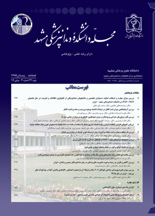
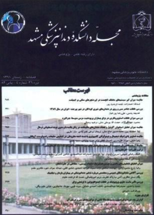
فهرست مطالب

مجله دانشکده دندانپزشکی مشهد
سال سی و چهارم شماره 1 (پیاپی 72، بهار 1389)
- تاریخ انتشار: 1389/01/20
- تعداد عناوین: 10
-
- مقالات پژوهشی
-
صفحات 1-6مقدمهتخمین عرض مزیودیستالی دندان سانترال فک بالا بویژه برای ساخت دندان مصنوعی در افراد بی دندان کار مشکلی می باشد. در این موارد یکی از راه های تعیین اندازه دندان استفاده از شاخص های خارج دهانی است. هدف ازاین تحقیق تعیین رابطه عرض مزیودیستالی دندان سانترال فک بالا با اندازه فاصله بین مردمک ها، که از ثبات خوبی در طول عمر برخوردار است، بود.مواد و روش هادر این مطالعه توصیفی-مقطعی در 100 فرد ایرانی (50 مرد و 50 زن با میانگین سنی 3/6±23 سال) عاری از ناهنجاری چشمی و دندانی، اندازه گیری عرض مزیودیستالی سانترال بالا و فاصله بین مردمک ها توسط گیج Boley با دقت 1/0 میلیمتر، برای هر فرد سه مرتبه انجام شده و میانگین آنها به دست آمد. برای ارزیابی ارتباط بین این دو پارامتر یک فرمول ریاضی محاسبه شد و در20 فرد دیگر (10 مرد و 10 زن با میانگین سنی 56/1±8/24 سال) کارایی فرمول جهت تعیین اندازه دندان مورد بررسی قرار گرفت و تفاوت بین اندازه محاسبه شده با اندازه واقعی دندان توسط آزمون آماری Pair t-test مقایسه گردید.یافته هادر مرحله اول این نتایج حاصل شد: میانگین عرض مزیودیستال دندان سانترال بالا 4/0±6/8 میلی متر و میانگین فاصله بین مردمک ها 8/2±1/62 میلیمتر بدست آمد. میزان همبستگی این دو پارامتر (05/0P<) (446/0r=) محاسبه گردید که براساس آن فرمول ریاضی زیر ارائه شد: (y عرض مزیودیستال دندان سانترال فک بالا و x فاصله بین مردمک ها می باشد.) (مردان x2 0002/0 -x 15/0 =Y)(زنان x2 0014/0 -x 22/0 = Y). در مرحله دوم تحقیق این فرمول ها در 90% افراد (18 مورد از 20 نفر) با دقت 5/0 میلی متر جهت تخمین عرض مزیودیستال دندان سانترال فک بالا صحت داشته و کاربردی بود.نتیجه گیریفاصله بین مردمک ها به عنوان یک لندمارک مناسب برای تخمین عرض دندان سانترال فک بالا پیشنهاد می گردد.
کلیدواژگان: فاصله بین مردمک ها، عرض مزیودیستال دندان سانترال فک بالا، بیماران بی دندان -
صفحات 7-14مقدمهژنژویت یک بیماری چندعاملی است که سیستم ایمنی میزبان و فاکتورهای ژنتیکی، نقش مهمی را در پاتوژنز آن ایفا می کنند. در تحقیقات اخیر ارتباط بین استعداد و شدت ابتلا به بیماری های پریودنتال و مارکرهای ژنتیکی بیان شده است. هدف از مطالعه حاضر بررسی پلی مورفیسم IL-10 در کودکان مبتلا به ژنژویت بود.مواد و روش هادر این مطالعه مورد-شاهدی، که مسایل اخلاقی آن مورد تصویب کمیته اخلاق دانشگاه همدان قرار گرفته است، 100 کودک 12-8 ساله که براساس شاخص های پریودنتال در دو گروه 50 نفری سالم و مبتلا به ژنژیویت مورد آزمایش قرار گرفتند. پس از تهیه نمونهاپی تلیوم مخاط گونه به روش اسکراب از هر فرد و استخراج DNA به روش Millers salting out method، پلی مورفیسم ژن IL-10 در نواحی 1082- و 819-، توسط روش RFLP PCR- تعیین و ارتباط آن با بیماری پریودنتال مورد بررسی قرار گرفت. سپس داده ها توسط آزمون Chi-square مورد تحلیل آماری قرار گرفتند.یافته هایافته ها نشان داد که در جایگاه 1082-، فراوانی الل H در گروه بیمار 34% و درگروه سالم برابر با 24% بود (27/0P=) و فراوانی الل L در هر دو گروه 98% مشاهده شد (1P=). در جایگاه 819-، فراوانی الل C در گروه بیمار 36% و در گروه سالم 42% بود (54/0P=) و فراوانی الل T درگروه بیمار 94% و در گروه سالم 90% بود (71/0P=). همچنین فراوانی ژنوتیپ ها نیز در گروه های مورد مطالعه تفاوت آماری نداشت.نتیجه گیرینتایج پژوهش نشان داد که بین پلی مورفیسم ژن IL-10 و ژنژویت در کودکان 12-8 سال ارتباطی وجود ندارد.
کلیدواژگان: پلی مورفیسم، اینترلوکین10، ژنژویت -
صفحات 15-24مقدمهدندان های مولر سوم شایع ترین دندان هایی هستند که دچار غیبت مادرزادیمی گردند. با در نظر گرفتن این مسئله که غیبت یا حضور این دندان ها می تواند طرح درمان های ارتودنسی، اطفال و پیوندهای جراحی را تحت تاثیر قرار دهد، تعیین وضعیت آنها قبل از انجام این درمان ها امری ضروری می باشد. هدف از این مطالعه بررسی فراوانی غیبت مادرزادی مولرهای سوم و تغییرات همراه با آن در بیماران25-14 ساله مراجعه کننده به دانشکده دندانپزشکی مشهد بود.مواد و روش هااین مطالعه از نوع مطالعات مقطعی، توصیفی- تحلیلی می باشد که در آن رادیوگرافی های پانورامیک بیماران 25-14 ساله مراجعه کننده به دانشکده دندانپزشکی مشهد (طی سال 1385) مورد بررسی قرار گرفتند و در ارتباط با غیبت مادرزادی مولرهای سوم و فاکتورهای وابسته به آن نظیر جنس، هیپودنشیا و سابقه خانوادگی برای هر بیمار پرسش نامه ای تکمیل شده و اطلاعات به دست آمده با استفاده از آزمون های Mann-Whitney و Wilcoxon مورد تجزیه و تحلیل قرار گرفتند.یافته ها1- شیوع غیبت مادرزادی مولرهای سوم 2/11% بود که اختلاف آماری معنی داری میان دو جنس وجود نداشت. 2- در 60% از افرادی که دارای غیبت مولر سوم بودند، غیبت در سایر دندان های دائمی و در 5/7% این افراد، آنومالی در سایر دندان های دائمی وجود داشت. 3- در 3/23% از افرادی که دارای کوچکی فک بالا بودند، دارای غیبت مادرزادی مولر سوم مشاهده شد. 4- تفاوت آماری معنی داری میان غیبت مولر سوم در فک بالا و فک پایین و نیز سمت راست و چپ وجود نداشت.نتیجه گیریفراوانی غیبت مادرزادی مولر سوم در جامعه ما مشابه با سایر جوامع بوده و ارتباط مستقیمی میان آژنزی مولر سوم و آژنزی و آنومالی در سایر دندان ها و نیز کوچکی فک بالا وجود داشت.
کلیدواژگان: غیبت مادرزادی، رادیوگرافی پانورامیک، مولر سوم -
صفحات 25-32مقدمهمطالعات زیادی در زمینه تاثیر تالاسمی ماژور بر استخوان های ناحیه کرانیوفاشیال انجام شده است اما تاثیر این بیماری بر تکامل دندان ها کمتر مورد مطالعه قرار گرفته است. هدف از این تحقیق بررسی میزان تکامل دندان ها و همچنین شیوع آنومالی های دندانی در بیماران بتاتالاسمی ماژور و مقاسیه آن با گروه شاهد بود.مواد و روش هادر این مطالعه مورد و شاهد که مسائل اخلاقی آن مورد تایید و تصویب کمیته اخلاق معاونت پژوهشی دانشگاه علوم پزشکی شیراز رسیده است، رادیوگرافی های پانورامیک 120 بیمار تالاسمی ماژور بررسی شد. بیماران شامل 68 پسر و 52 دختر با میانگین سنی 66/2±98/12 سال، در محدودهسنی 19-8 سال بودند و 120 نفرگروه شاهد از نظر سن و جنس، با گروه مورد یکسان سازی شدند. رادیوگرافی ها از نظر وقوع مواردآنومالی و همچنین تعیین سن دندانی بررسی شدند. آزمون دقیق فیشر برای مقایسه شیوع آنومالی های دندانی در دو گروه مورد و شاهد استفاده شد. آزمون Paired t-test، سن دندانی و سن تقویمی را در هر گروه مقایسه نمود. سن دندانی گروه مورد و شاهد نیز با استفاده از Independent t-test با یکدیگر مقایسه شدند.یافته هاشیوع میکرودونشیا، دندان های با ریشه کوتاه و میخی شکل، تارودونتیسم و کمبود دندانی در بیماران تالاسمی بیشتر از گروه شاهد بود. بین میانگین سن دندانی و سن تقویمی بیماران تالاسمی و همچنین بین میانگین سن دندانی بیماران تالاسمی و افراد گروه شاهد، اختلاف آماری قابل ملاحظه ای مشاهده نشد.نتیجه گیریبر اساس مطالعه حاضر آنومالی های دندانی در بیماران تالاسمی در مقایسه با افراد نرمال شایع تر است. اما سیر تکاملی دندان ها و زمان رویش آنها تحت تاثیر بیماری تالاسمی قرار نمی گیرد.
کلیدواژگان: تالاسمی ماژور، پانورامیک، آنومالی های دندانی، تکامل دندان ها -
صفحات 33-46مقدمهبرای تشخیص نهفتگی دندان های کانین معمولا از رادیوگرافی های کانونشنال استفاده می شود. اما این رادیوگرافی ها اطلاعات کاملی را در بعد سوم فراهم نمی کنند. در این مواقع از توموگرافی و توموگرافی کامپیوتری برای کسب اطلاعات جزئی در مورد موقعیت دندان نهفته استفاده می شود. هدف از انجام این مطالعه بررسی دقت توموگرافی اسپیرال در تعیین موقعیت دندان های نهفته فک بالا بود.مواد و روش هادر این مطالعه توصیفی 9 بیمار ارتودنسی که بدلیل دندان های نهفته فک بالایی که تشخیص موقعیت آنها از لحاظ کلینیکی مشکل بود تحت انجام توموگرافی اسپیرال قرار گرفته بودند مورد پیگیری قرار گرفتند. این بیماران شامل 5 مرد و 4 زن با میانگین سنی 2/16 سال بودند که در مجموع دارای 12 دندان نهفته (اعم از کانین یا دندان اضافی) بودند و جهت اسکپوژر دندان های نهفته تحت جراحی قرار گرفتند. تصاویر توموگرافی با دستگاه Cranex Tome با ضخامت 2میلیمتر (با فواصل 1 و 3 میلیمتر از نقطه تعریف شده به سمت مزیال و دیستال) با صفحات فسفری Digora تهیه شدند. بعد از انتقال تصاویر به کامپیوتر و کالیبراسیون تصویر، با حذف فاکتور بزرگنمایی (5/1×)، بر روی تصاویر ابتدا موقعیت تاج دندان نهفته (باکالی یا پالاتالی بودن و یا نداشتن تمایل خاص) و سپس فاصله بین نوک تاج دندان نهفته یا برجسته ترین سطح تاج تا خط مماس بر کرست به وسیله ابزار تعیین فاصله تا یک دهم میلیمتر اندازه گیری شد. نتایج با اندازه های به دست آمده از جراحی مقایسه شدند.یافته هادر تعیین موقعیت دندان نهفته،بین جراحی و توموگرافی توافق کامل (100%) وجود داشت. کمترین میزان اختلاف بین نتایج کمی توموگرافی و جراحی صفر و بیشترین میزان اختلاف 5/1 میلیمتر بود. میانگین اختلاف اندازه های حاصله از توموگرافی و جراحی 6/0 میلیمتر محاسبه شد.نتیجه گیریتوموگرافی اسپیرال نسبت به رادیوگرافی های معمولی اطلاعات بیشتری در رابطه با موقعیت دندان های نهفته فراهم می کند. این تکنیک تعیین موقعیت دقیق دندان های نهفته را آسان نموده و فاصله دندان نهفته از کرست آلوئول را با اختلاف جزئی نشان می دهد.
کلیدواژگان: دندان نهفته، توموگرافی، رادیوگرافی دندان پزشکی -
صفحات 47-64مقدمهموکواپیدرموئید کارسینوما ((MEC و آدنویید سیستیک کارسینوما (AdCC) شایع ترین تومورهای بدخیم غدد بزاقی هستند که رفتار بیولوژیک آنها کاملا مشخص نشده است. از عواملی که در رفتار بیولوژیک این تومورها موثر قلمداد شده است، تهاجم اطراف عصبی و عروقی می باشد. لذا هدف این مطالعه، بررسی تهاجم اطراف عصبی و عروقی AdCC و MEC غدد بزاقی اصلی و فرعی با استفاده از مارکرهای NCAM، S-100، GFAP به عنوان مارکرهای عصبی و PECAM-1 به عنوان مارکر عروقی بود.مواد و روش هادر این مطالعه مقطعی-توصیفی، 20 نمونه AdCC و 20 نمونه MEC غدد بزاقی که با فرمالین ثابت و در پارافین غوطه ور شده بودند، برای بررسی حضور پروتئین های S-100،GFAP،NCAM و PECAM-1 به روش Biotin-streptavidin novolink polymer، بازیافت آنتی ژنیک و رنگ آمیزی ایمونوهیستوشیمیایی شدند و داده های بدست آمده با استفاده از آزمون آماری مان-ویتنی و دقیق فیشر تحلیل آماری شدند.یافته هاتمام نمونه های AdCC و MEC به S-100 واکنش داده که بین 1+ تا 4+ متغیر بودند. میزان فراوانی پروتئین S-100 و شدت رنگ پذیری آن در AdCC بیشتر از MEC بود (05/0P<). دو مورد AdCC و 9 مورد MEC واکنش 1+ به GFAP نشان دادند که میزان فراوانی و شدت رنگ پذیری آن در MEC بیشتر از AdCC بود (013/0P=). هیچکدام از نمونه های AdCC و MEC به NCAM و PECAM-1 واکنش نشان ندادند (P=1).نتیجه گیریباتوجه به حضور پروتئین S-100 و GFAP در AdCC و MEC، احتمالا سلول های میواپی تلیال در هیستوژنز این دو تومور نقش دارند و با توجه به این که تهاجم اطراف عصبی در تومورهای غدد بزاقی به سلول میواپی تلیال نسبت داده شده است، الزاما خصوصیت مزبور نمایانگر پیش آگهی ضعیف تر این تومورها نمی باشد. در نتیجه گیری کلی، با توجه به فقدان بروز مارکر NCAM و PECAM- در AdCC و MEC و سایر یافته ها، به نظر نمی رسید تهاجم اطراف عصبی و عروقی این دو تومور، تفاوت معنی داری داشته باشد.
کلیدواژگان: موکواپیدرموئید کارسینوما، آدنویید سیستیک کارسینوما، S، 100، GFAP، NCAM، PECAM، 1، تهاجم اطراف عصبی، تهاجم عروقی -
صفحات 65-74مقدمهدر حال حاضر تناقضات زیادی درباره طراحی ارجح برای پروتزهای پارسیل انتهای آزاد به چشم می خورد. گرچه هدف، به حداقل رساندن تنش ها بر دندان ها و نسوج ساپورت کننده است ولی نتایج و نظریات متفاوتی گزارش شده اند. هدف این مطالعه واکاوی نیروهای وارد شده بر دندان پایه انتهای آزاد در طراحی های مختلف رایج کلاسپ بود.مواد و روش هااز جمجمه یک مرد 42 ساله، سی تی اسکن با مقاطع 6/0 میلی متری فراهم شد و با استفاده از اطلاعات به دست آمده یک الگوی سه بعدی از فک پایین به وسیله نرم افزار Mimics ساخته شد. سپس بر روی این الگو، اسکلت فلزی پروتز پارسیل کلاس I کندی با 3 طرح متفاوت کلاسپ Aker''s)، باری شکل و ترکیبی) طراحی گردید. نیروهای اکلوزال بر روی زین انتهای آزاد وارد شد و استرس ها به روش واکاوی اجزاء محدود و با استفاده از نرم افزار (Cosmos works 2008) بررسی گردید.یافته هادر طرح Aker''s الگوی تمرکز تنش ها دلالت بر اعمال نیروی دیستالی به دندان پایه داشت و کلاسپ سیم مفتولی الگوی مشابهی را ارائه می داد ولی تمرکز تنش ها به مراتب کمتر از کلاسپ Aker''s بود. در طرح کلاسپ باری شکل برخلاف دو طرح پیشین الگوی تمرکز تنش ها دلالت بر اعمال نیرویی به سمت مزیال از سوی پروتز پارسیل به دندان پایه داشت. در هر 3 طراحی مذکور از بعد باکولینگوالی اعمال نیروهای باکالی از سوی پروتز به دندان پایه مشاهده می شد.نتیجه گیریکاربرد کلاسپ Aker''s در پروتز پارسیل انتهای آزاد بر روی دندان پایه خلفی تنش هایی بیش از حد تحمل دندان و در جهتی نامناسب (دیستالی) به آن وارد می کند.
کلیدواژگان: کلاسپ، اجزای محدود، تنش، پروتز پارسیل انتهای آزاد -
صفحات 75-80مقدمهموفقیت درمان ایمپلنت بستگی به میزان کافی استخوان در محل قرارگیری ایمپلنت دارد. تهیه استخوان اتوژن چه داخل دهانی، چه خارج دهانی کاری ناخوشایند و تهاجمی برای بیمار است. لذا استفاده از جایگزین های استخوان برای این امر توصیه می شود. هدف مقاله حاضر مقایسه ثبات ایمپلنت های قرار گرفته در نواحی تحت درمان با بلوک های کورتیکوکنسلوس مینرالیزه با ایمپلنت های قرار گرفته در استخوان طبیعی با استفاده از آنالیز فرکانس انعکاسی بود.مواد و روش هادر این مطالعه کارآزمایی بالینی که مسایل اخلاقی آن مورد تصویب کمیته اخلاقی دانشگاه علوم پزشکی مشهد قرار گرفته بود، تعداد 9 بیمار که عرض ریج ناکافی در ناحیه قدامی فک بالا داشتند، انتخاب شدند و با استفاده از بلوکهای کورتیکوکنسلوس مینرالیزه عرض ریج در آنها افزایش داده شد. بعد از 6 ماه از قرار دادن بلوک آلوگرفت ناحیه مجددا باز گردید. تعداد 9 ایمپلنت در نواحی مورد نظر قرار گرفت. جهت گروه کنترل هم تعداد 9 بیمار انتخاب شدند که دارای عرض ریج کافی در ناحیه قدامی فک بالا و استخوان طبیعی بودند و ایمپلنت ها در نواحی معادل با نواحی دریافت کننده ایمپلنت در گروه اول قرار گرفتند. سپس شاخص ISQ، 3 و 6 ماه بعد از ایمپلنت گذاری اندازه گیری شد. تحلیل آماری توسط نرم افزار SPSS، با ویرایش 15 با کمک آزمون های آماری Paired t-test برای مقایسه در هر گروه و آزمون t-test Two sampleبرای مقایسه دو گروه با هم انجام گرفت.یافته هادر طی دوره مطالعه، 2 ایمپلنت در گروه آلوگرفت ها دچار شکست شدند. شاخص ISQ در گروه آلوگرفت بین دوره های زمانی 3 ماه و 6 ماه تفاوت آماری معنی داری داشت (008/0P=) ولی در گروه کنترل بین دوره های زمانی 3 ماه و 6 ماه تفاوت آماری معنی داری وجود نداشت (065/0P=). بین دو گروه آلوگرفت و کنترل در زمان 3 ماه تفاوت آماری معنی داری در شاخص ISQ وجود نداشت (317/0P=) اما بعد از 6 ماه شاخص ISQ در گروه کنترل به طور معنی داری بالاتر بود (000/0P=).نتیجه گیریشاخص ISQ در هر دو گروه با در نظر گرفتن حداقل 60 جهت بارگذاری ایمپلنت ها مناسب است. بلوک های کورتیکوکنسلوس مینرالیزه آلوگرفت می توانند به عنوان داربستی جهت رشد استخوان و افزایش عرض ریج باقیمانده جهت گذاشتن ایمپلنت ها بکار روند.
کلیدواژگان: ثبات ایمپلنت، بلوک آلوگرفت، اوسئواینتگریشن -
صفحات 81-94مقدمهدر سالیان اخیر ارتودنسی به سمت استفاده از تکنولوژی دیجیتال حرکت کرده است. هدف مطالعه حاضر، ارزیابی دقت اندازه گیری های سفالومتریک بدست آمده با نرم افزار تریسینگ سفالومتری در مقایسه با روش تریسینگ دستی جهت تعیین روش دقیق تر و مفیدتر بود.مواد و روش هادر این مطالعه توصیفی-تحلیلی نمونه های مورد استفاده شامل تصاویر رادیوگرافی سفالومتری جانبی دیجیتال و معمولی 30 بیمار بود که با روشSandwich تهیه شدند. در این روش برای هر بیمار یک فیلم رادیوگرافی و یک تصویر دیجیتال همزمان به دست می آید. 15 لندمارک سفالومتریک و 10 اندازه گیری زاویه ای و خطی در هر دو روش دیجیتال و دستی محاسبه و مورد مقایسه قرار گرفتند. خطای اندازه گیری برای هر روش با تکرار تریسینگ 15 سفالومتری از بین 30 سفالومتری اولیه، با استفاده از ضریب همبستگی Pearson محاسبه شد. برای مقایسه دو روش از Paired t test استفاده گردید.یافته هانتایج بدست آمده نشان دادند که اختلاف بین دو روش برای اندازه گیری SNA و U1-L1 و Wit’s از لحاظ آماری معنی دار بودند (05/0P<). در محاسبه خطا، تغییرات در اندازه گیری های دیجیتال اندکی بیشتر از روش دستی بود.نتیجه گیریبطور کلی هر دو روش نتایج کلینیکی مشابهی را در اختیار ما قرار می دهند. بنابراین سفالومتری دیجیتال می تواند انتخاب قابل اعتمادتری به عنوان یک وسیله تشخیصی متداول به دلیل مزایایی که نسبت به تصاویر معمولی از قبیل کاهش اشعه دریافتی بیمار، حذف مراحل ظهور و ثبوت و مواد شیمیایی و نیز ذخیره، پردازش و ارتقاء کیفیت تصاویر دیجیتال توسط کامپیوتر را دارد باشد.
کلیدواژگان: سفالومتری دیجیتال، روش ساندویج، تریسینگ دستی، سفالومتری جانبی - گزارش مورد
-
صفحات 95-100مقدمه
خارج کردن دندان یکی از مهمترین و شایع ترین اقداماتی است که در مطب دندانپزشکی انجام می شود. آشنایی با عوارض احتمالی این جراحی بدون تردید برای هر دندانپزشکی ضروری است. هدف از این گزارش معرفی یک مورد جابجا شدن تصادفی سومین مولر فک پایین به فضای اینفراتمپورال بعنوان یکی از عوارض جراحی دندان عقل می باشد.
یافته هاخانم 23 ساله با محدودیت شدید در باز کردن دهان از طرف یک دندانپزشک به بخش جراحی دانشکده دندانپزشکی ارجاع شده بود که در ارزیابی رادیوگرافی وجود دندان عقل فک پایین در فضای اینفراتمپورال مشهود بود. دندان جابجا شده بدلیل تداخل مکانیکی با زائده کرونوئید موجب محدودیت شدید در باز کردن دهان شده بود، دندان در اتاق عمل و تحت بیهوشی خارج و محدودیت حرکت دهان برطرف شد.
نتیجه گیریآگاهی از عوارض احتمالی خارج کردن دندان یک اصل پایه است و هر دندانپزشکی که اقدام به این جراحی می کند باید در مورد عوارض احتمالی آگاه و با نحوه کنترل آن نیز آشنا باشد.
کلیدواژگان: جابجایی دندان، فضای اینفراتمپورال، مولر سوم فک پایین
-
Pages 1-6IntroductionEstimating the width of maxillary central incisor may be difficult when an artificial tooth is selected for edentulous patients. One of the ways to choose a good size for this tooth is using extra-oral landmarks in face. This study examined the relationship between mesiodistal (MD) width of maxillary central incisor and interpupillary distance (IPD) which is a stable landmark in lifetime.Materials and MethodsIn this descriptive cross sectional study, 100 Iranian subjects (50 males and 50 females, mean age 23±6.3 years) without ocular and dental deformities were selected. The MD width of upper central incisor and IPD were measured three times for each subject using Boley Gauge with 0.1 mm accuracy and then, the mean values were calculated. A mathematic equation to evaluate correlation between MD width of upper central incisor and IPD was calculated and then examined in the other 20 subjects (10 males and 10 females, mean age 24.8±1.56 years) by statistically comparing the difference between calculated and real size of tooth with paired t-test.ResultsIn the first stage, these results were obtained: the mean MD width of maxillary central incisor was 8.6±0.4mm and the mean IPD was 62.1±2.8mm. The correlation coefficient between these two parameters was r=0.446 (P<0.05). The calculated equations for this relationship were: (Y=0.15x–0.0002x² in men and Y=0.22x–0.0014x² in women, “y” represents MD width of maxillary central incisor and “x” represents IPD). In the second stage, these equations were applicable in 90% of subjects (18 from 20 subjects) with 0.5mm of accuracy.ConclusionIPD is a good landmark for estimating MD width of maxillary central incisor in edentulous patients.
-
Pages 7-14IntroductionGingivitis is a multifactorial disease in which host immune system and genetic factors have an important role in its pathogenesis. Genetic polymorphisms in cytokines and their receptors have been proposed as potential markers for periodontal disease. Recent studies have suggested IL-10 gene polymorphism to be involved with gingivitis in children. The aim of this study was to evaluate IL-10 gene polymorphism association with gingivitis in 8-12 year old children.Materials and MethodsIn this case-control study, approved by Hamadan University of Medical Sciences, 100 children were divided into two groups of 50 controls and cases, according to the periodontal indices. An epithelial sample from buccal mucosa of each individual was prepared and DNA was extracted by Miller's salting out method. Then mutation at position -1082 (H/L), -819 (C/T) in the IL-10 gene was detected by PCR-RFLP method and the data were analyzed by chi-square test.ResultsResults showed that at position-1082, distribution of H allele in case group was 34% and for control group it was 24% (P=0.27). Distribution of L allele in two groups was 98% (P=1). Callele distribution in-819 position in case group was 36% and for the control group was 42% (P=0.54) and T allele distribution for case group was 94% and for control group was 90% (P=0.71). There was no significant difference in genotype distribution between the groups.ConclusionThis study suggests that there is no correlation between IL-10 polymorphism with gingivitis in 8-12 year old children.
-
Pages 15-24IntroductionThird molars have the highest prevalence of congenital missing. Missing or presence of third molars is one of the key factors that can change the orthodontic, pediatric and surgical graft treatment. The aim of this study was to evaluate the frequency and risk factors of congenitally missing of third molars in 14-25 year old patients in Mashhad dental school.Materials and MethodsIn this cross-sectional analytical study, panoramic radiographs of 120 14-25 year old patients evaluated in 2006 and the frequency of congenital missing of third molars and risk factors such as gender, hypodontia and family history was checked. The data were analyzed by Mann-Whitney and Wilcoxon tests.Results1. Missing of third molars was 11.2% prevalent. No statistical difference was found between male and female. 2. In patients with third molar agenesis, 60% had agenesis in other permanent teeth and 7.5% had anomaly in permanent teeth. 3. In 23.3 % of patients with small size of maxilla, third molar agenesis was detected. 4. No statistical difference was found between maxilla & mandible or right & left sides in third molar agenesis.ConclusionFrequency of congenitally missing of third molar in our society was similar to other societies and a direct relationship was found between either small size of the maxilla, anomaly or agenesis in other teeth, and agenesis of third molar.
-
Pages 25-32IntroductionSeveral studies have been done on the effect of Thalassemia Major on craniofacial skeleton. Its effect on dental development however, has rarely been studied. The aim of this study was to evaluate dental development as well as prevalence of dental anomalies in patients with beta Talassemia Major in comparison with control group.Materials and MethodsIn this case control study, apprived by ethical committee of Shiraz University of Medical Sciences, panoramic radiographs of 120 Talassemic patients were assessed. The case group consisted of 68 males and 52 females ranging in age from 8-19 with mean age of 12.98±2.66 years. 120 control individuals matched for age and sex. Radiographs were assessed for prevalence of dental anomalies and dental age. Fisher's exact test was used to compare prevalence of dental anomalies between case and control groups. Dental and chronological ages were compared using paired t-test. Independent t-test was used to compare dental ages between case and control groups.ResultsMicrodontia, missing, tarudontism and short spiky roots were more prevalent in Talassemic patients. There was not either a significant difference between the mean of dental and chronological ages in Talassemic patients or between mean of dental age of Talassemia and control individuals.ConclusionAccording to the present study, dental anomalies are more common in Thalassemic patients in comparison with normal individuals. Dental developments as well as time of eruption however, were not affected by Thalassemia.
-
Pages 33-46IntroductionConventional radiographs are usually used for diagnosis of dental impaction, but these radiographs do not provide the dentist with complete information in 3rd dimension. In this situation, tomography or computed tomography is used for acquisition of more detailed information for localization of impacted tooth. The aim of this study was to evaluate the efficiency of tomography in localization of impacted maxillary tooth.Materials and MethodsIn this descriptive study, nine orthodontic patients (5 males, 4 females, mean age 16.2 Years) with 12 impacted teeth (canine or supernumerary teeth) with diagnostically difficult impacted maxillary tooth who had undergone spiral tomography, were followed and received surgical treatment. Spiral tomography (2mm thickness with 1 and 3mm interval in mesial and distal directions) from marked point by cranex tome sordex machine (Helsinki, Finland) and Digora PSP (photostimulable phosphorus plates) were provided. After image transfer to computer, tomographic measurements were corrected by an enlargement factor×1.5. On the images, location of coronal portion of impacted tooth and the distance between tip or prominent point of coronal portion to tangent line of crestal bone in digital program to 0.1 mm accuracy was measured. The results of radiographic measurements were compared with surgical results.ResultsThere was a 100% agreement between tomographic and surgical findings in localization of impacted teeth. The minimum and maximum differances between measurements of surgery and tomography were 0 and 1.5 mm respectively. The mean difference was 0.6 mm.ConclusionSpiral tomography gives us additional information about location of impacted tooth compared with conventional radiography. This method facilitates the localization of impacted tooth and also measures the distance between impacted tooth and alveolar crest with minimum difference.
-
Pages 47-64IntroductionMucoepidermoid carcinoma (MEC) and Adenoid cystic carcinoma (AdCC) are the most common salivary malignancies, and their biologic behavior has not been completely determined. Perineural and vascular invasion are effective on the biologic behavior. Therefore, the aim of this study was to evaluate perineural and vascular invasion in AdCC and MEC of major and minor salivary glands by expression of neural markers such as S-100, GFAP, NCAM and vascular marker such as PECAM-1.Materials and MethodsFormalin-fixed and parafin-embedded tissue sections of 20 cases of AdCC and 20 cases of MEC were immunohistochemically analyzed for the presence of S-100, GFAP, NCAM, PECAM-1by biotin streptavidin Novolink polymer procedure after antigen retrieval. Data were analyzed by Mann-Whitney and Fisher's exact tests.ResultsAll AdCCs and MECs were positive (+1→+4) for S-100. S-100 positive frequency and severity ratios in AdCC were higher than MEC (P<0.05). Two specimens of AdCC and nine specimens of MEC were positive (+1) for GFAP. GFAP positive frequency and severity ratios in MEC were higher than AdCC (P=0.013). All AdCCs and MECs were negative for NCAM and PECAM-1 (P=1).ConclusionThe presence of S-100 and GFAP in AdCC and MEC probably indicates the role of myoepithelial cells in histogenesis of theses tumors. Considering that perineural invasion in salivary gland tumors has been ascribed to myoepithelial cell, this characteristic does not necessarily indicate the poor prognosis of these tumors. As a result, according to the lack of expression of NCAM and PECAM-1 in AdCC and MEC and other findings, perineural and vascular invasion in these tumors do not appear to be different significantly.
-
Pages 65-74IntroductionToday, many controversies about the most beneficial design of distal extension base partial dentures exist. Although the purpose is minimizing the stress on abutment teeth and supporting tissues, different results and theories have been reported. The purpose of this study was to analyze the stress applied on distal extended abutment in different types of conventional clasp designs.Materials and MethodsThe skull of a 42 year old man was CT-Scanned with 0.6mm sections and data were used to make a 3D model of the mandible in mimics software. Then a frame work of Kenedy's class I partial denture with 3 different clasp designs (Akers, Bar type and combination) was designed on this model. Occlusal force was applied on the saddle and stress was measured with a finite element method analysis using "cosmos works 2008" soft ware.ResultsIn Aker's design, stress concentration pattern showed applying distal load on abutment. The combination clasp presented identical pattern, but stress concentration was significantly lower than that of the Akers. On the contrary, stress concentration pattern showed that bar type clasp applied mesial load to abutment. In all designs, buccal load was applied from partial denture to abutment in buccolingual dimension.ConclusionAker's clasp in distal extension base partial dentures applies extra stress on distal extended abutment in unfavorable (distal) direction, which is greater than tooth tolerance.
-
Pages 75-80IntroductionLateral ridge augmentations are traditionally performed using autogenous bone grafts. However, the bone harvesting procedure, whether intra orally or extra orally, is accompanied by patient morbidity. So, applying bone substitute has been suggested. The aim of the present study was to compare the stability of implants in ridges augmented with mineralized cortical cancellous allograft blocks with implants in native bone using Resonance Frequency Analysis.Materials and MethodsIn this clinical trial study, approved by ethical committee of Mashhad university of Medical Sciences, nine subjects with insufficient ridge width were selected. Their ridges were augmented with mineralized cortical cancellous allograft blocks. After 6 months, implants were inserted. In control group, 9 subjects with sufficient ridge width were selected. Implants were inserted in the same place as the test group. ISQ was used for measurement of implants stability on third and sixth month after implants placement. Data were analyzed by paired and two sample t tests using SPSS version 15 software.ResultsDuring the study period, two implants in allograft group failed. In the test group, the difference between mean ISQ at third and sixth month was statistically significant (P=0.008), but in the control group, it was not significant (P=0.065). At third month, the mean ISQ between 2 groups was not statistically significant (P=0.317), but at sixth month, the mean ISQ in control group was significantly higher than the test group (P=0.000).ConclusionThe mean ISQ in both groups were enough to load the implants. This means that mineralized cortical cancellous blocks could be used as a scaffold for growth of the bone and ridge augmentation for implant placement.
-
Pages 81-94IntroductionThe purpose of this study was to evaluate the accuracy of cephalometric measurements obtained from digital tracing software compared with equivalent hand traced measurements to determine the more accurate and useful method.Materials and MethodsDigital and analogue radiographic images of 30 orthodontic patients were obtained through Sandwich technique (a conventional radiographic film and a digital image are produced simultaneously). Fifteen cephalometric landmarks were identified and 10 linear and angular measurements calculated, both manually and with digital tracing software. Measurement errors were assessed for each method by duplicating measurements of 15 randomly selected radiographs using Pearson’s correlation coefficient. A paired t- test was used to detect differences between the manual and digital methods.ResultsDifferences between the two methods for SNA, U1-L1and Wit’s appraisal were statistically significant (P<0.05). In error measurements, an overall greater variability in the digital cephalometric measurements was found.ConclusionThe two tracing methods provided similar clinical results; therefore, efficient digital cephalometric software could be reliably chosen as a routine diagnostic tool due to its advantages (lower exposure to X-ray, image development bypass, image quality enhancement through computer).
-
Pages 95-100Introduction
Lateral ridge augmentations are traditionally performed using autogenous bone grafts. However, the bone harvesting procedure, whether intra orally or extra orally, is accompanied by patient morbidity. So, applying bone substitute has been suggested. The aim of the present study was to compare the stability of implants in ridges augmented with mineralized cortical cancellous allograft blocks with implants in native bone using Resonance Frequency Analysis.
Materials and MethodsIn this clinical trial study, approved by ethical committee of Mashhad university of Medical Sciences, nine subjects with insufficient ridge width were selected. Their ridges were augmented with mineralized cortical cancellous allograft blocks. After 6 months, implants were inserted. In control group, 9 subjects with sufficient ridge width were selected. Implants were inserted in the same place as the test group. ISQ was used for measurement of implants stability on third and sixth month after implants placement. Data were analyzed by paired and two sample t tests using SPSS version 15 software.
ResultsDuring the study period, two implants in allograft group failed. In the test group, the difference between mean ISQ at third and sixth month was statistically significant (P=0.008), but in the control group, it was not significant (P=0.065). At third month, the mean ISQ between 2 groups was not statistically significant (P=0.317), but at sixth month, the mean ISQ in control group was significantly higher than the test group (P=0.000).
ConclusionThe mean ISQ in both groups were enough to load the implants. This means that mineralized cortical cancellous blocks could be used as a scaffold for growth of the bone and ridge augmentation for implant placement.


