فهرست مطالب

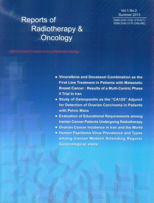
Reports of Radiotherapy and Oncology
Volume:2 Issue: 2, Jun 2015
- تاریخ انتشار: 1394/07/18
- تعداد عناوین: 8
-
-
Page 1BackgroundDepending on the location and depth of tumor, the electron or photon beams might be used for treatment. Electron beam have some advantages over photon beam for treatment of shallow tumors to spare the normal tissues beyond of the tumor. In the other hand, the photon beam are used for deep targets treatment. Both of these beams have some limitations, for example, the dependency of penumbra with depth, and the lack of lateral equilibrium for small electron beam fields.ObjectivesIn this study, improvement of the penumbra and Dmax changes will be investigated. Also the effects of cut-outs on the beam parameters prepared as well. Patients andMethodsIn first, we simulated the conventional head configuration of Varian 2300 for 16 MeV electron, and the results approved by benchmarking the percent depth dose (PDD) and profile of the simulation and measurement. In the next step, a perforated Lead (Pb) sheet with 1 mm thickness placed at the top of the applicator holder tray. This layer producing bremsstrahlung x-ray and a part of the electrons passing through the holes, in result, we have a simultaneous mixed electron and photon beam. For making the irradiation field uniform, a layer of steel placed after the Pb layer. The simulation was performed for 10 × 10, and 4 × 4 cm2 field size.ResultsThe measured R50 and RP for 10 × 10 cm2 field were 6.5 and 7.8 cm, respectively. The photon percentage for 1 mm thickness with 0.2, 0.3, and 0.5 cm holes diameter Lead layer target was about 33%, 32%, and 28% and for 2 mm targets punched with 0.2, 0.3, and 0.5 cm holes, the x-ray percentages were 43%, 41%, and 35%.ConclusionsThis study showed the advantages of mixing the electron and photon beam by reduction of pure electron’s penumbra dependency with the depth, especially for small fields, also decreasing of dramatic changes of PDD curve with irradiation field size.Keywords: Monte Carlo Methods, Photons, Electrons
-
Page 2BackgroundSerum creatinine level is frequently used as a measure for renal function assessment. However, there are some situations in which patients may suffer significant renal impairment but serum creatinine levels remain within normal ranges.ObjectivesWe conducted this study to evaluate the discrepancy between serum creatinine (SCr) level and glomerular filtration rate (GFR) in determining the eligibility for cisplatin-based chemotherapy among cancer patients. Patients andMethodsA total of 198 cancer patients had received cisplatin-based chemotherapy at Jorjani Cancer Center, Emam Hossein Hospital, Tehran, Iran were retrospectively investigated. The discordance between SCr level and calculated GFR by Cockcroft-Gault equation was analyzed.Results130 patients (66%) were men and 68 (34%) were women with mean age of 54.5 years. Squamous cell carcinoma (SCC) and head and neck were the most common primary tumor histology and site respectively. Of 165 patients with available data to calculate eGFR, 45 (27.3%) had normal kidney function based solely on SCr levels, but their GFR was less than 60 mL/min (renal dysfunction). The discordance between SCr and GC calculated GFR values were most pronounced in the older age, transitional cell carcinoma histology and bladder primary site.ConclusionsThis study shows that SCr level alone may not be a reliable measure of normal kidney function to determine eligibility for cisplatine-based chemotherapy.Keywords: Serum, Creatinine, Glomerular Filtration Rate, Cisplatin, Cancer
-
Page 3BackgroundRadical prostatectomy is an established treatment modality for prostate cancer. Following radical prostatectomy, patients with positive surgical margins have increased risk of biochemical, and subsequently, clinical relapse. However, not all patients with positive margins will suffer disease recurrence. The aim of this study was to assess the factors that might predict the higher risk of disease recurrence in prostate cancer patients with positive surgical margins.ObjectivesThe aim of this study was to assess the factors that might predict the higher risk of disease recurrence in prostate cancer patients with positive surgical margins. Patients andMethodsFrom March 2009 till October 2013, seventy seven patients who had pathologically proven positive surgical margins after radical prostatectomy were followed and serum PSA levels were measured every three months. In case of biochemical failure, they were treated with salvage radiotherapy. Apart from pre-op and serial post-op PSA levels, number of positive margins based on anatomical classification of prostate, lymphovascular and perineural invasion, Gleason score and T-stage of the cancer were documented accurately.ResultsFifty one patients (66.2%) had a single positive margin, while 26 (33.8%) had multiple positive margins. Among all 77 patients, 67 (87%) had biochemical failure. Cox regression analysis showed that among various parameters, only pre-op PSA>20ng/ml and having more than one positive margins were able to predict the likelihood of biochemical failure in the patients; while Gleason score, perineural invasion and lymphovascular invasion did not seem to have an important role in this regard.ConclusionsAmong patients with positive surgical margins after radical prostatectomy, those with pre-op PSA>20ng/ml or more than one positive margins are at greater risk of biochemical or/and clinical failure. In these patients, starting salvage radiotherapy after surgery might be considered as a logic option.Keywords: Prostate Cancer, Relapse, Patients
-
Page 4BackgroundPositron emission tomography computerized tomography (PET-CT) is useful in radiotherapy planning for lung cancer. However, its role in malignant pleural mesothelioma (MPM) is unknown.ObjectivesThis exploratory study investigated the possible role for PET-CT in radiotherapy planning for MPM. Patients andMethodsPatients receiving radiotherapy for the treatment of pain in MPM, had fluorodeoxyglucose (FDG) PET-CT scanning in addition to their standard CT scan. PET-CT images were then fused with CT planning images, termed Planning-PET-CT. Target volume delineation was undertaken first using CT and subsequently incorporated Planning-PET-CT. Planning treatment volume (PTV), conformity index (CI), mean distance to conformity (MDC), center of gravity distance (CGD) and standard uptake values (SUV) were examined.ResultsSixteen patients were recruited into the study. PET-CT upstaged nine patients. No association between SUV max and either survival or pain response was seen. Volumes contoured using Planning-PET-CT differed markedly from those outlined using CT alone as shown by the following parameters: CI = 0.3 (0.24 - 0.38). MDC = 21.47 (16.73 - 33.70) and CGD = 16.40 (11.80 - 33.87). The median percentage of over contouring was 44.00% (34.33 - 72.50) with 46.67% under contoured (34.00 - 55.00).ConclusionsPET-CT alters the position of the PTV in MPM and upstaged a proportion of patients. Further work to elucidate the role of Planning-PET-CT in target volume definition is justified.Keywords: Positron Emission Tomography Computerized Tomography (PET, CT), Radiotherapy, Mesothelioma
-
Page 5BackgroundIn spite of all complications and disabilities due to advanced head and neck malignancies, treatment of oral mucositis following radiotherapy is mainly supportive and oral hygiene is the mainstay of treatment. In this context, mouthwash containing sodium bicarbonate can have a major role in the treatment of this complication.ObjectivesThe present trial assessed the value of this type of mouthwash for preventing oral mucositis following radiotherapy in patients suffered locally advanced head and neck squamous cell carcinoma. Patients andMethodsTwenty nine patients suffered locally advanced squamous cell carcinoma were consecutively entered into the study. All patients had stage III to IV head and neck malignancies. The patients were randomly assigned to receiving bicarbonate mouthwash (20 ml every 6 hours per day from the beginning to the end of the treatment) (n = 18) or placebo (n = 11). The mouthwash assessed was containing 1 vial of sodium bicarbonate, 500 ml of normal saline, and diphenhydramine 25 mg/kg).ResultsA significant difference was revealed in the mucositis scoring between the intervention and placebo group so that mucositis grade III or more was observed in 7 patients in intervention group (38.9%) and in 9 patients in control group (81.8%) that the observed discrepant was significant (p = 0.024). The first evidences of oral mucositis in intervention group was found in forth weeks of treatment (sessions of 16 to 17 of treatment schedule), while the appearance of mucositis in placebo group was occurred in the middle of the third week (sessions of 13 to 14 of treatment).ConclusionsOral care by mouthwash containing sodium bicarbonate for head and neck cancer patients undergoing radiotherapy is an effective interventional option to prevent oral mucositis.Keywords: Mouthwash, Sodium Bicarbonate, Radiotherapy, Mucositis
-
Page 6BackgroundTuberculosis is an old disease that is still one of the main causes of death in the world. Nearly 60-67% of TB patients are from developing countries. Proper treatment results resolution in almost all cases. Pelvic TB occurs in about 10% of pulmonary TB patients. The present case reviews a TB case misdiagnosed in a young woman with a pelvic mass and ascetic fluid. Case Report: A 35-year-old woman living in an area exposed to immigrants from TB endemic regions presented with ascetic fluid and a pelvic mass and high titer of sacrum CANS (350). She was misdiagnosed with ovarian malignancy due to a positive malignancy report of ascetic fluid. After chemotherapy she was referred for definite operation. In the surgical field, a TB diagnosis was made.ConclusionsReview of preoperative reports and careful history of the case revealed that had an awareness about TB existed in this case, a proper diagnosis might have been made before the erroneous chemotherapy. She was young, suffering from pelvic pain, fever, and weight loss coming from an exposed area to TB cytology of ascetic fluid included dominancy of lymphocytes. Relying on a positive cytology finding for malignancy might be questionable.Keywords: Tuberculosis, Pelvic Inflammatory Disease, Ovarian Neoplasms
-
Page 7IntroductionRadiotherapy is considered the main part of the curative and preventive regimes in the pediatric cancer especially has been highly used in acute lymphocytic leukemia (ALL) as prophylaxis. A large number of the radiotherapy secondary delayed effects have been well known.Case PresentationIn present study, we has introduced a 15-year-old boy with ALL underwent chemotherapy and radiotherapy. Who had generalized tonic-colonic seizure which has been lasted 10 minutes (without any previous history). The patient with ALL ten years ago has been treated by chemotherapy and whole brain prophylactic radiotherapy with dose 1200 cGy/120 cGy/10. In his MRI a heterogeneous mass with the dimensions 3 × 2 cm in the left frontal lobe with the peripheral edema and after complete the diagnose process the final report of this mass was Cavernoma.ConclusionsThere has been identified a clear link between radiotherapy and brain cavernoma; therefore, it should be considered in differential diagnosis of brain hemorrhagic lesions in each patient with the history of radiotherapy of the central nervous system. This risk is higher when the patient has the history of the brain radiotherapy in childhood.Keywords: Brain Cavernomas, Radiotherapy, Acute Lymphocytic Leukemia (ALL)
-
Page 8Context: According to Association of Academic Health Centers definition, integrative medicine is as a healing-oriented approach of medicine that takes account of the whole person, including all aspects of lifestyle by employing both conventional and unconventional as traditional or complementary and alternative medicine to achieve the best treatment and recovery which is the focus of the present study. Evidence Acquisition: Main oncology resources pointing to complementary and alternative medicine (CAM) modalities in cancer care, evidence-based articles, data obtained from integrative oncology clinics and government organizations associated with cancer were the materials used in this study.ResultsStudies indicate the growing trend in using CAM worldwide. Generally, 38% of adults and 12% of children use CAM in United States referring national researches in 2007; however, other studies show 83 to 92 percent of cancer patient have made use of it. Most complementary modalities are noninvasive and useful in reducing symptoms and side effects of conventional treatments and improving quality of life. Some high quality scientific research has produced evidence that acupuncture, massage, music, and mind-body therapies effectively and safely reduce physical and emotional affects. Integrative oncology relates to an emerging conversation between CAM scholars, oncology care professionals, and family practitioners imagining a holistic patient-centered approach to oncology care.ConclusionsAn increasing recognition in health care systems has emerged, especially in oncology of the importance of maintaining or improving patients’ quality of life all over the disease course. Cancer patients with lower quality of life are more likely to terminate therapy so this should be as an alarm to oncologist the risk for not completing therapy. Complementary methods have played a significant role in this situation for enhancing quality of life, reduced severity of symptoms and side effects, and developed general wellbeing in patients.Keywords: Integrative Medicine, Cancer


