فهرست مطالب

Journal of Dentistry, Shiraz University of Medical Sciences
Volume:16 Issue: 4, Dec 2015
- تاریخ انتشار: 1394/09/25
- تعداد عناوین: 13
-
-
Pages 296-301Statement of the Problem: Immediate application of bonding agent to home- bleached enamel leads to significant reduction in the shear bond strength of composite resin due to the residual oxygen. Different antioxidant agents may overcome this problem.PurposeThis study aimed to assess the effect of different antioxidants on the shear bond strength of composite resin to home-bleached.Materials And MethodSixty extracted intact human incisors were embedded in cylindrical acrylic resin blocks (2.5×1.5 cm), with the coronal portion left out of the block. After bleaching the labial enamel surface with 15% carbamide peroxide, they were randomly divided into 6 groups (n=10). Before performing composite resin restoration by using a cylindrical Teflon mold (5×2 mm), each group was treated with one of the following antioxidants: 10% sodium ascorbate solution, 10% pomegranate peel solution, 10% grape seed extract, 5% green tea extract, and aloe vera leaf gel. One group was left untreated as the control. The shear bond strength of samples was tested under a universal testing machine (ZwickRoell Z020). The shear bond strength data were analyzed by one-way ANOVA and post hoc Tukey tests (p< 0.05).ResultsNo significant difference existed between the control and experimental groups. Moreover, there was no statistically significant difference between the effects of different antioxidants on the shear bond strength of bleached enamel.ConclusionDifferent antioxidants used in this study had the same effect on the shear bond strength of home-bleached enamel, and none of them caused a statistically significant increase in its value.Keywords: Antioxidant, Shear Bond Strength, Tooth Bleaching, Composite Restoration
-
Pages 302-309Statement of the Problem: Root resorption (RR) after orthodontic tooth movement (OTM) is known as a multifactorial complication of orthodontic treatments. Hormonal deficiencies and their effect on bone turnover are reported to have influences on the rate of tooth movement and root resorption.PurposeThis study was designed to evaluate the effect of female and male steroid sex hormones on tooth movement and root resorption.Materials And MethodOrthodontic appliances were placed on the right maxillary first molars of 10 ovariectomized female and 10 orchiectomized male Wistar rats as experimental groups and 10 female and 10 male healthy Wistar rats as control groups. NiTi closed-coil springs (9mm, Medium, 011"×.030", Ortho Technology®; Tampa, Florida) were placed between the right incisors and the first right maxillary molars to induce tipping movement in the first molars with the application of a 60g force. After 21 days, the rats were sacrificed and tooth movement was measured by using a digital caliper (Guanglu, China). Orthodontic induced root resorption (OIRR) was assessed by histomorphometric analysis after hematoxylin and eosin staining of sections of the mesial root.ResultsThe rate of tooth movement was significantly higher in all female rats, with the root resorption being lower in the experimental group. The rate of tooth movement in experimental male rats was significantly higher than the control group (p= 0.001) and the rate of root resorption was significantly lower in the experimental group (p= 0.001).ConclusionIt seems that alterations in plasma levels of estrogen, progesterone, and testosterone hormones can influence the rate of OTM and RR. The acceleration in tooth movement increased OTM and decreased RR.Keywords: Orthodontic Tooth Movement, Root Resorption, Testosterone, Estrogen, Progesterone
-
Pages 310-313Statement of the Problem: Soldering is a process commonly used in fabricating dental prosthesis. Since most soldered prosthesis fail at the solder joints; the joint strength is of utmost importance.PurposeThe purpose of this study was to evaluate the effect of gap angle on the tensile strength of base metal solder joints.Materials And MethodA total number of 40 Ni-Cr samples were fabricated according to ADA/ISO 9693 specifications for tensile test. Samples were cut at the midpoint of the bar, and were placed at the considered angles by employing an explicitly designed device. They were divided into 4 groups regarding the gap angle; Group C (control group) with parallel gap on steady distance of 0.2mm, Group 1: 10°, Group 2: 20°, and Group3: 30° gap angles. When soldered, the specimens were all tested for tensile strength using a universal testing machine at a cross-head speed of 0.5 mm/min with a preload of 10N. Kruskal-Wallis H test was used to compare tensile strength among the groups (p< 0.05).ResultsThe mean tensile strength values obtained from the study groups were respectively 307.84, 391.50, 365.18, and 368.86 MPa. The tensile strength was not statistically different among the four groups in general (p≤ 0.490).ConclusionMaking the gap angular at the solder joints and the subsequent unsteady increase of the gap distance would not change the tensile strength of the joint.Keywords: Base Metal, Solder Gap angle, Solder Joint, Tensile Strength
-
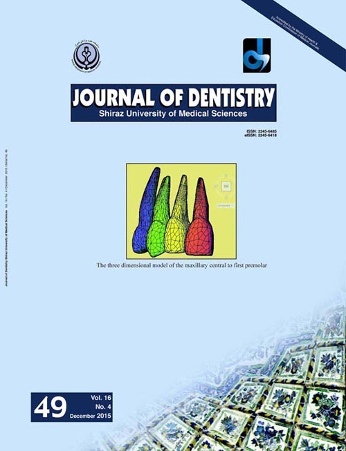
Pages 314-322Statement of the Problem: The use of miniscrews has expedited the true maxillary incisor intrusion and has minimized untoward side effects such as labial tipping.PurposeThe aim of this study was to assess the stress distribution in the periodontal ligament of maxillary incisors when addressed to different models of intrusion mechanics using miniscrews by employing finite element methods. The degree of relative and absolute intrusion of maxillary incisors in different conditions was also evaluated.Materials And MethodFinite element model of maxillary central incisor to first premolar was generated by assembling images obtained from a three-dimensional model of maxillary dentition. Four different conditions of intrusion mechanics were simulated with different placement sites of miniscrews as well as different points of force application. In each model, 25-g force was applied to maxillary incisors via miniscrews.ResultsIn all four models, increased stress values were identified in the apical region of lateral incisor. Proclination of maxillary incisors was also reported in all the four models. The minimum absolute intrusion was observed when the miniscrew was placed between the lateral incisor and canine and the force was applied at right angles to the archwire, which is very common in clinical practice.ConclusionFrom the results yield by this study, it seems that the apical region of lateral incisor is the most susceptible region to root resorption during anterior intrusion. When the minimum flaring of maxillary incisors is required in clinical situations, it is suggested to place the miniscrew halfway between the roots of lateral incisor and canine with the force applied to the archwire between central and lateral incisor. In order to achieve maximum absolute intrusion, it is advised to place miniscrew between the roots of central and lateral incisors with the force applied at a right angle to the archwire between these two teeth.Keywords: Finite Element Method, Intrusion, Miniscrew
-
Pages 323-328Statement of the Problem: P63 gene is a member of TP53 and its homologous gene family. Its expression was observed in some odontogenic lesions, more expression in aggressive lesions.PurposeThis study aimed to investigate the possible diagnostic impact of P63 protein on dentigerous cysts and various types of ameloblastoma. Its expression with Ki-67 proliferation marker was also compared.Materials And MethodThis cross-sectional retrospective study was enrolled on 25 cases of dentigerous cyst including 21 unicystic ameloblastomas and 17 conventional ameloblastomas. The expression of P63 and Ki-67 was assessed by immunohistochemical (IHC) examinations. Data were analyzed by employing MannWhitney and correlation coefficient tests.ResultsP63 expression was significantly higher in ameloblastoma than unicystic ameloblastoma and dentigerous cysts. There was no significant difference between unicystic ameloblastoma and dentigerous cyst in P63 expression. A 90% cut-off point was obtained for basal layer which gave 88% sensitivity and 78% specificity to distinguish more invasive lesions from others. There was not any correlation between P63 and Ki-67 immunostaining in the three study groups.ConclusionMore aggressiveness and more invasiveness of odontogenic lesions depicted higher rate and also more intensive expression of P63. Moreover, the expression of P63 protein had not any correlation with Ki-67 protein in dentigerous cysts and ameloblastomas.Keywords: P63, Ki, 67, Dentigerous Cyst, Ameloblastoma, Unicystic
-
Pages 329-334Statement of the Problem: Prediction of child cooperation level in dental setting is an important issue for a dentist to select the proper behavior management method. Many psychological studies have emphasized the effect of birth order on patient behavior and personality; however, only a few researches evaluated the effect of birth order on child’s behavior in dental setting.PurposeThis study was designed to evaluate the influence of children ordinal position on their behavior in dental setting.Materials And MethodA total of 158 children with at least one primary mandibular molar needing class I restoration were selected. Children were classified based on the ordinal position; first, middle, or last child as well as single child. A blinded examiner recorded the pain perception of children during injection based on Visual Analogue Scale (VAS) and Sound, Eye and Movement (SEM) scale. To assess the child's anxiety, the questionnaire known as “Dental Subscale of the Children's Fear Survey Schedule” (CFSS-DS) was employed.ResultsThe results showed that single children were significantly less cooperative and more anxious than the other children (p<0.001). The middle children were significantly more cooperative in comparison with the other child's position (p< 0.001).ConclusionSingle child may behave less cooperatively in dental setting. The order of child birth must also be considered in prediction of child’s behavior for behavioral management.Keywords: Behavior, Birth order, Anxiety
-
Pages 335-340Statement of the Problem: Jaw bone lesions are common pathologic conditions. The role of ultrasonography in evaluation of the extra-osseous lesions is confirmed, however, this imaging modality is not the diagnostic routine for the intraosseous jaw lesions.PurposeThe purpose of this study was to evaluate the efficiency of ultrasonography in diagnosis of intra-osseous jaw lesions concerning their size and content and also to study its correlation with the histopathological findings.Materials And MethodFor this study, 15 patients with intra-osseous jaw lesions in the maxilla and mandible were selected from those referred to the Department of Oral Surgery. Panoramic imaging, computed tomography (CT) or cone beam computed tomography (CBCT) and ultrasonography (USG) were performed for all the lesions. The size of the lesions was measured by USG and then compared with CT or CBCT. Moreover, the correlation amongst the echographic patterns and histopathologic results was evaluated.ResultsIn 12 cases, size values were in complete agreement with CT or CBCT. The size of 3 lesions could not be measured by the radiologist due to the thickness of buccal cortical plate.ConclusionFindings of this study suggested that USG might be feasible in estimating the size of intra-osseous jaw lesions with little underestimation. This study also confirmed that ultrasound imaging was a very useful imaging technique which could provide significant diagnostic information regarding the content of jaw bone lesions where the buccal bone thickness was thin enough.Keywords: Ultrasonography, Tomography, X-ray Computed, Odontogenic Tumors, Odontogenic Cyst, Jaw
-
Pages 341-348Statement of the Problem: The use of miniscrew as an absolute anchorage device in clinical orthodontics is growing increasingly. Many attempts have been made to reduce the size, to improve the design, and to increase the stability of miniscrew.PurposeThe purpose of this study was to determine the effects of different thread shapes and force directions of orthodontic miniscrew on stress distribution in the supporting bone structure.Materials And MethodA three-dimensional finite element analysis was used. A 200-cN force in three angles (0°, 45°, and 90°) was applied on the head of the miniscrew. The stress distribution between twelve thread shapes was investigated as categorized in four main groups; buttress, reverse buttress, square, and V-shape.ResultsStress distribution was not significantly different among different thread shapes. The maximum amount of bone stress at force angles 0°, 45°, and 90° were 38.90, 30.57 and 6.62 MPa, respectively. Analyzing the von Mises stress values showed that in all models, the maximum stress was concentrated on the lowest diameter of the shank, especially the part that was in the soft tissue and cervical cortical bone regions.ConclusionThere was no relation between thread shapes and von Mises stress distribution in the bone; however, different force angles could affect the von Mises stress in the bone and miniscrew.Keywords: Miniscrew, Thread, Finite Element Analysis, Force Direction
-
Pages 349-355Statement of the Problem: Hydrogen peroxide (H2O2) has been suggested to be used in sequence or in combination with chlorhexidine (CHX) to enhance the antibacterial activity against Enterococcus faecalis, but there is no research in the literature on the safety and effectiveness of this irrigation protocol.PurposeThis study aimed to assess the cytocompatibility and antibacterial activity of different concentrations of CHX combined with H2O2 in comparison with the activity of 5.25 and 2.5% sodium hypochlorite (NaOCl).Materials And MethodDifferent concentrations of H2O2 (10, 5, 3 and 1%) were exposed to the PDL cells. Then, the solution with minimal cytotoxicity was selected (3% H2O2). The cytocompatibility and antibacterial activity of 0.1, 0.2, 1 and 2% CHX combined with 3% H2O2 were evaluated and compared with 5.25 and 2.5% NaOCl. The differences in the mean viability of PDL cells were evaluated by one-way ANOVA. Kruskal-Wallis and post-hoc Dunn's tests were adopted to compare the antibacterial activity of the solutions against E.faecalis.ResultsThe viability of PDL cells was lower when treated with 5.25 or 2.5% NaOCl than all combinations of CHX and H2O2. There was no significant difference in the antibacterial activity of the solutions against E.faecalis, except for the 0.1% CHX + 3% H2O2 combination, which had significantly lower efficacy than other groups.ConclusionAll combinations of CHX and H2O2 (used in this study) except 0.1% CHX + 3% H2O2 were efficient irrigants against planktonic E.faecalis and had a better cytocompatibility with PDL cells than 5.25 and 2.5% NaOCl.Keywords: Chlorhexidine, Cytotoxicity, Hydrogen peroxide, Periodontal ligament cells, Sodium hypochlorite
-
Pages 356-361Statement of the Problem: Pulp stones are calcifications found in the pulp chamber or pulp canals of the teeth. Its different prevalence in different population is a matter of concern.PurposeThis study aimed to assess the prevalence of pulp stones in a sample of Iranian population and to report its occurrence regarding gender, dental arch, tooth type and dental status.Materials And MethodsDental records of patients who attended Shiraz Dental School were selected randomly. Only bitewing and periapical radiographs of maxillary and mandibular permanent posterior teeth were studied. Teeth were classified in the case of presence or absence of pulp stones, and the prevalence was analyzed in different gender, tooth types, dental arch, and dental status (intact, carious, or restored) groups. Statistical analysis was performed using X2 test.ResultsOf the 652examined subjects, 306 (46.9%) had one or more teeth with pulp stones. Of the 8244 posterior teeth examined, 928 (11.25%) had pulp stones in the pulp chamber. These pulp stones were detected in 76(37.6%) of males and 230 (51%) of females. The frequency of pulp stones among different teeth between maxillary and mandibular arches had almost a similar pattern. Among teeth demonstrating the condition, first molars were the most prevalent, followed by second molars. In maxillary molars the frequency of occurrence (26%) was higher than mandibular molars (18.7%). No Significant difference was found between dental status and pulp stones occurrence.ConclusionThe occurrence of pulp stones noted in this study was significantly higher in female, molar teeth than premolar and 1st maxillary molar than mandibular. There was no significant association between pulp stone and condition of the crown.Keywords: Pulp stones, Prevalence, Radiographic assessment
-
Pages 362-370Statement of the Problem: Early childhood caries is an important oral health issue. Finding its prevalence would predict the need for oral health promotion disciplines for specific age groups.PurposeThe aim of this study was to assess the caries experience of children living in Tehran, Iran. It also would evaluate the impact of gender, ethnicity, and socioeconomic status (SES) on this oral condition.Materials And MethodThis epidemiological cross-sectional study was based upon stratified cluster random sampling. The samples consisted of 239 children (2- to 3- years old) registered in Tehran’s public healthcare centers for “Healthy Child Program”. Mothers of the recruited children were interviewed for the background data; then children were examined for the oral health status according to ICDAS-II (International Caries Detection and Assessment System) and WHO (World Health Organization) criteria. Statistical analyses were conducted using STATA.11 for SES classification considering six socioeconomic variables, and SPSS.21 for descriptive/analytical analyses.ResultsPrimary Component Analysis (PCA) demonstrated five classes of SES ranging from the lowest to the highest. The distribution of caries-free (CF) children was 10.87%, non-cavitated enamel caries (codes 01-02) were 28.03%, and about 61.1% had cavitated caries (codes 03-06). There was no significant difference in caries experience between the two genders. Cavitated lesions were more prevalent among Kurdish, who also had the least CF children. Caries prevalence, especially code 02, was more among children from 3rd class SES (moderate level). Gender, ethnicity, or SES had no impact on the CF status of the children; however, ethnicity showed significant impact on the prevalence of extensive caries (codes 05-06).ConclusionThe result of the present study is indicative of high caries prevalence among 2 to 3 years old children residing in Tehran. It highlights the need for comprehensive oral health promotion disciplines for this age group.Keywords: ICDAS, II, Early childhood caries, Caries, free, Young children, Dental caries, Prevalence, Oral health, Iran
-
Pages 371-373Careful understanding of internal anatomy of root canal system is crucial for successful endodontic treatment. The presence of two palatal canals in maxillary second molar is unusual but noteworthy as an aid to appropriate diagnosis and treatment. This paper reported a case of a maxillary right second molar with two root canals in the palatal root. The root canal treatment and case management were also explained.Keywords: Maxillary Second Molar, Two Palatal Canals, Root canal therapy
-
Pages 374-379The calcifying cystic odontogenic tumor (CCOT) is a rare cystic odontogenic neoplasm frequently found in association with odontome. This report documents a case of CCOT associated with an odontome arising in the anterior maxilla in a 28-year-old man. Conventional radiographs showed internal calcification within the lesion but were unable to visualize its relation with the adjacent structures and its accurate extent. In this case cone beam computed tomography (CBCT) could accurately reveal the extent and the internal structure of the lesion which aided the presumptive diagnosis of the lesion as CCOT. This advanced imaging technique proved to be extremely useful in the radiographic assessment and management of this neoplasm of the maxilla.Keywords: Cone Beam Computed Tomography, Conventional Radiographs, Calcifying Cystic Odontogenic Tumor, Compound Odontome

