فهرست مطالب

Iranian Journal of Kidney Diseases
Volume:10 Issue: 1, Jan 2016
- تاریخ انتشار: 1394/12/01
- تعداد عناوین: 10
-
-
Pages 1-9Chronic allograft dysfunction is the most common cause of allograft lost. Chronic allograft dysfunction happens as a result of complex interactions at the molecular and cellular levels. Genetic and environmental factors both influence the evolution and progression of the chronic allograft dysfunction. Epigenetic modification could be considered as a therapeutically modifiable element to pause the fibrosis process through novel strategies. In this review, the PubMed database was searched for English-language articles on these new areas.Keywords: chronic allograft dysfunction, epigenetic, genetic, fibrosis
-
Pages 11-16IntroductionMyocardial infarction is a common cause of mortality in patients with chronic kidney disease (CKD). Since troponins I and T levels rise in CKD patients without any myocardial cause, diagnostic value of cardiac troponins is not high in these patients. This study aimed to evaluate the value of troponin I and other cardiac biomarkers to differentiate acute coronary syndrome in CKD patients.Materials And MethodsIn this cross-sectional study, patients with stage 3 to 5 of CKD with typical chest pain were enrolled. Troponins I and T and other biomarkers were measured, and angiography was carried out in these patients. Cardiac biomarkers and other variables were evaluated in patients and compared with angiography results.ResultsNinety CKD patients with a mean age of 61.67 ± 15.87 years were enrolled. Angiography results were normal in 48.9% of the patients, while it showed single-vessel disease in 14.5%, two-vessel disease in 23.3%, and three-vessel disease in 13.3%. Serum creatinine level, glomerular filtration rate, troponin I level, and creatine kinase level were not significantly different in patients with normal and abnormal angiography findings. The serum troponin I, creatine kinase, and creatine kinase-myocardial bound levels had no significant diagnostic values to differentiate abnormal angiography in CKD patients.ConclusionsSerum levels of cardiac troponin I and creatine kinase-myocardial bound were not suitable to diagnose ACS in CKD patients (stages 3 to 5); therefore, we suggest using other diagnostic attempts in similar conditions. More evaluation is needed to confirm these findings.Keywords: handgrip strength, malnutrition, protein, energy wasting, hemodialysis
-
Pages 17-21IntroductionIn 2009, the Oxford classification of immunoglobulin A (IgA) nephropathy was proposed by the working group of the International IgA Nephropathy Network and Renal Pathology Society. It established specific pathologic features that predict the risk of progression of disease. This study aimed to evaluate the interobserver reproducibility of the Oxford classification of IgA nephropathy between Iranian nephropathologists.Materials And MethodsWe included 100 patients with primary IgA nephropathy diagnosed between 2001 and 2011. Histologic slides were circulated among 4 pathologists. A score sheet was answered by each individual pathologist for each biopsy, according to the instruction of the Oxford classification. Reproducibility was determined for each variable, using intraclass correlation coefficient (ICC).ResultsThe ICC values calculated for each major category of the Oxford classification were as follows: the highest score of 0.94 for tubular atrophy and interstitial fibrosis; 0.8 for glomerular basement membrane duplication, extracapillary proliferation, and segmental endocapillary proliferation; and 0.1 to 0.3 for arterial lesions, especially for hyalinosis of arterioles and intimal thickening of arcuate vessels and interlobar arteries.ConclusionsThe Oxford classification of IgA nephropathy is a useful tool and evidenced-based method with high interobserver reproducibility in pathology reporting. Our data suggest that Oxford classification may be used as a model for classification of other renal pathologies in the future.Keywords: IgA nephropathy, Oxford classification, Interobserver reproducibility
-
Pages 22-25IntroductionAortic root stiffness is a highly prevalent condition in patients with chronic kidney disease (CKD), which independently associates with increased risk of mortality and morbidity in this group. This study aimed to investigate the relationship between glomerular filtration rate (GFR) level as a marker of kidney function and elastic properties of aortic root obtained with echocardiography.Methods and Materials: Eighty-two patients (53 men and 29 women; mean age, 57.9 ± 15.0 years) with CKD stages 3 and 4 were enrolled in this study. Echocardiography and tissue Doppler imaging of the upper and lower aortic walls were done. Systolic wave (S wave) was obtained from each walls. Aortic distensibility and aortic root stiffness index were calculated using blood pressure measurement and aortic dimensions in systole and diastole by M-mode echocardiography.ResultsIn all of the patients, aortic stiffness index (14.24 ± 9.82), aortic distensibility (0.018 ± 0.015), aortic upper wall S velocity (0.10 ± 0.03), and aortic lower wall S velocity (0.08 ± 0.02) were severely impaired. There was no correlation between GFR and aortic distensibility (r = -0.253, P =. 20), aortic root stiffness index (r = 0.193, P =. 09), aortic upper wall S velocity (r = -0.106, P =. 17), and aortic lower wall S velocity (r = -0.150, P =. 09).ConclusionsAortic stiffness was seen in all patients with kidney failure in stage 3 or 4 of CKD; however, we could not find any direct relationship between GFR and this phenomenon. Thus, aortic root stiffness may be a consequence of hypertension and other risk factors.Keywords: chronic kidney disease, aortic stiffness, tissue Doppler imaging
-
Pages 26-29IntroductionThe increased susceptibility to infection in patients with end-stage renal disease is probably secondary to the impaired immune defense in uremia. Mannose-binding lectin (MBL) has an important role in host defense through activation of the lectin complement pathway. The aim of this study was to measure serum MBL level in peritoneal dialysis patients and compare it with a healthy group.Materials And MethodsSeventy peritoneal dialysis patients and 70 healthy individuals were enrolled in this study. Serum MBL levels were measured by an enzyme-linked immunosorbent assay kit using the mannan molecule. In addition, serum C-reactive protein and albumin levels were measured to determine whether there is a correlation between serum MBL level and these two parameters.ResultsThe mean serum MBL level was 2.32 ± 2.54 µg/mL (range, zero to 6.93 µg/mL) in the patients group and 1.80 ± 2.14 µg/mL (range, zero to 6.97µg/mL) in the control group (P =. 19). No significant correlation was detected between age and serum MBL level in either the groups. In the patients group, no significant correlation was found between serum MBL and C-reactive protein levels or MBL and albumin levels. There were no correlation between duration of peritoneal dialysis and MBL or dialysis adequacy and MBL, either.ConclusionsThis study did not find MBL deficiency in peritoneal dialysis patients as compared to the healthy individuals.Keywords: mannose, binding lectin, C, reactive protein, albumin, peritoneal dialysis, chronic kidney disease
-
Pages 30-35IntroductionProtein-energy wasting (PEW) is very common in patients with chronic kidney disease and those undergoing maintenance dialysis. Reduced handgrip strength is associated with PEW and considered as a reliable nutritional parameter that reflects loss of muscle mass. This study aimed to evaluate the handgrip strength and its relationship with the Malnutrition-Inflammation Score (MIS) among Iranian dialysis patients.Materials And MethodsThe study population consisted of 83 randomly selected hemodialysis patients from the dialysis centers in Kerman, Iran. Handgrip strength was measured using a dynamometer according to the recommendations of the American Society of Hand Therapists. All the patients were interviewed and the MIS of the patients were recorded.ResultsThe PEW was prevalent in Kerman hemodialysis patients, with 83% and 17% having mild and moderate PEW based on MIS, respectively. Handgrip strength was significantly associated with age, sex, height, weight, and diabetes mellitus. After adjustment for age, handgrip strength was significantly associated with nutritional assessment markers on the basis of the MIS.ConclusionsHandgrip strength can be incorporated as a reliable tool for assessing nutrition status in clinical practice. However, further research is needed to determine the reference values and cutoff points both in healthy people and in hemodialysis patients to classify muscle wasting.Keywords: handgrip strength, malnutrition, protein, energy wasting, hemodialysis
-
Pages 36-43IntroductionCarnitine deficiency is commonly seen in dialysis patients. This study assessed the association dialysis and pediatric patient's characteristics with plasma carnitines levels.Materials And MethodsPlasma carnitine concentrations were measured by tandem mass spectrometry in 46 children on hemodialysis or peritoneal dialysis. The total carnitine, free carnitine (FC), and L-acyl carnitine (AC) levels of 40 µmol/L and less, less than 7 µmol/L, and less than 15 µmol/L were defined low, respectively. An FC less than 20 µmol/L and an AC/FC ratio greater than 0.4 were considered as absolute and relative carnitine deficiencies. The correlation between carnitines levels and AC/FC ratio and age, duration of dialysis, characteristics of dialysis, and blood urea nitrogen and serum albumin concentrations were assessed.ResultsAbsolute carnitine deficiency, low total carnitine, and low AC concentrations were found in 66.7%, 82.6%, and 51% of the patients, respectively. All of the patients had relative carnitine deficiency. Carnitine measurements were not significantly different between the hemodialysis and peritoneal dialysis groups. More severe relative carnitine deficiency was found in those with lower blood urea nitrogen levels and those on peritoneal dialysis. No linear correlation was found between carnitine levels and age, duration of dialysis, characteristics of dialysis, serum albumin level, or blood urea nitrogen level.ConclusionsAbsolute and relative carnitine deficiencies are common among children on dialysis. Patients with lower blood urea nitrogen levels and peritoneal dialysis patients are more prone to severe relative carnitine deficiency.Keywords: carnitine deficiency, hemodialysis, peritoneal dialysis
-
Pages 44-47Cystinuria is an inherited disease characterized by the formation of cystine calculi in the kidneys, ureters, and bladder. Cystinuria is associated with mutation in the SLC3A1 and SLC7A9 genes. These defects prevent appropriate reabsorption of dibasic amino acids lysine, ornithine, and arginine. Cystinuria is classified as type I (silent heterozygotes) and non-type I (heterozygotes with urinary hyperexcretion of cystine). In molecular term, cystinuria is classified as type A (mutations on SLC3A1 gene) and type B (mutations on SLC7A9 gene). This report describes 7 patients with early onset of cystine calculus formation. We are report a new mutation in SLC3A1 gene in exon 1. A novel nucleotide substitution c.-29A>G was found in exon 1 of the SLC3A1 gene, which had not been reported elsewhere previously.Keywords: cystinuria, urinary calculi, gene mutations
-
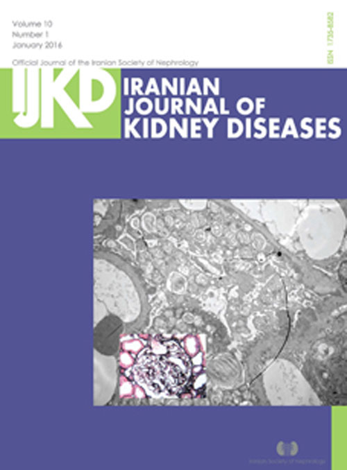
Pages 48-50A 21-year-old man with no family history or characteristic symptoms of Fabry disease presented with proteinuria. Histological and immunofluorescent analysis of kidney tissue collected revealed stage 1 membranous nephropathy. Electron microscopy of the same tissue revealed a large number of myeloid bodies (zebra bodies) in the glomerular epithelial cytoplasm and a mild irregular thickening of basement membrane. A diagnosis of Fabry disease was supported by the low α-galactosidase A activity detected in the patient’s plasma, and confirmed by the detection of a pathogenic homozygous mutation in the α-galactosidase A gene. Therefore, the final diagnosis was of coexistent Fabry disease and stage 1 membranous nephropathy. This is the first case study reporting the coexistence of Fabry disease and membranous nephropathy. Our results emphasize the importance of electron microscopy in Fabry disease diagnosis.Keywords: Fabry disease, membranous nephropathy, alpha, galactosidase A, genetics


