فهرست مطالب

Journal of Dentistry, Shiraz University of Medical Sciences
Volume:17 Issue: 3, Sep 2016
- تاریخ انتشار: 1395/06/20
- تعداد عناوین: 12
-
-
Pages 164-170Statement of the Problem: Treatment with salivary substitutes and stimulation of salivary flow by either mechanical or pharmacologic methods has side effects and only provides symptomatic relief but no long-lasting results.PurposeTo assess the effectiveness of extraoral transcutaneous electric nerve stimulation (TENS) as a mean of stimulating salivary function in healthy adult subjects; as well as to determine the gender and age-dependent changes in salivary flow rates of unstimulated and stimulated parotid saliva.Materials And MethodHundred patients were divided into two groups; Group I aged 20-40 and Group II aged ≥ 60 years. The TENS electrode pads were externally placed on the skin overlying the parotid glands. Unstimulated and stimulated parotid saliva was collected for 5 minutes each by using standardized collection techniques.ResultsEighty seven of 100 subjects demonstrated increased salivary flow when stimulated via the TENS unit. Ten experienced no increase and 3 experienced a decrease. The mean unstimulated salivary flow rate was 0.01872 ml/min in Group I and 0.0088 ml/min in Group II. The mean stimulated salivary flow rate was 0.03084 ml/min (SD= 0.01248) in Group I, and 0.01556 ml/min (SD 0.0101) in Group II. After stimulation, the amount of salivary flow increased significantly in both groups (pConclusionThe TENS unit was effective in increasing parotid gland salivary flow in healthy subjects. There was age-related but no gender-related variability in parotid salivary flow rate.Keywords: Electrostimulation, Stimulated Salivary Flow, Transcutaneous Electric Nerve Stimulation (TENS), Age, Gender, Parotid Saliva Flow
-
Pages 171-176Statement of the Problem: Trichomonas tenax and Entamoeba gingivalis are commensal protozoa which inhabit the human oral cavity. These parasites are found in patients with poor oral hygiene and might be a reason for progressive periodontal diseases.PurposeThe aim of this study was to evaluate the effect of nonsurgical periodontal treatment on the frequency of these protozoa in saliva and plaque samples.Materials And MethodIn this clinical trial, samples of saliva and dental plaque were collected from 46 patients with moderate to severe chronic periodontitis before and after periodontal therapy. The samples were assessed for the frequency of parasites.ResultsThe frequency of Entamoeba gingivalis was reduced in saliva (p= 0.007) and plaque (p= 0.027) three weeks after the treatment. Likewise, the frequency of Trichomonas tenax reduced in saliva (p= 0.030); however, the decrease was not significant in plaque (p= 0.913). Trichomonas tenax frequency in dental plaque directly related to the severity of periodontitis (r= 0.565, p≤ 0.000). In contrast, the number of Entamoeba gingivalis in both saliva (r= -0.405, p≤ 0.005) and plaque (r= -0.304, p= 0.040) was inversely related with the severity of the periodontal disease.ConclusionNonsurgical periodontal treatment could reduce the number of Trichomonas Tenax and Entamoeba gingivalis in the oral environment of patients with chronic periodontitis.Keywords: Trichomonas Tenax, Entamoeba Gingivalis, Periodontitis, Parasite
-
Pages 177-184Statement of the Problem: Impaction of foreign bodies in the soft tissues is a sequela of traumatic and penetrating injuries. Such foreign bodies should be removed due to the complications they cause. Patients history, clinical evaluation and imaging examinations aid in the proper detection and localization of the foreign bodies.PurposeThe aim of the present study was to compare the sensitivity of computed tomography (CT) and ultrasonography for detecting foreign bodies in in-vitro models simulating facial soft tissues.Materials And MethodFifty foreign particles with five different compositions including wood, glass, metal, plastic, and stone were embedded in five calf tongues at 1, 2, 3, 4 and 5 cm depths. CT and ultrasonography were compared regarding their capability of detecting and localizing the foreign bodies.ResultsWood and plastic foreign bodies were demonstrated more clearly on ultrasonography images. High density materials such as metal, stone, and glass were detected with almost the same accuracy on CT and ultrasonography examinations. Visibility of the foreign bodies deteriorated on ultrasonography images as their depth increased; however, CT appearances of the foreign particles were not influenced by their depths.ConclusionUltrasonography is an appropriate technique for detection of foreign bodies especially the ones with low density. Therefore, it seems logical to perform ultrasonography in combination with CT in cases with the suspicion of foreign body impaction.Keywords: Foreign body, Computed tomography, Ultrasonography
-
Pages 185-192Statement of the Problem: Oral cancer is among the ten most common cancers worldwide. It affects the life quality of patients in many ways.PurposeThe aim of this study was to compare the effects of two different systemic doses of Viola Odorata syrup on the prevention of 4-Nitroquinoline-1-oxide (4-NQO) induced tongue dysplasia in rats.Materials And MethodForty-eight male Wistar rats were divided into four groups of A, B, C and D. Group A served as the control group. The rats in groups B to D received 30 ppm of 4-NQO in drinking water for 12 weeks. Additionally, the rats in groups B and C received Viola Odorata syrup at doses of 15 and 5 ml/kg, respectively, 3 times a week. Body weights were measured three times a week. At the end, the rats were euthanized and the tongue was removed. Histological evaluations for carcinogenesis were carried out under a light microscope.ResultsThe mean body weight of the rats in groups B, C, and D were lower than that in group A (pConclusionViola Odorata extract has dose-dependent inhibitory effects on the development of tongue induced dysplasia.Keywords: 4, nitroquinoline, 1, oxide, Viola Odorata, Tongue, Dysplasia
-
Pages 193-200Statement of the Problem: Gingival recession has been considered as the most challenging issue in the field of periodontal plastic surgery.PurposeThe purpose of this study was to evaluate the clinical efficacy of root coverage procedures by using partial thickness double pedicle graft and compare it with full thickness double pedicle graft.Materials And MethodEight patients, aged 15 to 58 years including 6 females and 2 males with 20 paired (mirror image) defects with class I and II gingival recession were randomly assigned into two groups. Clinical parameters such as recession depth, recession width, clinical attachment level, probing depth, and width of keratinized tissue were measured at the baseline and 6 months post-surgery. A mucosal double papillary flap was elevated and the respective root was thoroughly planed. The connective tissue graft was harvested from the palate, and then adapted over the root. The pedicle flap was secured over the connective tissue graft and sutured. The surgical technique was similar in the control group except for the prepared double pedicle graft which was full thickness.ResultsThe mean root coverage was 88.14% (2.83 mm) in the test group and 85.7% (2.75 mm) in the control group. No statistical differences were found in the mean reduction of vertical recession, width of recession, or probing depth between the test and control groups. In both procedures, the width of keratinized tissue increased after three months and the difference between the two groups was not statistically significant in this respect.ConclusionConnective tissue with partial and full thickness double pedicle grafts can be successfully used for treatment of marginal gingival recession.Keywords: Root Coverage, Connective Tissue Graft, Pedicle Flap, Gingival Recession
-
Pages 201-206Statement of the Problem: Mechanical properties of interim restorations are considered as important factors specially when selecting materials for long-term application or for patients with para-functional habits. Flexural strength is one of the most important components of these restorations.PurposeThe purpose of this study was to compare the flexural strength of five interim restorative materials.Materials And MethodFifty identical samples sized 25×2×2-mm were made from five interim materials (TempSpan; Protemp 4, Unifast III, Trim, and Revotek LC) according to ADA specification #27. The specimens were stored in artificial saliva for 2 weeks and then thermocycled for 2500 cycles (5-55˚C). A standard three-point bending test was conducted on the specimens with a universal testing machine at a crosshead speed of 0.75mm/min. Data were analyzed by using one-way ANOVA and Tamhanes post-hoc tests to measure the flexural strength of temporary materials.ResultsOne of the bis-acryl resins (TempSpan) showed the highest, and the light polymerized resin (Revotek LC) showed the lowest flexural strength. The mean values of flexural strength (MPa) for the examined materials were as follow: TempSpan=120.00, Protemp 4=113.00, Unifast III=64.20, Trim= 63.73 and Revotek LC=47.16. There were significant differences between all materials except Trim and Unifast III which did not show any statistical significant difference.ConclusionBis-acryl resins were statistically superior to traditional methacrylate and light-cured resins. Therefore, application of bis-acryl resins should be deliberated in patients with heavy occlusion and in cases that need long-term use of interim restorations.Keywords: Flexural Strength, Interim, Fixed Prosthesis
-
Pages 207-212Statement of the Problem: Neoadjuvant chemotherapy (NCH) is controversial in the treatment of oral squamous cell carcinoma (OSCC).PurposeThe aim of this study was to evaluate the efficacy of NCH on OSCC prognosis.Materials And MethodIn this retrospective cohort study, 94 patients were studied in two groups. The patients in group 1 received NCH before the surgery, and those in group 2 underwent resection without any chemotherapy prior to surgery. The employed NCH agents consisted of cisplatin in combination with 5-fluorouracil in two treatment courses. Tumor size, lymph node involvement, age, and follow-up time were considered as variable factors of the study. Local recurrence (LR) and distant metastasis (DM) were outcomes of the study.ResultsComparison of LR and DM in various tumor sizes demonstrated no significant difference between the two groups (p> 0.05). Analysis of the data did not show any statistically significant difference between the groups for LR in subjects with N0, N1 and N2. Each one-year increase in age was associated with 10% increase in the hazard ratio (HR) (HR distance metastasis Y/N = 1.10, p= 0.05). In the same analysis, when considering LR as a dependent factor, LR risk in N2 was 3 times more than in N1 (p= 0.02). LR risk in N3 was 5 times more than in N1 [HR local recurrence (p= 0.006).ConclusionBased on our results, neoadjuvant chemotherapy with combination of cisplatin and 5-fluorouracil may not improve prognosis of OSCC. However, further studies are suggested to assess other neoadjuvant chemotherapy protocols in OSCC patients.Keywords: Squamous Cell Carcinoma Oral, Metastasis, Recurrence, Chemotherapy
-
Pages 213-218Statement of the Problem: Dental caries is one the most prevalent diseases that affects humans throughout their lives. Streptococcus mutans (S. mutans) is recognized as the most important microorganism during tooth cariogenicity. Reducing this germ in oral cavity can reduce the rate of tooth decays in humans.PurposeThe present study compared the antimicrobial activity of ethanolic extract of Peganum harmala L. seeds and 0.2% chlorhexidine on S. mutans.Materials And MethodAgar diffusion technique and micro broth dilution method were employed to test the antimicrobial effects of these two agents on S. mutans. Moreover, the cytotoxicity of ethanolic extract of P. harmala was studied on Vero cells by MTT (thiazolyl blue tetrazolium dye) colorimetric method. The data were analyzed with descriptive methods.ResultsConcentrations of 50, 25, and 12.5 mg/mL of the extract made inhibition zones of bacterial growth around the wells; but, lower concentrations could not inhibit the growth of S. mutans. Besides, the antimicrobial effect of 0.2% chlorhexidine was more than 50 mg/mL of the extract. Minimum inhibitory concentration (MIC) of the extract on S. mutans was 1.83±0.6 mg/mL and minimum bactericidal concentration (MBC) was 4.3±1 mg/mL. The MIC and MBC for 0.2% chlorhexidine were reported to be 0.19 mg/mL, and 0.78 mg/mL, respectively. The extract concentrations more than 0.5 mg/mL were toxic and caused more than 50% Vero cell death.ConclusionDespite the remarkable antimicrobial effects of high concentrations of P. harmala on S. mutans, high cell toxicity of this plant would restrict its in vivo therapeutic use.Keywords: Peganum Harmala L., Streptococcus Mutans, Chlorhexidine, Cytotoxicity
-
Pages 219-225Statement of the Problem: Use of cyclooxygenase inhibitors as chemotherapy agents has attracted the attention of a large number of investigators in recent years. Given the importance of cancer therapy, only a limited number of studies have been carried out to investigate the effects of cyclooxygenase inhibitors on specific cell lines.PurposeThis research aimed to determine the in vitro cytotoxic effects of cyclooxygenase inhibitors (COX-1 and COX-2 inhibitors) on KB, Saos-2, 1321N, U-87MG, SFBF-PI 39 cell lines.Materials And MethodPowders of celecoxib, mefenamic acid, aspirin and indometacin were dissolved in the appropriate solvent. The viability of cell lines was carried out by MTT (3-(4,5-Dimethylthiazol-2-yl)-2,5-Diphenyltetrazolium Bromide) assay technique. Data gathered from four separate experiments were expressed as mean±SD. Statistical significance was defined at pResultsCelecoxib showed marked cytotoxic effects on KB, Saos-2, and 1321N cells, which was significant in comparison with the control group. Celecoxib was not effective in killing U-87MG cell line. Mefenamic acid exerted cytotoxic effects on KB, Saos-2, and 1321N cells, where the viability was approximately 75%. U-87MG cells were resistant to mefenamic acid. Indometacin had the highest rate of activity on U-87MG cells, which was significant in comparison with the control group. Aspirin did not exhibit any activity on these cell lines and was not effective in killing U-87MG, KB, Saos-2, and 1321N cells.ConclusionThis research showed that celecoxib, indometacin, and mefenamic acid have the cytotoxic effects on KB, Saos-2, 1321N and U-87MG cell lines. Therefore, it appears that these drugs can be considered as anti-neoplastic agents in the experimental phase.Keywords: In Vitro, Cytotoxicity, Drug, Celecoxib, Mefenamic Acid, Aspirin, Indometacin, Cell lines
-
Pages 226-232Statement of the Problem: The presence of bacterial biofilms is the major cause of gingivitis and periodontitis, their mechanical removal is not often enough. Therefore, laser therapy and photodynamic therapy can be effective as adjunctive treatment.PurposeThis study aimed to evaluate the impact of these treatments on the level of gingival crevicular fluid (GCF), inflammatory mediators, and periodontal clinical status.Materials And MethodIn this clinical trial, three quadrants were studied in 12 patients with chronic periodontitis aged 30-60 years. The clinical parameters were recorded and GCF samples were taken. After the first phase of periodontal treatment, one of the three quadrants was determined as the control group, one was treated by diode laser, and one underwent photodynamic therapy. The clinical parameters were recorded 2 and 6 weeks later. The data were statistically analyzed by using Friedman, ANOVA, and LSD post-test.ResultsSignificant reduction was observed over time in the level of Interleukin-1β (IL-1β), Interleukin-17 (IL-17), clinical attachment loss, and pocket depth in the three treatment groups (pConclusionLaser and photodynamic therapy reduced the inflammatory mediators (IL-1β and IL-17) and improved the clinical symptoms.Keywords: Cytokines, Laser Therapy, Photosan
-
Pages 233-237Incontinentia pigmenti is a rare genodermatosis in which the skin involvement occurs in all patients. Additionally, other ectodermal tissues may be affected such as the central nervous system, eyes, hair, nails and teeth. The disease has an X-linked dominant inheritance pattern. But in our case, there was a mutation in the body cells due to incontinentia pigmenti. The dermatological findings occur in four successive phases. We report the case of a 10-year-old female presented cutaneous, dental and ophthalmic characteristic with 3 years follow-up. Dental anomalies such as hypodontia, peg-shaped anterior teeth, malformed primary and permanent teeth, and delayed eruption were seen in our patient.Keywords: Genetic Diseases, X, Linked, Incontinentia Pigmenti, Dental Anomalies
-
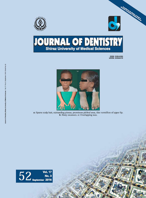
Pages 238-241Langer-Giedion syndrome is a very uncommon autosomal dominant genetic disorder caused by the deletion of chromosomal material. It is characterized by multiple bony exostosis, short stature, mental retardation, and typical facial features. The characteristic appearance of individuals includes sparse scalp hair, rounded nose, prominent philtral area and thin upper lip. Some cases with this condition have loose skin in childhood which typically resolves with age. Oral and dental manifestations include micrognathia, retrognathia, hypodontia, and malocclusion based on cephalometric analysis. This report presents a case of Langer-Giedion syndrome in a 10-year-old child.Keywords: Trichorhinophalangeal Syndrome type 2, Exostosis, Hypodontia, Langer, Giedion Syndrome

