فهرست مطالب

Journal of Dentistry, Shiraz University of Medical Sciences
Volume:17 Issue: 4, Dec 2016
- تاریخ انتشار: 1395/09/21
- تعداد عناوین: 12
-
-
Pages 297-300Statement of the Problem: Prevention is the key factor in acquiring dental and oral health. Community health workers, as a part of health care networks in Iran, play an important role in delivering primary care and their knowledge and attitude directly affect the population whom they interact with in their service scope.PurposeThe aim of this research was to evaluate the knowledge and attitude level of community health workers regarding oral health.Materials And MethodThis descriptive analytical study was carried out on 1170 community health workers who were employed in health offices in East Azerbaijan to evaluate their knowledge and attitude level about oral health. Data were acquired through filled out questionnaires and were analyzed by SPSS software.ResultsThere was no significant statistical relationship between knowledge and gender (p= 0.063), level of education (p= 0.08) and the period spent from the last continuing education course (p= 0.148).However, by increasing age (p= 0.016), work experience (p=0.083) and number of attended continuing education courses (p= 0.023), the knowledge scores were reduced. No statistically significant relationships were found between attitude and any of research variables.ConclusionThe level of knowledge and attitude of community health workers in East Azerbaijan regarding oral health was good. There was a reverse relationship between age, work experience, and frequency of participation in continuing education courses and knowledge scores which emphasizes the necessity of continuous training and revising the method of training in education of community health workers and other staffs of health care system.Keywords: Community Health Worker, Health, Oral, Knowledge, Attitude
-
Pages 301-308Statement of the Problem: Marginal fitness is the most important criteria for evaluation of the clinical acceptability of a cast restoration. Marginal gap which is due to cement solubility and plaque retention is potentially detrimental to both tooth and periodontal tissues.PurposeThis in vitro study aimed to evaluate the marginal and internal fit of cobalt- chromium (Co-Cr) copings fabricated by two different CAD/CAM systems: (CAD/ milling and CAD/ Ceramill Sintron).Materials And MethodWe prepared one machined standard stainless steel master model with following dimensions: 7 mm height, 5mm diameter, 90˚ shoulder marginal finish line with 1 mm width, 10˚ convergence angle and anti-rotational surface on the buccal aspect of the die. There were 10 copings produced from hard presintered Co-Cr blocks according to CAD/ Milling technique and ten copings from soft non- presintered Co-Cr blocks according to CAD/ Ceramill Sintron technique. Marginal and internal accuracies of copings were documented by the replica technique. Replicas were examined at ten reference points under a digital microscope (230X). The Student's t-test was used for statistical analysis. pResultsStatistically significant differences existed between the groups (pConclusionHard presintered Co-Cr copings had significantly higher marginal and internal accuracies compared to the soft non-presintered copings.Keywords: Marginal, Internal fit, CAD, CAM, Ceramill Sintron, Base Metal Alloy
-
Pages 309-317Statement of the Problem: The usage of glass ionomer cements (GICs) restorative materials are very limited due to lack of flexural strength and toughness.PurposeThe aim of this study was to investigate the effect of using a leucite glass on a range of mechanical and optical properties of commercially available conventional glass ionomer cement.Materials And MethodBall milled 45μm leucite glass particles were incorporated into commercial conventional GIC, Ketac-Molar Easymix (KMEm). The characteristics of the powder particles were observed under scanning electron microscopy. The samples were made for each experimental group; KMEm and lucite- modified Ketac-Molar easy Mix (LMKMEm) according to manufacturers instruction then were collected in damp tissue and stored in incubator for 1 hour. The samples were divided into two groups, one stored in distilled water for 24 hours and the others for 1 week.10 samples were made for testing biaxial flexural strength after 1 day and 1 week, with a crosshead speed of 1mm/min, calculated in MPa. The hardness (Vickers hardness tester) of each experimental group was also tested. To evaluate optical properties, 3 samples were made for each experimental group and evaluated with a spectrophotometer. The setting time of modified GIC was measured with Gillmore machine.ResultThe setting time in LMKMEm was 8 minutes. The mean biaxial flexural strength was LMKMEm/ 1day: 24.13±4.14 MPa, LMKMEm/ 1 week: 24.22±4.87 MPa KMEm/1day:28.87±6.31 MPa and KMEm/1 week: 26.65±5.82 MPa which were not statistically different from each other. The mean Vickers hardness was LMKMEm: 403±66 Mpa and KMEm: 358±22 MPa; though not statistically different from each other. The mean total transmittance (Tt) was LMKMEm: 15.9±0.7, KMEm: 22.3±1.2, the mean diffuse transmittance (Td) was LMKMEm: 12.2±0.5, KMEm: 18.0±0.5 which were statistically different from each other.ConclusionLeucite glass can be incorporated with a conventional GIC without interfering with setting time. Yet, it did not improve the mechanical and optical properties of the GIC.Keywords: Dental material, Glass Ionomer Cement, Glass, Mechanical Phenomena, Optical Phenomena
-
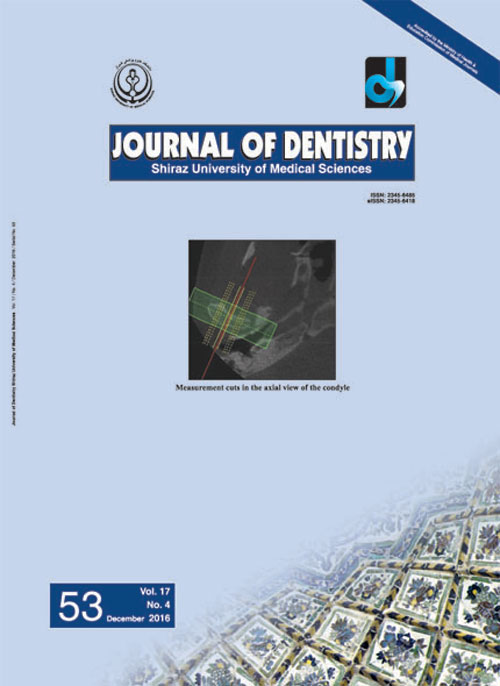
Pages 318-325Statement of the Problem: In orthognathic surgeries, proper condylar position is one of the most important factors in postoperative stability. Knowing the condylar movement after orthognathic surgery can help preventing postoperative instabilities.PurposeThe aim of this study was to evaluate the condylar positional changes after Le Fort I maxillary superior repositioning along with mandibular advancement by using cone beam computed tomography (CBCT).Materials And MethodThis cross-sectional study was conducted on 22 subjects who had class II skeletal malocclusion along with vertical maxillary excess. Subjects underwent maxillary superior repositioning (Le Fort I osteotomy) along with mandibular advancement. The CBCT images were taken a couple of days before the surgery (T0), and one month (T1) and 9 months (T2) after the surgery. The condyles positions were determined from the most superior point of the condyle to three distances including the deepest point of the glenoid fossa, the most anterior-inferior point of the articular eminence, and the most superior point of the external auditory meatus in the sagittal plane.ResultsThe mean mandibular advancement was 4.33±2.1 mm and the mean maxillary superior repositioning was 4.66±0.3 mm. The condyles displaced inferiorly, anteriorly, and laterally between T0 and T1. They were repositioned approximately in the initial position in T2. No correlation was observed between the mandibular and maxillary movement and the condylar positions.ConclusionThe condyles displaced in the inferior-anterior-lateral position one month after the bilateral sagittal split osteotomy for mandibular advancement in combination with the maxillary Le Fort I superior repositioning. It seems that the condyles adapted approximately in their initial position nine months after the surgeries.Keywords: Mandible, Condyle, CBCT, Sagittal Osteotomy, Vertical Maxillary Excess
-
Pages 326-334Statement of the Problem: Similar to conventional amalgam, high-copper amalgam alloy may also undergo corrosion, but it takes longer time for the resulting products to reduce microleakage by sealing the micro-gap at the tooth/amalgam interface.PurposeThe aim of this study was to evaluate the effect of self-etch adhesives with different pH levels on the interfacial corrosion behavior of high-copper amalgam restoration and its induction potential for self-sealing ability of the micro-gap in the early hours after setting by means of Electro-Chemical Tests (ECTs).Materials And MethodThirty cylindrical cavities of 4.5mm x 4.7mm were prepared on intact bicuspids. The samples were divided into five main groups of application of Adhesive Resin (AR)/ liner/ None (No), on the cavity floor. The first main group was left without an AR/ liner (No). In the other main groups, the types of AR/ liner used were I-Bond (IB), Clearfil S3 (S3), Single Bond (SB) and Varnish (V). Each main group (n=6) was divided into two subgroups (n=3) according to the types of the amalgams used, either admixed ANA 2000 (ANA) or spherical Tytin (Tyt). The ECTs, Open Circuit Potential (OCP), and the Linear Polarization Resistance (LPR) for each sample were performed and measured 48 hours after the completion of the samples.ResultsThe Tyt-No and Tyt-IB samples showed the highest and lowest OCP values respectively. In LPR tests, the Rp values of ANA-V and Tyt-V were the highest (lowest corrosion rate) and contrarily, the ANA-IB and Tyt-IB samples, with the lowest pH levels, represented the lowest Rp values (highest corrosion rates).ConclusionSome self-etch adhesives may increase interfacial corrosion potential and self-sealing ability of high-copper amalgams.Keywords: Electrochemical Test, Dental Amalgam, Corrosion, Self, etch adhesive
-
Pages 334-342Statement of the Problem: Oral mucositis (OM) is a common side effect of anti-cancer drugs and needs significant attention for its prevention.PurposeThis study aimed to evaluate the healing effects of olive leaf extract on 5-fluorouracil-induced OM in golden hamster.Materials And MethodOM was induced in 63 male golden hamsters by the combination of 5-fluorouracil injections (days 0, 5 and 10) and the abrasion of the cheek pouch (days 3 and 4). On day 12, hamsters were received topical olive leaf extract ointment, base of ointment, or no treatment (control) for 5 days. Histopathology evaluations, blood examinations, and tissue malondialdehyde level measurement were performed 1, 3 and 5 days after treatments.ResultsHistopathology score and tissue malondialdehyde level were significantly lower in olive leaf extract treated group in comparison with control and base groups (p= 0.000). Significant decreases in white blood cell, hemoglobin, hematocrit , and mean corpuscular volume and an increase in mean corpuscular hemoglobin concentration were observed in olive leaf extract treated group in comparison with control and base groups (pConclusionOur findings demonstrated that daily application of olive leaf extract ointment had healing effect on 5-fluorouracil induced OM in hamsters. Moreover, the beneficial effect of olive leaf extract on OM might be due to its antioxidant and anti-inflammatory properties.Keywords: 5, fluorouracil, Anti, inflammatory, Antioxidant, Olive Leaf, Oral Mucositis
-
Pages 343-347Statement of the Problem: Over the past three decades, significant improvements have been achieved in the survival of children with cancer. However, the considerable morbidity which occurs as a result of chemotherapy often restricts the treatment intensity. One of the important dose-limiting and costly adverse effects of cancer therapy is mucositis. Children with hematological malignancies are greatly at risk of developing mucositis.PurposeThis study aimed to assess the effectiveness of palifermin in preventing mucositis in children with acute lymphocytic leukemia (ALL) who undergo chemotherapy.Materials And MethodIn this clinical trial, 90 children with ALL were randomized to receive chlorhexidine (n=45) or palifermin (n=45). One group received 60 μg/ kg/ day palifermin as an intravenous bolus once daily for 3 days before and 3 days after the chemotherapy. Chlorhexidine mouthwash was administered once daily for 3 days before and 3 days after the chemotherapy. The world health organization (WHO) oral toxicity scale was employed for grading the mucositis. The data were analyzed by using two-way ANOVA.ResultsThe two groups were matched for age and gender. The study groups were significantly different in terms of mucositis grading (P values after 1 and 2 week therapy were 0.00). Palifermin decreased the incidence and severity of chemotherapy-induced mucositis.ConclusionPalifermin reduces the oral mucositis in children with ALL. Several mechanisms of action are suggested for keratinocyte growth factor (such as palifermin) including promotion of cell proliferation and cytoprotection, restraining the apoptosis, and changing the cytokine profile.Keywords: Oral Mucositis, Palifermin, Leukemia
-
Pages 348-353Statement of the Problem: Stem cells from human exfoliated deciduous teeth (SHEDs) are a population of highly proliferative cells, being capable of differentiating into osteogenic, odontogenic, adipocytes, and neural cells. Vitamin D3 metabolites such as 1α, 25-dihydroxyvitamin D3 are key factors in the regulation of bone metabolism.PurposeThe aim of this study was to investigate the effect of 1α, 25-dihydroxyvitamin D3 on osteogenic differentiation (alkaline phosphatase activity and alizarin red staining) of stem cells of exfoliated deciduous teeth.Materials And MethodDental pulp was removed from freshly extracted primary teeth and immersed in a digestive solution. Then, the dental pulp cells were immersed in α-MEM (minimum essential medium) to which 10% fetal bovine serum was added. After the third passage, the cells were isolated from the culture plate and were used for osteogenic differentiation. As a control group, the cells were cultured in osteogenic cell culture medium. As the case group, the cells were cultured in osteogenic culture medium supplemented with 100 nM 1α,25 (OH)2D3. The alkaline phosphatase (ALP) activity and alizarin red staining were analyzed to evaluate the osteogenic differentiation at day 21. The results were analyzed by using t-test.ResultsCompared with the control group, significant increase was observed in ALP activity of SHEDs after being treated with 1α,25(OH)2D3 (p= 0.002). Alizarin red staining demonstrated that the cells exposed to 1α,25(OH)2D3 induced higher mineralized nodules (pConclusionOsteoblast differentiation in SHEDs was stimulated by 1α,25(OH) 2D3. It can be concluded that 1α,25(OH)2D3 can improve osteoblastic differentiation.Keywords: Stem Cells, Dental Pulp, Deciduous Tooth, 1α, 25, dihydroxyvitamin D3
-
Pages 354-360Statement of the Problem: Oral candidiasis is the most common opportunistic infection affecting the human oral cavity. Photodynamic therapy, as one of its proposed treatment modalities, needs a distinct dye for achieving the best effect.PurposeThe purpose of this study was to evaluate photosensitization effects of four distinct dyes on standard suspension of Candida albicans (C. albicans) and Candida dubliniensis (C. dubliniensis) and biofilm of C. albicans considering the obtained optimum dye concentration and duration of laser irradiation.Materials And MethodIn this in vitro study, colony forming units (CFU) of two sets of four groups of Laser plus Dye (L), Dye (L-D), Laser (L) and No Laser, No Dye (L-D-) were assessed individually with different methylene blue concentrations and laser irradiation period. The photodynamic therapy effect on standard suspension of Candida species (using methylene blue, aniline blue, malachite green and crystal violet) were studied based on the obtained results. Similar investigation was performed on biofilm of C. albicans using the spectral absorbance. Data were imported to SPSS and assessed by statistical tests of analysis of variance (ANOVA) and Tukey test (α= 0.05).ResultsCFU among the different dye concentration and irradiation time decrease in dose- and time-dependent manner (p> 0.05), all of which were significantly lower than the control groups (p 0.05) though all of them were significantly decrease CFU compared with the control groups (pConclusionPhotodynamic therapy might be used as an effective procedure to treat Candida associated mucocutaneous diseases and killing biofilm in the infected surfaces such as dentures.Keywords: Candida albicans, Candida dubliniensis, Laser, Photodynamic Therapy
-
Pages 361-366Statement of the Problem: The masseter is generally involved in myofascial pain, myositis, oral submucous fibrosis (OSMF), bruxism, and in subjects with habitual tobacco/arecanut chewing. In all the above conditions, changes in the internal echogenic pattern on ultrasonography of the muscle may be observed.PurposeThe present study aimed at evaluating the internal echogenic pattern of masseter by ultrasonography in subjects with various conditions affecting masster muscle.Materials And MethodThe study subjects were categorized into 5 groups consisting of 20 subjects each with the following conditions; Group 1: myofascial pain or myositis, Group 2: oral submucous fibrosis (OSMF), Group 3: habitual chewing of tobacco/arecanut without OSMF, Group 4: bruxism. Group 5 consisted of 20 healthy subjects. An ultrasonographic examination of masseter was performed in all subjects and the echogenic pattern was classified into Types I, II and III. The images were examined by two observers and inter-observer variability was assessed. Differences in internal echogenic pattern between study groups and control group was evaluated using Chi- square test.ResultsA good inter observer agreement was noted (k value= 0.8). An equal distribution of Types II and III echogenic pattern was noted in myofascial pain/myositis group. Type II was predominant in subjects with OSMF, habitual tobacco/arecanut chewing and bruxism. Type I was predominant in controls. The echogenic pattern differed significantly from controls in subjects with myofascial pain/myositis and OSMF (p=0.00001*, 0.0237* respectively), whereas in subjects with habitual tobacco/ arecanut chewing and bruxism, it did not differ significantly from controls (p=0.2482, 0.1223 respectively).ConclusionUltrasonographic examination of the echogenic pattern may help in understanding the nature of the disease process affecting the masseter muscle in various conditions.Keywords: Ultrasonography, Masseter, Echogenic Pattern, Myofascial Pain, Bruxism
-
Pages 367-369Ameloblastic fibroma is a rare mixed odontogenic tumor mostly occurring in the posterior region of the mandible. The peripheral variant is very rare and to the best of our knowledge, only three cases have been reported in the English literature. In this report, we describe a case of peripheral ameloblastic fibroma in a 54-year-old woman with two years of follow-up.Keywords: Ameloblastic fibroma, Gingiva, Peripheral
-
Pages 370-674Adenomatoid odontogenic tumor (AOT) is an uncommon tumor of odontogenic origin and often misdiagnosed as an odontogenic cyst. It is predominantly found in young female patients, located more often in maxilla, and in most cases associated with an unerupted permanent tooth. There are three variants of AOT namely follicular, extra follicular, and peripheral. We report an unusual case of extrafollicular AOT in maxilla of a 50-year old male patient.Keywords: Adenoameloblastoma, Extrafollicular, Odontogenic Tumors

