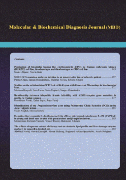
فهرست مطالب
Journal of Molecular and Biochemical Diagnosis
Volume:1 Issue: 1, Spring 2014
- تاریخ انتشار: 1393/10/30
- تعداد عناوین: 7
-
Pages 1-11BackgroundDifferentiation ofmesenchymal stem cells (MSCs) to hepatocyte-like cells could be associated with development of liver function factors. The impact of differentiation-dependent changes on DNA integrity is not well understood. In this study, hepatocytes and their progenitor stem cells were treated with aflatoxin B1 (AFB1) and amplification of selected genes linked to DNA damage was examined.MethodsMSCs and CD34 cells isolated from umbilical cord blood (UCB) were treated with AFB1 (0, 2.5, 10 and 20 µM) in selective media supporting the hepatocyte differentiation. After 24 htreatment the DNA damage (Comet assay) and amplification rates ofP53 and β-globin genes were measured using real time polymerase chain reaction (QPCR).ResultsThe results show that AFB1 treatments resulted in a concentration- dependent increase in the DNA damage and suppression of the specific gene amplification. The extent of DNA damage was significantly greater in hepatocytes differentiated from MSCs when compared to those obtained from CD34 cells. The effects of AFB1 on the rate of selected gene amplification in QPCR showed that the lesions (expressed as lesions/10 kb) in P53 and β-globin genes was significantly greater in hepatocytes derived from MSCs as compared to the cells derived from CD34 cells.ConclusionsThese data together with the results of cytochrome P450 (CYP3A4) expression in the cells suggest that the non-differentiated stem cells are probably less vulnerable to genotoxic agents as compared to hepatocytes differentiated from them.Keywords: Aflatoxin B1, Hepatocytes, Stem cells, Real time PCR, DNA damage, CYP3A4
-
Pages 13-20BackgroundDiabetic retinopathy (DR) is a sight-threatening microvascular complication of diabetes in which the vascular endothelium is damaged due to oxidative stress and inflammation, and vitreous VEGF concentration becomes elevated. The aim of the present study was to assess the association of DR with genetic variations of the MnSOD, a major antioxidant enzyme, and VEGF, an important mediator of neovascularisation, in northern Iran.Methods70 patients with DR and 70 healthy control subjects matched for age and sex was recruited for this study. PCR-based RFLP assay was used to determine the genotypes of MnSODA16V and VEGF C/G polymorphisms.
Results andConclusionsA higher frequency of the AV genotype (71.43%) of the MnSODA16V polymorphism was found in the patients compared with controls which had a 8.33-fold increase in risk of DR (OR= 8.33, 95% CI= 2.56-27.13, P= 0.0004). The frequency of GG, GC, and CC genotypes of VEGF C/G polymorphism in controls were 42.86%, 45.71% and 11.43%, respectively, while in DR patients were 18.57%, 48.57%, and 32.86%, respectively.The allele was considered as a high risk factor of DR (OR= 2.55, 95% CI= 1.57-4.14, P= 0.0001). In conclusion, It is suggested that the MnSODA16V and the VEGF C/G polymorphisms may be associated with the risk of DR in northern Iran.Keywords: Diabetic retinopathy, gene polymorphism, VEGF andMnSOD -
Pages 21-33BackgroundEstrogens play a substantial role in the proliferation, progression and treatment of breast cancer by binding with two estrogen receptors, alpha and beta (ERα and ERβ). Resistance to endocrine therapy is a major problem in the treatment of breast cancers and, in some cases, may be related to loss of ER gene expression. We have already showed that ERα methylation occurs in high frequency and may be one of the important mechanisms for ERα gene silencing in a subset of Iranian primary sporadic breast cancers. In the other hand, the CpG Island methylation status of ERβ and the relationship between clinicopathological features and the pattern of ERβ methylation in sporadic breast cancer are still unknown, especially in Iranian women.MethodsIn this study, we examined the exact role of DNA methylation in the estrogen receptors, alpha and beta genes using Combined Bisulfite Restriction enzyme Analysis (COBRA) and Methylation specific polymerase chain reaction (MSP) methods in 34 tissue and 40 peripheral white blood cells in the breast cancers.
Results andConclusionsERα promoter methylation was identified in 29(72.5%) tissue samples and 35(87.5%) peripheral blood. Among these ERα-methylated cases, the co-occurrentmethylation of ER promoter in peripheral blood and tissue samples was evident in 25 (71.4%) patient (P=0.56). Furthermore, ERβ promoter methylation was detected in 13(32.5%) tissue samples and 4(10.0%) peripheral blood specimens. Of these ERα-methylated cases, the co-ocurrent methylation of ERβ promoter in the peripheral blood and tissue samples was evident in 1(7.7%) patient (P= 0.11). Based on COBRA analysis the percentage of DNA methylation at methylation-sensitive BstUI restriction site of the ERα promoter A ranged from 1% to 91%. The percentages at promoters A region showed a borderline associations with lymph node involvement (P=0.079, r=0.55) and a significant correlation with the grade of tumors (p= 0.27, r=0.65). No significant relation was found between ERα promoter and ERβ promoter methylation (Odds ratio =2.82, 95%, CI =0.2828.5, P=0.36). The methylation of promoter ON was observed in only a subset of tumors without ER by IHC. In addition, we did not find any significant correlationbetween the prognostic factors such as grade, tumor size, lymph node involvement, and methylation status of this promoter. Our results indicate that methylation of ERβ promoter ON is not responsible for the loss of gene expression in of all breast tumors.Keywords: Estrogen receptor, CpG Island, COBRA, Breast Tumors -
Pages 35-40Background And AimsThe E2F family of transcription factors is encoded by at least eight genes, E2F1-8. These proteins are important targets of the retinoblastoma protein (RB) contributed in regulation of transcription, cell cycle and apoptosis. The aim of this study was to investigate the expression levels of E2F protein family including E2F1, E2F2, E2F7, E2F8 and RB1 in the lineage negative (Lin-) hematopoietic stem cells (HSCs) of young and aged black mice using Real Time RT-PCR and western blot techniques.MethodsLin- HSCs of 4 young (7-10 weeks old) and 3 aged (76 weeks old ) mice have been isolated from their bone marrow cells using MACS column and after RNA extraction of culturing cells and cDNA preparation, samples were then analyzed by Real Time RT- PCR and western blot techniques.ResultsThe E2F7 and E2F8 expression levels of the Lin- HSCs of old mice were only the transcriptional factors significantly decreased when compared with young mice.
InConclusionIt seems the functional roles of important E2F7 and E2F8 transcription factors in moderating potentially destructive activity of E2F1 and regulation of cell cycle have been diminished in Lin- HSCs of aged mice. Hence, the apoptotic activity of the E2F1 would affect to the most HSCs, reinforcing bone marrow to proliferate the HSCs in old mice. However, Real Time RT-PCR data showed that the expression level of the E2F1 in those cells was not increased significantly as expected. This is the first report in this regard and further investigation with more samples need to reconfirm these data.Keywords: E2Fs, lineage negative, HSCs, Expression levels -
Pages 41-49BackgroundAccumulative research is in progress to clarify clinical aspects of GBV-C. The possibility of interaction between HCV and GBV-C as well as its consequence on development of liver diseases is the most important clinical aspect which encourages researchers to develop a rapid and cost effective technique for simultaneous detection of both viruses.MethodsIn this study, a SYBR Green real time multiplex RT-PCR technique as a new economical and sensitive method was designed and validated for simultaneous detection of HCV/GBV-C in HCV positive plasma samples. SYBR green real time RT-PCR technique optimization was performed separately for each virus. Multiplex PCR was established next. Standard sera with known concentrations of HCV RNA and dual HCV/GBV-C positive control samples along with negative control samples were used to validate the assay.
Results andConclusionsFifty six non cirrhotic HCV positive plasma samples [29 of genotype 3a and 27 of genotype 1a] were collected from patients before receiving treatment. 20.6% of genotype 3a and 18.7% of genotype 1a showed HCV/GBV-C co-infection. As a result, 19.6% of 56 samples had HCV/GBV-C co-infection that was compatible with other results from all over the world. SYBR Green real time multiplex RT-PCR technique can be used to detect HCV/GBV-C co-infection in plasma samples. Furthermore, with application of this method more time and cost could be saved in clinical-research settings.Keywords: HCV, GBV-C, SYBR Green real time, Rapid detection, multiplex RT- PCR -
Pages 51-58Background And ObjectivesHBV and HTLV-I are life threatening infectious agents in patients who receive blood and blood products. Although serological methods have been proved to be useful, detection of these viruses has remained a challengingissue due to the many obstacles. By the advent of Nucleic Acid Testing methods, especially in multiplex format, more precise detection is possible.The objective of this study was to develop a reliable, rapid and cost- effective method tosimultaneously detect HBV and HTLV-I.Materials And MethodsWe have developed a multiplex Real time-PCR assay for simultaneous detection of HBV and HTLV-I. Primer sets were designed for highly conserved regions of genome of each virus. Using these primers and standard plasmids, we determined the limit of detection, clinical and analytical specificity and sensitivity of the assay. Monoplex and multiplex Real-time PCRs were performed.ResultsAnalytical sensitivity was considered to be 1000 and 100 copies/ml for HBV and HTLV-I, respectively. High concentration of one virus had no adverse effect on detection of t low concentrations of the other one. By analyzing 30 samples, clinical sensitivity of the assay was determined to be 87% and 96% for HBV and HTLV-I, respectively. Using different viral and human genome samples, the specificity of the assay was verified to be 100%.ConclusionsWe have developed a reliable, rapid and cost effective method tosimultaneously detect HBV and HTLV-I.Our results indicatedthe high capability of this simple and rapid method for detecting these viruses in clinical samples.Keywords: Multiplex Real-time PCR, HBV, HTLV-I


