فهرست مطالب

نشریه التیام
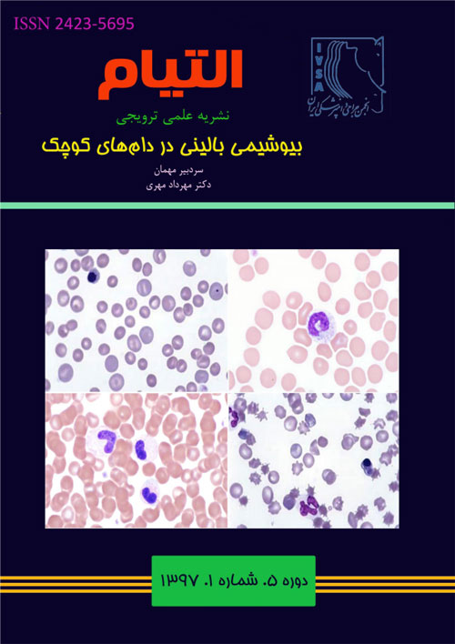
سال پنجم شماره 1 (بهار و تابستان 1397)
- تاریخ انتشار: 1397/04/08
- تعداد عناوین: 8
-
-
صفحات 4-13امروزه تشخیص دقیق بیماری های کبدی نیازمند به کارگیری روش های مختلف تشخیصی و پاراکلینیکی است. از گذشته تشخیص دقیق بیماری های کبدی چالشی جدی در طب انسان و حیوان بوده است. روش های بیوشیمی بالینی به عنوان بخش مهمی از روش های تشخیصی بیماری های کبدی مطرح است. ارزیابی عملکرد کبد از طریق اندازهگیری متغیرهای مرتبط با فعالیت ترشحی کبد، اعمال متابولیسمی و اندازهگیری فعالیتهای آنزیمهای کبدی صورت میگیرد. متاسفانه به علت ظرفیت کارکردی بالای کبد نشانه های مرتبط با با بیماری های کبدی زمانی ظاهر می شود که بخش عمدهای از بافت کبد آسیب دیده باشد. لذا به کارگیری روش های آزمایشگاهی حساس و در عین حال اختصاصی میتواند کمک شایانی در تشخیص به هنگام بیماری های کبدی داشته باشد. در حال حاضر تقریبا هیچ یک از آزمایشهای موجود از ویژگی های یاد شده برخوردار نیستند. به نظر میرسد استفاده از سایر روش های تشخیصی اعم از سونوگرافی، سیتولوژی و… رویکرد مناسبی در جهت تشخیص بیماری های کبدی باشد. مقاله حاضر تلاشی در جهت به کارگیری صحیح آزمایشهای عملکردی کبد در تشخیص بیماری های کبد است.کلیدواژگان: کبد، تشخیص آزمایشگاهی، آنزیم، زردی
-
صفحات 14-23تستهای متداول برای ارزیابی عملکرد کلیه شامل اندازه گیری اوره و کراتینین میباشد که البته مارکرهای غیر مستقیم برای تعیین GFR مثل Cystatin C و Symmetrical Dimethyl Arginine در حال گسترش هستند. با استفاده از این تست ها میتوان به وقوع ازوتمی در بیمار پی برد. در قدم بعدی برای تشخیص و درمان باید ازوتمی را در یکی از گروه های پیش کلیوی، کلیوی و پس کلیوی طبقه بندی کرد. در بیماری های کلیوی قدم بعدی در جهت اطلاع از پیش آگهی طبقه بندی اختلال و نارسایی به حاد و مزمن است. تشخیص بیماری حاد از مزمن براساس تاریخچه مرید و معاینه فیزیکی انجام میشود. کاهش وزن و کم خونی غیر جبرانی ممکن است از نشانه های بیماران مبتلا به CKD باشد. آزمایش مفید دیگر در جهت اطلاع از وضعیت عملکرد کلیه ها و مجاری ادرارای آنالیز کامل ادرار است. آزمایش ادرار کامل اطلاعات ارزشمندی در مورد ازوتمی و دلایل آن میدهد. آزمایشات تکمیلی دیگری نیز جهت تشخیص نارسایی کلیه مثل اندازه گیری فسفر، کلسیم، پتاسیم، وضعیت اسید و باز، کلسترول، آلبومین ادرار، نسبت GGT به کراتینین ادرار وجود دارند. دستهای از مارکرها هم در جهت تشخیص زود هنگام بیماری های کلیوی در حال پژوهش و تجاری سازی میباشند اما هنوز کاربرد بالینی پیدا نکردهاند. از جمله این بیومارکرها میتوان به Cystatin C، Neutrophil gelatinase-associated Lipocalin اشاره کرد.کلیدواژگان: کلیه، بیوشیمی بالینی، ازوتمی، آزمایش ادرار
-
صفحات 24-33پانکراتیت شایعترین بیماری بخش اگزوکرین پانکراس در سگ و گربه است. پانکراتیت بیماری التهابی حاد است که با نکروز و ادم مشخص میشود. فعال شدن زود هنگام تریپسین در سلولهای آسینی پانکراس منجر به بروز آبشاری از واکنشهایی می شود که به آسیب و هضم بافت پانکراس ختم میگردد. علایم بالینی در سگها و گربه های مبتلا غیر اختصاصی است. سگها اغلب علایم گوارشی را بروز میدهند در حالیکه بی اشتهایی و بیحالی از یافته های متداول پانکراتیت حاد در گربه ها میباشد. پانکراتیت حاد ممکن است شوک قلبی عروقی، انعقاد داخل عروقی منتشر، یا از کار افتادن چندین عضو و مرگ را نشان دهند. تشخیص پانکراتیت حاد در سگ و گربه دشوار است. روش های تشخیصی متعددی در طی سالیان گذشته برای تشخیص پانکراتیت مطرح شده است که اکثر آنها به دلیل عملکرد ضعیف، در دسترس نبودن و یا تهاجمی بودن قابل استفاده نیستند. روش های تصویربرداری متعددی نیز استفاده می شود که به جز اولتراسونوگرافی هیچکدام برای تشخیص پانکراتیت کاربردی نیستند. تستهای آزمایشگاهی متعددی شامل اندازه گیری فاکتورهای هماتولوژی و بیوشیمیایی وجود دارد که متاسفانه اختصاصی پانکراتیت نمیباشد و تنها در رد سایر بیماری ها کمک کننده است. هدف از مطالعه حاضر بررسی تستهای تشخیصی اختصاصیتر بیماری پانکراتیت حاد در دامهای کوچک است.کلیدواژگان: پانکراتیت، دام های کوچک، PLI، TLI، TAP
-
صفحات 34-50دیابت ملیتوس یک هیپرگلیسمی پایدار ناشی از کاهش تولید یا اختلال در فعالیت انسولین است. تقریبا تمام دام های اهلی و آزمایشگاهی به دیابت مبتلا میشوند. شیوع دیابت در سگ ها و گربه ها به ترتیب 0/6-0/3 و 1/2-0/43 درصد است. دیابت ملیتوس در سگ ها بیشتر در ماده های بالغ یا مسن رخ میدهد، در حالیکه گربه های نر بیشتر از گربه های ماده به این بیماری مبتلا میشوند. چهار علامت بالینی مشخص دیابت ملیتوس پرادراری، پرنوشی، پرخوری و کاهش وزن هستند. در برخی سگها و گربه های مبتلا عوارض شدید دیابت از قبیل کتواسیدوز یا هیپراسمولالیتی رخ میدهد که در این موارد علائم بالینی همچون بیحالی، کاهش مصرف آب و استفراغ دیده میشود. تشخیص دیابت ملیتوس بر اساس علائم بالینی، هیپرگلیسمی پایدار و گلوکزاوری صورت میگیرد. به منظور مستند نمودن دیابت ملیتوس نیاز به اندازهگیری مکرر گلوکز خون وجود دارد.
البته در صورتیکه در یک نمونه خون و ادرار هیپرگلیسمی همراه با کتونمی، گلوکزاوری و کتوناوری وجود داشته باشد. احتمال وجود دیابت ملیتوس بسیار زیاد است. آزمایش های دیگری از قبیل اندازه گیری فروکتوز آمین (Fructosamine) و هموگلوبین گلیکوزیله خون (Glycated hemoglobin, GHb) و آزمایش های تحمل گلوکز (Glucose tolerance tests) نیز برای تائید یا رد بیماری کمک کننده خواهند بود. در صورتیکه نیاز به تجویز انسولین جهت درمان بیماری وجود دارد، در شروع درمان می توان از منحنی سریالی گلوکز (Serial glucose curve) جهت تعیین نوع و دوز مناسب انسولین استفاده نمود. با گذشت مدت زمانی مشخصی از شروع درمان، برای بررسی روند پاس به درمان و کنترل بیماری در کنار توجه به علائم بالینی و وضعیت عمومی ادرار میتوان از آزمایش های اندازه گیری گلوکز خون و ادرار، فروکتوز آمین و هموگلوبین گلیکوزیله خون استفاده نمود.کلیدواژگان: دیابت ملیتوس، هیپرگلیسمی، تشخیص آزمایشگاهی، سگ، گربه -
صفحات 66-85غدد فوق کلیه از اندام های درون ریز مهم در بدن دام های کوچک هستند که اختلالات مرتبط بتا عملکرد آنها به دلیل عدم تشخیص به موقع در بیشتر موارد خسارات جبران ناپذیر و یا مرگ حیوان را به دنبال دارد. ساختار بافتی این غدد منحصر بته فرد است و بخشهای قشری و داخلی این غدد هر کدام مسئول ترشح هورمونهای مهمی در بدن دامها میباشد. بیماری های عملکردی مهم در میان بیماری های غدد فوق کلیه در دامهای کوچک را میتوان در دو دسته تقسیم بندی کرد. دسته اول بیماری های ناشی از پرکاری و افزایش بیش از حد معمول هورمونهای غدد فوق کلیه هستند که به نامهای هایپرآدرنوکورتیسیسم یا سندرم/بیماری کوشینگ شناخته میشوند و دسته دیگر بیماری های ناشی از کمکاری غدد فوق کلیه هستند که به نامهای هایپوآدرنوکورتیسیسم یا سندرم/بیماری آدیسون شناخته میشوند. بیماری کوشینگ در سگهای مسن و موش خرما شایع است اما به ندرت در گربه ها رخ میدهد. پر ادراری، پرنوشی، بیحالی، شکم متسع و آویزان و مو ریختگی از جمله شایعترین علائم بالینی کوشینگ هستند. لوکوگرام استرس (شامل نوتروفیلی، لنفوپنی، ائوزنوپنی و مونوسیتوز) همراه با افزایش آلکالین فسفاتاز و کاهش وزن مخصوص ادرار (کمتر از 020 /1) مهمترین یافته های آزمایشگاهی کوشینگ هستند که در تشخیص آن کمک کننده خواهند بود. افزایش نسبت کورتیزول به کراتینین ادرار در سگی که علائم بالینی و آزمایشگاهی مرتبط با کوشینگ را نشان میدهد، احتمال وجتود بیماری را تقویت مینماید. در چنین مواردی جهت تشخیص قطعی بیماری میتوان از آزمایش سرکوب دگزامتازون با دوز پایین و یا آزمایش تحریک ACTH استفاده کرد. در صورت تشخیص قطعی کوشینگ، در مرحله بعد با اندازه گیری ACTH خون یا آزمایش سرکوب دگزامتازون با دوز بالا میتوان نوع کوشینگ (وابسته به هیپوفیز یا وابسته بته آدرنال) را نیز تعیین نمود. آدیسون معمولا در سگ های جوان تا میانسال (3-6 ساله) و با علائم بالینی از قبیل بی حالی، ضعف، استفراغ، اسهال، درد شکمی و بی اشتهایی به دلیل کمبود گلوکوکورتیکوئیدها و برادیکاردی، میکروکاردی، کاهش فشار خون، پرادراری و پرنوشی به دلیل کمبود مینرالوکورتیکوئیدها رخ میدهد. مشاهده یافته های آزمایشگاهی از قبیل ازتمی، کاهش وزن مخصوص ادرار، عدم وجود لوکوگرام استرس و به ویژه تغییرات الکترولیت های خون (کاهش سدیم، افزایش پتاسم و نسبت سدیم به پتاسم خون کمتر از 23 احتمال وجود آدیسون را تقویت می کند. در چنین مواردی با اندازه گیری غلظت کورتیزول خون و یا آزمایش تحریک ACTH میتوان وجود بیماری را تائید نمود و در مرحله بعد با اندازه گیری غلظت ACTH خون نوع آن (وابسته به هیپوفیز یا وابسته به آدرنال) را تعیین نمود.کلیدواژگان: غدد فوق کلیه، بیماری آدیسون، بیماری کوشینگ، تشخیص آزمایشگاهی
-
صفحات 86-93آسیبهای عضلانی در دام های کوچک میتواند به دو صورت ارثی و یا اکتسابی و با واسطه عوامل مختلفی نظیر عوامل عفونی، داروها، توکسین ها، سیستم ایمنی، اختلالات اندوکرین و متابولیک بروز یابد. انجام تستهای هماتولوژی، بیوشیمیایی، ایمنولوژی، مولکولی، پاتولوژی و غیره جهت تشخیص و مانیتورینگ این بیماری ها پیشنهاد میگردد. از جمله تستهای بیوشیمیایی میتوان به اندازهگیری فعالیت سرمی آنزیم های آسپارتات آمینوترنسفراز، لاکتات دهیدروژناز، کراتینین کیناز و غلظت سرمی لاکتات، میوگلوبین، تروپونین ها، ناتریورتیک پپتیدها و غیره اشاره نمود. هدف از مطالعه حاضر، معرفی بیومارکرهایی است که امروزه در تشخیص آسیب عضلات اسکلتی و میوکارد در دامهای کوچک مورد استفاده قرار میگیرد و همچنین اطلاعاتی در ارتباط با ساختار، عملکرد، متابولیسم، مقادیر مرجع و کاربرد این بیومارکرها ارائه می شود تا با انتخاب مناسب بیومارکر درک بهتری از وضعیت سلامت عضلات اسکلتی و میوکارد حاصل گردد.کلیدواژگان: میوکارد، عضلات اسکلتی، ناتریورتریک پپتیدها، تروپونین
-
صفحات 94-103آهن یکی از عناصر ضروری برای حیات اغلب موجودات زنده میباشد. اختلال در تنظیم میزان آهن بدن میتواند منجر به بروز تظاهرات بالینی مختلف گردد. اختلالات مرتبط با متابولیسم آهن در پستانداران شامل آنمی فقر آهن، آنمی بر اثر بیماری های مزمن و سرباری آهن یا هماکروماتوز می باشد. مطالعه حاضر به طور خلاصه به متابولیسم آهن در دام های کوچک، اختلالات مرتبط با آن و همچنین روش های ارزیابی وضعیت آهن بدن پرداخته است.کلیدواژگان: آنمی فقر آهن، هموکروماتوز، ترانسفرین، هپسیدین، دام های کوچک
-
Pages 4-13Today’s accurate diagnosis of disease requires using of different diagnostic and paraclinical methods. Diagnosis of liver disease was a serious challenge both in medicine and veterinary medicine from the past. Clinical biochemistry is one of the main parts of diagnostic methods. Liver function is evaluated by measuring the variables such as excreting and metabolic functions and enzymes. Because of large functional reserve of liver, symptoms of liver disease appear after loss of huge number of hepatocytes, therefore using of laboratory methods with high specificity and sensitivity could be helpful. None of existing laboratory methods has all characteristics mentioned above. It seems that using different laboratory methods of liver function beside other diagnostic methods such as sonography, cytology and … could be an appropriate approach for reaching a diagnosis of hepatobiliary disease. Current article reviews the perfect utility of liver function tests for general diagnosis of liver disease.Keywords: Liver, Laboratory Diagnosis, Enzyme, Icterus
-
Pages 14-23Common tests for evaluating renal function include the measurement of urea and creatinine. However, indirect markers for the determination of GFR, such as Cystatin C and Symmetrical Dimethyl Arginine, are in developing. In the next step, for diagnosis and treatment, azotomia should be classified into one of the pre-renal, renal and post-renal groups. In the next step, it is necessary of categorizing the disorder to acute or chronic failure. Diagnosis of chronic or acute illness is done based on the history of the patient and physical examination. Weight loss and non-regenerative anemia may be signs of patients with CKD.Another useful test is urine analysis. A urine test prepared valuable information about azotemia and its causes. Additional tests are also available to diagnose kidney failure such as phosphorus, calcium, potassium, acid-base status, cholesterol, urine albumin, and GGT to urine creatinine ratio. Newbiomarkers such as Cystatin C, a Kidney injury molecule, and Neutrophil gelatinase-associated Lipocalinare also being studied and commercialized for early diagnosis of kidney disease, but they have not yet been clinically available for veterinary use.Keywords: Kidney, Clinical biochemistry, Azotemia, Urine analysis
-
Pages 24-33Pancreatitis is the most common exocrine pancreatic disease in both dogs and cats. Acute pancreatitis is an inflammation with acute onset and characterized by necrosis and edema. Premature activation of trypsin in the acinar cells starts a cascade of reactions that result in autodigestion. Dogs are often presented with gastrointestinal signs, whereas lethargy and anorexia are the most commonly observed symptoms in cats. Acute pancreatitis may cause cardiovascular shock, disseminated intravascular coagulation or disability of multi organs and/or death. Diagnosing acute pancreatitis in dogs and cats is difficult. Several diagnostic methods have been proposed for the diagnosis of pancreatitis over the past few years, most of which are not applicable due to poor performance, inaccessibility or aggressiveness. Besides, many radiographic methods are used yet none of them are efficient except ultrasonography. Although several laboratory tests including measurement of hematology and biochemistry factors are available, none of them are specific for pancreatitis and they are merely beneficial in rejecting other diseases. The aim of this study was to investigate the more specific diagnostic tests for acute pancreatitis in small animals.Keywords: Pancreatitis, Small animal, PLI, TLI, TAP
-
Pages 34-50Diabetes mellitus is a persistent hyperglycemia caused by a deficiency of insulin production or an interference with the action of insulin in target tissues. Although diabetes mellitus has been reported in virtually all animals it is most frequently found in dogs and cats. Estimate of the incidence of diabetes is 0.3%-0.6% for dogs and 0.43%-1.2% for cats. The disease in dogs occurs most frequently in the mature or older female, while male cats appear to be more commonly affected than females. Polyuria, polydipsia, polyphagia and weight loss are the main clinical signs observed in diabetic patients. Sever complications such as ketoacidosis and hyperosmolality might be occurred in some cases resulting in lethargy, reduced water intake and vomiting. Diagnosis of diabetes mellitus should be considered based on related clinical signs, persistent hyperglycemia and glycosuria. Repeating measurements of blood glucose level is necessary for diabetes mellitus diagnosis. However, hyperglycemia along with glycosuria and ketonemia in one sampling could also be diagnostic for the disease. Other laboratory tests including blood fructosamine and glycated hemoglobin and glucose tolerance tests can also be helpful in rule in or rule out of the disease. At the beginning of diabetes mellitus treatment, stablishing a serial glucose curve might be beneficial for finding the best dosage of insulin therapy. Blood and urine glucose, blood fructosamine and glycated hemoglobin are effective indices for evaluating successful insulin therapy.Keywords: Diabetes Mellitus, Hyperglycemia, Laboratory diagnosis, Dog, Cat
-
Pages 51-65Thyroid diseases are among the most common endocrine disorders in small animals. Hypothyroidism is a common disease in dogs, but spontaneous hypothyroidism is very rare in adult cats. Hyperthyroidism is one of the most common diseases of cats and is uncommon in dogs. Hypothyroidism is primarily a disease of middle aged to old dogs with clinical signs including weight gain to obesity, lethargy, dull haircoat, cold intolerance detected as heat-seeking behavior, decreased libido, reproductive failure, alopecia with no pruritus, and hyperpigmentation in areas of alopecia. Laboratory abnormalities may include mild anemia, increased liver enzymes and increases in muscle enzymes (CPK). Hypertriglyceridemia and hyperlipidemia occurs in a majority of cases. Hypercholesterolemia is seen in approximately 80% of hypothyroid dogs and a serum cholesterol concentration greater than 500 mg/dL is very suggestive of hypothyroidism. Basal concentration of total T4 should be the initial endocrine diagnostic test utilized when hypothyroidism is suspected. However, approximately 20% of dogs without hypothyroidism may also have decreased TT4. In addition, total T4 may be in the normal range in about 10% of dogs with hypothyroidism. Therefore, it is important to measure other endocrine tests (free T4 and TSH concentrations). The challenging cases may require more intensive diagnostic procedures such as repeat testing in 4 weeks and/or stimulation tests (TSH or TRH). Hyperthyroidism is the most common endocrine disease of cats. Hyperactivity, weight loss, and polyphagia in a middle aged to old cat are the most frequent clinical problems. Increase in one or more liver enzymes, azotemia, hyperphosphatemia and erythrocytosis are the most consistent lab abnormalities of the hyperthyroid cats. If a cat has some of the physical and clinical laboratory abnormalities characteristic of hyperthyroidism, and an increased TT4 concentration, it is diagnostic of hyperthyroidism and fT4 or any additional tests are not needed. When faced with conflicting clinical signs and lab data while trying to confirm a diagnosis of hyperthyroidism, other endocrine tests such as repeating total T4 in 1-2 weeks, free T4 concentration, T3 suppression test and/or stimulation tests (TSH or TRH) should be considered.Keywords: Hypothyroidism, Hyperthyroidism, Laboratory findings, Diagnosis
-
Pages 66-85The adrenal glands are important organs in the body of small animals, which disruptions associated with their performance due to lack of timely detection in most cases, result in irreparable damage or death of the animal. The tissue structure of these glands is unique and the cortical and internal parts of these glands are responsible for secreting important hormones in the animal's body. In general, important functional diseases among adrenal gland diseases in small animals can be classified into two categories. The first category is the diseases caused by excessive proliferation and excessive growth of the adrenal gland, called hyperadrenocorticism, or cushing's syndrome/disease, and other diseases caused by low activity of adrenal glands that are known as Hypoadrenocorticism or Addison's syndrome/disease. Hyperadrenocorticism is primarily a disease of old dogs and ferrets, but it also occurs rarely in cats. Polyuria and polydipsia, lethargy, pot-belly and alopecia, are among the most frequent clinical signs. A stress leukogram (leukocytosis, mature neutrophilia, lymphopenia, eosinopenia, and monocytosis), increased alkaline phosphatase (ALP) and urine specific gravity <1.020 are good indicators of suspicion to Cushing’s disease. Increased urine cortisol: creatinine ratio (UCCR) in a dog that has classical signs and lab data of Cushing’s is very suggestive of hyperadrenocorticism. If the history, clinical signs, and routine laboratory data are suggestive for hyperadrenocorticism then the diagnosis at this stage is a two-step process: first rule in or rule out hyperadrenocorticism with screening tests [Low Dose Dexamethasome Suppression Test (LDDST) and ACTH stimulation test] and then try to differentiate pituitary and adrenal dependent hyperadrenocorticism with confirmatory tests [blood ACTH concentration and High Dose Dexamethasome Suppression Test (HDDST)]. Addison’s disease usually occurs in young to middle aged (3–6 years) dogs. Lethargy, weakness, vomiting, diarrhea, abdominal pain, anorexia, bradycardia, microcardia, decreased blood pressure, polyuria and polydipsia are the most constant clinical signs of the disease. Laboratory findings including azotemia, decreased urine specific gravity, absence of a stress leukogram and especially a Na: K ratio <23:1 are the key abnormalities to indicate primary hypoadrenocortisim. In such cases, blood cortisol concentration and ACTH stimulation test could be used for diagnosis of Addison’s disease. To differentiate pituitary and adrenal dependent hypoadrenocorticism, the concentration of blood ACTH should be measured.Keywords: Addison’s disease, Cushing’s disease, Laboratory findings, Diagnosis
-
Pages 86-93Muscle diseases can be either inherited or acquired that result from several different disease processes including; infectious, drug- and toxin-induced, and immune mediated, endocrine and metabolic disorders. Standard hematological and biochemical, immunologic, molecular, pathological tests are indicated to diagnosis and monitoring these diseases. From the biochemical tests, the serum activities of aspartate aminotransferase enzymes, lactate dehydrogenase, creatinine kinase, and serum concentration of lactate, myoglobin, troponins, natriuretic peptides, are measured. The aim of this study is to introduce biomarkers that used nowadays to detect skeletal muscle and myocardial damage in small animals. It also provides information on the structure, function, metabolism, reference values and applicability of these biomarkers to provide a better understanding of the health status of skeletal and myocardial muscles by choosing an appropriate biomarker.Keywords: Myocardium, Skeletal muscle, Natriuretic peptides, Troponins
-
Pages 94-103Iron is essential to virtually all living organisms and is integral to multiple metabolic functions. Disorders of iron in the body include iron deficiency anemia, anemia of inflammatory disease, and iron overload. This article summarizes iron metabolism and disorders associated with iron metabolism in small animals and the diagnostic tests currently in use for assessing iron status are discussed.Keywords: Iron deficiency anemia, Hemocromatose, Transferrin, Hepcidin, Small animal


