فهرست مطالب

مجله دانش آزمایشگاهی ایران
سال پنجم شماره 3 (پیاپی 19، پاییز 1396)
- تاریخ انتشار: 1396/08/20
- تعداد عناوین: 6
- اخبار
- استاندارد
- مقالات
-
Page 5
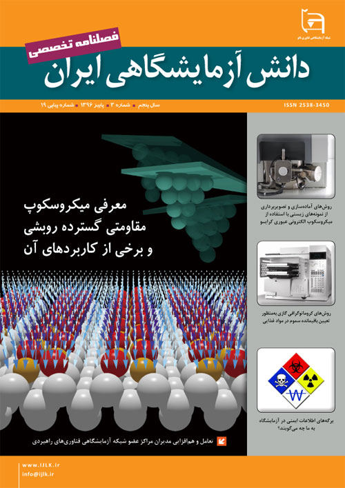
In 16th century, Zacharias Janssen invented the first optical microscope that its operation was based on visible light and a number of lenses. Since then scientists and doctors used this kind of microscope to view their small scale biological samples at 10 to 1000x magnifications. Soon biology scholars noticed the magnification limits of this device doesn’t allow them to view smaller organelles reside in a single cell. After so many experiments, researchers established that acquiring higher magnifications with visible light source is not possible. So about 300 years later in 1930s, two German engineers Max Knoll and Ernst Ruska tested the idea of using electrons instead of light for a microscope and they called their invention ‘Transmission Electron Microscope (TEM)’. A TEM compared to an optical microscope provide a much higher magnification and resolution. The reason for this is that almost every TEM equipped with: a source of electrons (called ‘Electron Gun’), different apertures, lenses, magnetic fields and etc. Image formations in TEMs depend on large amount of electrons passing through a very thin specimen (here thickness plays an important role in final TEM image quality). Choosing the right thickness for specimens (specially: biological tissues and suspensions) will result in images with much better quality and resolution. With respect to recent developments in fields of nanotechnology and medicine, researchers are continuously looking for new ways to keep internal structures of a cell as close as possible to its native state without using harmful chemicals for procedures such as: dehydration, solvent and resin infiltration, positive and negative staining, and etc. in 1980s, a group of scientists and researchers successfully prepared a virus in frozen hydrated state for imaging under a TEM. This ground breaking experiment gave researchers the idea of designing TEMs capable of preserving frozen state of specimens under electron beam. These types of microscopes are called ‘Cryo-Transmission Electron Microscope (Cryo-TEM)’. Cryo is a Greek word that means ‘icy cold’. After going through a short preparation procedure (omitting dehydration) Most of the biological specimens are ready to be imaged inside Cryo-TEM. In this article, available methods for preparing and imaging of different biological specimens inside Cryo-TEM are presented in detail.
Keywords: Cryo-transmission electron microscope, High Pressure Freezing, Plunge Freezing -
Page 15
Gas chromatography (GC) is used widely in applications involving food analysis. Typical applications pertain to the quantitative and/or qualitative analysis of food composition, natural products, food additives, flavor and aroma components, a variety of transformation products, and contaminants, such as pesticides, fumigants, environmental pollutants, natural toxins, veterinary drugs, and packaging materials. The aim of this article is to give a brief overview of the uses of GC/MS in food analysis from sample preparation to final report and GC/MS instrument.
Keywords: Residue pesticide, Sample preparation, Extraction, Column, Detectors, Derivation -
Page 21
In accordance with the international regulations, a safety data sheet (SDS) should be supplied with any hazardous chemical. Safety data sheets (SDSs) provide useful information on chemicals, describing the hazards the chemical presents, and giving information on handling, storage and emergency measures in case of an accident. Over the coming years, SDSs may include further information on safe handling, in the form of exposure scenarios. International regulations requires users of hazardous chemicals to follow the advice on risk management measures given in the exposure scenario, where provided.
Keywords: Material classification systems, Globally Harmonized System, Safety Data Sheet, safety information sheets in the lab -
Page 30
Scanning spreading resistance microscopy is a kind of the atomic force microscopy (AFM), techniques which is widely used for the characterization of semiconducting materials such as silicon materials .In this technique, using commercial diamond coated silicon probe, the resolution is obtained in the range of to 10-20nm. Scanning spreading resistance microscopy (SSRM) has been demonstrated to have attractive sensitivity, and easy measuring quantification of local resistance. So, use of this technique during the recent years has been increased .In this article we present a number of measurement procedures, applications of the scanning spreading resistance and measuring quantification with using this technique.
Keywords: Scanning ion conductance microscopy, living cell, micro pipetteprobe, Electrophysiology


