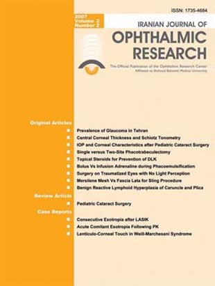
فهرست مطالب

Journal of Ophthalmic and Vision Research
Volume:2 Issue: 2, Automn and Winter 2007
- 8 صفحه، بهای روی جلد: 1,200ريال
- تاریخ انتشار: 1385/10/11
- تعداد عناوین: 12
-
-
Page 93PurposeTo determine the prevalence of glaucoma in adults 40 years of age or older in Tehran, Iran.MethodsThis stratified random-sampling cross-sectional population survey was performed on residents of Tehran, the capital of Iran, aged 40 years and older in the year 2001. Refraction, best-corrected visual acuity, slitlamp biomicroscopy, Goldmann applanation tonometry, funduscopy, and gonioscopy were performed in all subjects. Automated perimetry was performed in selected cases.ResultsOut of 4418 sampled subjects, 2184 individuals (49.4%) participated in the survey. Eventually data from 2160 individuals including 814 (38%) male and 1346 (62%) female subjects with mean age of 55.1±10.2 (range 40-92) years were analyzed. The overall prevalence of glaucoma was 1.44% (95% confidence interval, 0.94-1.94) including primary open angle glaucoma 0.46%, chronic angle closure glaucoma 0.33%, normal tension glaucoma 0.28%, pseudoexfoliation glaucoma 0.23%, and other types of glaucoma 0.14%. More than 80% of affected subjects were unaware of their condition.ConclusionThe prevalence of glaucoma in adults 40 years of age or older in Tehran is 1.44%, which is in the lower range reported in other populations. The large majority of cases are unaware of their condition.
-
Page 101PurposeTo investigate the correlation between central corneal thickness (CCT) and intraocular pressure (IOP) measured by Schiotz and Goldmann tonometers.MethodsCCT and IOP were determined by ultrasonic pachymetry and with the Goldmann and Schiotz tonometers, respectively. The correlation between IOP measured by Goldmann and Schiotz tonometers on one hand and CCT on the other, was determined by regression analysis.ResultsOverall, 168 eyes of 168 glaucoma patients including 85 male (50.6%) and 83 female (49.4%) subjects with mean age of 54.6±19.8 (range 7-85) years were evaluated. Mean CCT was 547.6±53.02 (range: 446-848)? m without any significant difference between men and women (P=0.811). IOP was correlated with CCT with both types of tonometry: every 20? m change in CCT was associated with 1.9±1.4 mmHg and 1.54±0.95 mmHg change in IOP as measured by the Goldmann and Schiotz tonometers respectively with no statistically significant difference between the two devices (P=0.6).ConclusionIOP measurement by Schiotz tonometry is affected by CCT to the same extent as Goldmann applanation tonometry
-
Page 107PurposeTo investigate central corneal thickness (CCT), endothelial cell characteristics and intraocular pressure (IOP) in eyes with prior pediatric cataract surgery and to compare them with eyes of normal age and sex matched controls.MethodsSpecular microscopy CCT and IOP measurements were performed in 31 eyes of 17 patients with prior congenital cataract extraction and 40 eyes of 20 age and sex matched subjects. The mean of three pachymetric and specular microscopic measurements were recorded. IOP was measured using Goldmann applanation tonometry.ResultsMean CCT was 632±45 µm in eyes with prior pediatric cataract surgery vs 546±33 µm in control eyes (P < 0.001, independent t test and Mann Whitney U-test). Mean IOP was 22.1±3.9 mmHg in eyes with prior pediatric cataract surgery and 14.0±1.6 mmHg in the control group (P < 0.001, independent t-test). There was no significant difference between the two groups in cell count, polymegethism and mean cell area of corneal endothelial cells.ConclusionsAlthough the corneas were clinically clear and there was no significant difference in endothelial characteristics in eyes with prior pediatric cataract surgery as compared to normal controls, central corneal thickness in the operated eyes was significantly greater. To differentiate actual glaucoma from artifactual IOP increase, CCT measurement should be performed in these patients.
-
Page 111PurposeTo compare the outcomes of single-site versus two-site mitomycin-C (MMC) augmented phacotrabeculectomy.MethodsThis matched randomized clinical trial included 34 eyes of 30 patients with visually significant cataracts and poorly controlled glaucoma. Equal numbers of eyes were randomly assigned to the single-site and two-site groups. In the single-site approach, phacoemulsification was performed under a superior scleral tunnel followed by trabeculectomy. The two-site approach included a temporal clear corneal phaco-emulsification combined with a separate superior trabeculectomy. MMC 0.2 mg/ml was similarly applied for one minute in both groups.ResultsPatients were followed for a mean period of 13±1.4 (range, 12 to 15) months. Mean best corrected visual acuity one year after surgery was 0.6±0.4 LogMAR in the single-site group and 0.4±0.28 LogMAR in the two-site group (P=0.12). In the single-site group, mean preoperative intraocular pressure (IOP) was 26.4±6.6 mmHg which was decreased to 14.8±2.5 mmHg, one year after the operation (P < 0.001). Corresponding figures for the two-site group were 22.9±3.3 and 13.6±1.7 mmHg respectively (P < 0.001). At final follow up no significant difference in IOP existed between the study groups. Mean number of anti-glaucoma medications was 0.06±0.24 in the two-site group vs 0.43±0.5 in the single-site group (P=0.014). The rate of complications was not different between the study groups (P=1).ConclusionsBoth single-site and two-site phacotrabeculectomy improved visual acuity which was slightly, but not significantly, better with two-site surgery. Final IOP was comparable with both techniques; however, the two-site group required fewer antiglaucoma medications.
-
Page 119PurposeTo determine the role of prophylactic topical steroids in the prevention of diffuse lamellar keratitis (DLK) after laser in situ keratomileusis (LASIK).MethodsThis randomized double blind clinical trial included consecutive LASIK candidates aged 18 to 55 years with stable (1 year or more) myopia ranging from -2 to -12 diopters. The day before surgery, eyes were randomly allocated to topical betamethasone 0.1% or placebo every four hours. One hour preoperatively, the dosage was increased to every five minutes for at least six times. DLK was graded according to the Linebarger-Lindstorm classification. Patients were examined one week and one and three months after surgery. Best-corrected visual acuity (BCVA), manifest and cycloplegic refraction and severity of DLK were documented at each visit by a masked examiner.ResultsOverall, 198 eyes (100 in the treatment group and 98 in the control group) of 101 patients (97 bilateral and 4 unilateral cases) were operated. Pre- and post-LASIK refraction and BCVA were comparable in the study groups (P > 0.05). There were no significant complications in either group during or after LASIK except for DLK which developed in 55 eyes (55%) of the treatment group including 44 eyes with grade I and 11 eyes with grade II, versus 36 eyes (36.7%) of the control group including 29 eyes with grade I and 7 eyes with grade II DLK (P= 0.81).ConclusionAlthough steroids play a key role in the treatment of DLK, pretreatment with topical steroids 24 hours prior to LASIK does not seem prevent this complication.
-
Page 124PurposeTo compare early postoperative corneal endothelial cell density and morphology after phacoemulsification using bolus versus infusion intracameral adrenaline.MethodsIn this randomized clinical trial, 71 eyes of 71 patients scheduled for phaco-emulsification were randomly assigned to two groups: one group (31 eyes) received bolus intracameral adrenaline (1:10,000) and the other group (30 eyes) received adrenaline infusion (1:1,000,000). Pre- and one month postoperatively, a complete ophthalmologic examination as well as endothelial evaluation using ConfoScan III was performed; effective phaco time (EPT) and mydriasis during surgery were also compared between the study groups.ResultsThe two study groups were not significantly different in terms of demographic characteristics, lens opacity and EPT. Endothelial cell density was 2737±321 cell/mm2 in the bolus group vs 2742±426 cell/mm2 in the infusion group preoperatively (P=0.1). One month postoperatively, the rate of cell loss was 7.21% in the infusion group versus 8.87% in the bolus group (P= 0.13). Pupil diameter was > 6 mm in 48% of eyes in the infusion group vs 33% of eyes in the bolus group (P=0.5).ConclusionAdrenaline was safe at the studied concentrations and there was no significant difference between bolus and infusion routes of administration in terms of pupil dilation and endothelial cell loss.
-
Page 129PurposeTo evaluate the anatomical and functional outcomes of surgical intervention in severely traumatized eyes with no light perception (NLP).MethodsIn this prospective interventional case series, 18 eyes of 18 patients with severe ocular trauma whose vision was documented as NLP and with relative afferent pupillary defect (RAPD) of 3-4+ underwent deep vitrectomy and other necessary procedures once to three times.ResultsVision was NLP in all eyes at the time of surgery which was performed 3-14 days after the initial trauma. During a mean follow up period of 20.5±5.2 (range 11 to 49) months, except for one case of phthisis, other eyes achieved acceptable anatomic and functional outcomes. Postoperative vision was NLP in two eyes (11.1%), light perception in three eyes (16.7%), hand motions in four eyes (22.2%), counting fingers in three eyes (16.7%) and 20/200 or better in six eyes (33.3%).ConclusionFollowing eye trauma, NLP vision and RAPD of 3-4+ alone may not be an indication for enucleation. Performing exploratory surgery within 14 days after the injury may salvage the globe and improve vision; this approach may be more acceptable psychologically for patients and relatives.
-
Page 135PurposeTo compare mersilene mesh and autogenous fascia lata for upper lid sling procedure in the management of ptosis with poor levator function.MethodsThis randomized clinical trial included 9 patients with unilateral and 11 patients with bilateral congenital ptosis and poor levator function. All subjects underwent upper lid sling procedure with a random choice of two different materials: mersilene mesh in 16 eyelids and autogenous fascia lata in 15 eyelids.ResultsOverall, 31 eyelids underwent the upper lid sling procedure. There was no difference between the two groups in terms of final functional (lid fissure height stability) and cosmetic (lid margin contour) results. Dermatochalasis was more common in the fascia lata group (10 eyes) compared to the mersilene mesh group (2 eyes). Mersilene mesh extrusion occurred in two eyelids.ConclusionMersilene mesh has favorable long-term functional results and a low rate of complications. This material may be considered as an alternative to autogenous fascia lata for frontalis suspension surgery
-
Page 141PurposeTo report the clinical and histopathologic features of five cases of benign reactive lymphoid hyperplasia (BRLH) of the caruncle and plica semilunaris. PATIENTS ANDFindingsOver a period of ten years, five patients with fish-flesh pinkish mass lesions in the caruncle and plica semilunaris were referred for evaluation. After performing systemic work-up, all lesions were treated by surgical excision. The appearance and clinical course was compatible with BRLH; histopathologic and immunohistochemical evaluations confirmed this diagnosis. The patients were followed for 2-108 months and no recurrence or complications were observed during this period.ConclusionBRLH must be considered in the differential diagnosis of pinkish mass lesions of the caruncle and plica semilunaris. Histopathologic and immunohistochemical evaluation should be performed to rule out lymphoma. Surgical excision seems to be appropriate and does not entail complications in the short-term.
-
Page 154PurposeTo report consecutive exotropia after hyperopic laser in situ keratomileusis (LASIK) in a patient with accommodative esotropia.MethodsA 22-year-old female patient with hyperopia and mild to moderate amblyopia in her right eye was referred for accommodative esotropia. She had undergone right medial rectus recession 4 years ago. Examination revealed right esotropia of 25 and 6 prism diopters (PD) without and with correction, respectively. She underwent LASIK in both eyes to correct the refractive accommodative esotropia. Eight months after LASIK, her refraction was plano-0.75@160 and +0.5-0.75@180 in the right and left eyes respectively, but she developed right exotropia of 20 PD without correction.ConclusionsAlthough hyperopic LASIK may be considered as an alternative treatment for refractive accommodative esotropia, the presence of amblyopia and absence of fusion preoperatively increase the risk of postoperative exotropia.
-
Page 157PurposeTo report consecutive exotropia after hyperopic laser in situ keratomileusis (LASIK) in a patient with accommodative esotropia.MethodsA 22-year-old female patient with hyperopia and mild to moderate amblyopia in her right eye was referred for accommodative esotropia. She had undergone right medial rectus recession 4 years ago. Examination revealed right esotropia of 25 and 6 prism diopters (PD) without and with correction, respectively. She underwent LASIK in both eyes to correct the refractive accommodative esotropia. Eight months after LASIK, her refraction was plano-0.75@160 and +0.5-0.75@180 in the right and left eyes respectively, but she developed right exotropia of 20 PD without correction.ConclusionsAlthough hyperopic LASIK may be considered as an alternative treatment for refractive accommodative esotropia, the presence of amblyopia and absence of fusion preoperatively increase the risk of postoperative exotropia.
-
Page 161PurposeTo report a case of Weill-Marchesani syndrome (WMS) with spontaneous sequential bilateral lenticulo-corneal touch, a previously unreported finding in this condition. CASE REPORT: A 26-year-old woman, a known case of WMS, presented with central lenticulo-corneal touch and corneal edema but normal intraocular pressure (IOP) and a patent laser iridotomy in her left eye. She underwent lensectomy, anterior vitrectomy and iris claw-fixated aphakic intraocular lens implantation. Seven days later a similar presentation developed in her right eye which underwent a similar procedure. Seven months postoperatively, best corrected visual acuity was 20/25, IOP was normal and the corneas were clear in both eyes; central corneal thickness was 555 and 573 µm in the right and left eyes, respectively. Ocular examinations were otherwise unremarkable.ConclusionsSpontaneous lenticulo-corneal touch is a previously unreported finding in Weill-Marchesani syndrome.

