فهرست مطالب

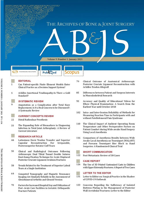
Archives of Bone and Joint Surgery
Volume:10 Issue: 1, Jan 2022
- تاریخ انتشار: 1400/10/13
- تعداد عناوین: 17
-
-
Pages 1-2
The first journal that formalized the peer review process is The Philosophical Transactions of the Royal Society, the first and longest-running journal launched in 1665 by Henry Oldenburg (1618- 1677) (1). The review of a research paper starts with the ‘Internal Review’ process. All authors must read the article and reconcile their comments before submission. The external review process begins after article submission, and the editor assigns the paper to the outside reviewers unrelated to the work of the study. External reviewers evaluate the submitted article for quality, accuracy, and completeness based on the journal’s requirements. Reviewers’ feedback includes accepting, rejecting, or requesting a revision to the article. The editor determines the final decision, but the reviewers’ comments and recommendations show if the article is amenable to improvement by revision.
Keywords: Peer Review, McMaster Online Rating of Evidence, Publones, The Royal Society, Elsevier -
Pages 3-16
Distal radioulnar joint (DRUJ) instability and triangular fibrocartilage complex (TFCC) tears are more usual than estimated and are frequently overlooked. Diagnosis is often clinical, which can be confirmed using computed tomography (CT) scan and magnetic resonance imaging (MRI). In doubtful cases, bilateral computed tomography in neutral forearm rotation, supination, and pronation should also be performed. Wrist arthroscopy can be diagnostic and therapeutic for ulnar-sided wrist pain. Two systematic reviews showed equivalent outcomes between open and arthroscopic repair of the TFCC. There is scant proof to advise one technique over the other in clinical practice. TFCC repair and reconstruction are contraindicated when there is a bony deformation of the radius or ulna or osteoarthritis of the DRUJ. With the advancement of implant arthroplasty, salvage procedures are less desirable. Constrained distal radioulnar arthroplasty is stable, and the longevity is encouraging. Level of evidence: III
Keywords: Distal radioulnar joint, Instability, Triangular fibrocartilage complex, Treatment -
Pages 17-22BackgroundRecent studies have shown that human bone marrow-derived mesenchymal stem cells (hBM-MSCs) have several drawbacks in treating critical-sized bone defect (CSD). Secretome may offer considerable advantages over living cells in terms of potency, manufacturing and storing easiness, and potential as a ready-to-go osteoinductive agent. However, thus far, there are no studies regarding the efficacy of secretome in bone healing. The objective of this study is to investigate the effect of the secretome in rat models with CSD.MethodsThis was an experimental study with post-test only control group design using 60 skeletally mature Sprague Dawley rat which was divided evenly into 5 treatment groups (MSC only, Secretome only, MSC + Secretome, MSC + Secretome + BMP-2, Control group using Normal Saline). We used Bone Marrow derived MSC in this research. The critical-sized bone defect was created by performing osteotomy and defect was treated according to the groups. Rats were sacrificed on 2nd and 4th week and we measured the radiological outcome using Radiographic Union Score for Tibia (RUST) and histomorphometric (callus, osseous, cartilage, fibrous, and void area) evaluation using Image J.ResultsThere was no difference in the weight of rats between groups before and after the intervention. RUST score in all intervention group is significantly higher than the control group, however, the MSC-only group was not statistically significant higher than the control group. There is no statistically significant difference in RUST Score between intervention groups. Histomorphometric evaluation showed that total callus formation is the widest in the MSC+Secretome+BMP-2 combination group while the osseous area is found highest on the secretome-only group.ConclusionSecretome, whether used solely or combined with BM-MSC and BMP-2, is a novel, potent bone-healing agent for CSD in rat models. Level of evidence: VKeywords: Secretome, Mesenchymal stem cell, radiographic, Histomorphometric, Bone Healing, Sprague Dawley Rat
-
Pages 23-31BackgroundThis study assessed the impact of the COVID-19 pandemic on acute upper limb referrals and operative case-mix at the beginning and ease of British lockdown.MethodsA longitudinal multicentre observational cohort study was conducted for both upper limb trauma referrals and operative case-mix over a 12-week period (6 weeks from the beginning and 6 weeks from the ease of the national lockdown). Statistical analysis included median (± median absolute deviation), risk and odds ratios, and Fisher’s exact test to calculate the statistical significance, set at p ≤ 0.05.ResultsThere was a 158% (n = 456 vs. 177) increase in upper limb referrals and 133% (n = 91 vs. 39) increase in the operative trauma caseload at the ease of lockdown compared with its commencement. An increase in sporting injuries was demonstrated (p=0.02), specifically cycling (p=0.004, OR=2.58). A significant increase in COVID-19 testing was demonstrated during the ease of lockdown (p=0.0001) with more patients having their management changed during the early pandemic (9.6% vs. 0.7%, p=0.0001). Of these patients, 47% went on to have delayed surgery within 6 months. No patients who underwent surgery tested positive for COVID-19 infection within 14 days post-operatively and no mortalities were recorded at 30 days.ConclusionThe ease of lockdown has seen upper limb referrals and operations more than double compared to early lockdown. With no patients testing positive for COVID-19 within 14 days of the procedure, this demonstrates that having upper limb surgery during the current pandemic is safe. Level of evidence: IIIKeywords: Trauma, Coronavirus, COVID-19, Upper Limb, lockdown
-
Pages 32-37BackgroundDifferent causative factors for revision total knee arthroplasty (TKA) surgeries are elucidated in the arthroplasty registry data of different countries and the patient records at tertiary care centers. We aimed to determine the changes in the causes for revision TKAs before and after 2011 (The year that the Musculoskeletal Infection Society proposed a new definition for periprosthetic joint infection) and the changes in the demographics of patients who underwent revision TKAs during the same time intervals.MethodsPatients who underwent revision TKAs between 2004 and 2017 were evaluated. A total of 291 patients operated before (period 1, n = 139) and after (period 2, n = 152) 2011 were included, while 53 patients with inconclusive diagnoses were excluded. The demographic data of patients and the causes for revision TKAs were collected and compared between the two periods.ResultsInfection was the most common cause of revision TKAs during periods 1 (58%) and 2 (48%). Aseptic loosening (46%) and infection (37%) were the 2 most common causes for late revisions during period 2. Aseptic loosening almost doubled during period 2 compared with that in period 1. Age, sex, and body mass index distribution in patients were similar across both the periods.ConclusionAlthough the incidence of aseptic loosening has significantly increased since 2011, infection is still the most common cause for revision knee arthroplasty surgery. Level of evidence: IIIKeywords: Revision knee arthroplasty, Periprosthetic joint infection, Aseptic loosening
-
Pages 38-44BackgroundThis study aimed to evaluate the sensitivity and specificity of the leukocyte esterase (LE) band in two groups of patients receiving and not receiving antibiotics and compare the results.MethodsThis prospective cross-sectional study was conducted on 105 joints with clinical suspicion of infectious arthritis (based on Kocher criteria) admitted in Shohada Hospital, Tabriz, Iran, within 2017-2018. Patients were divided into two groups, including receiving antibiotics (n=29; group 1) and not receiving antibiotics (n=76; group 2). Articular fluid aspiration was performed under sterile conditions with an 18-gauge angiocath with at least 1 ml volume of the hip, knee, ankle, elbow, and shoulder joints. Polymorphonuclear cell percentage count, cell count, Gram staining (GS), culture, and leukocyte esterase test were performed immediately after the aspiration of the specimens.ResultsLevels of synovial fluid white blood cell count, serum white blood cell count, PMN, serum glucose, erythrocyte sedimentation rate, C-reactive protein, and time of aspiration (TOA) were significantly higher in the group receiving antibiotics (P<0.05). Synovial glucose levels were significantly lower in the group receiving antibiotics. Furthermore, the positive frequency of glucose esterase, blood culture, GS, serum culture, and ultimate diagnosis of septic arthritis tests were significantly lower in the antibiotic receiving group (P<0.05). The sensitivity, specificity, and positive predictive value of the leukocyte esterase test were obtained at 100%, 0%, and 96.55% in the antibiotic receiving group, respectively. Moreover, in the group not receiving antibiotics, the sensitivity, specificity, positive predictive value, and negative predictive value of the leukocyte esterase test were estimated at 72.22%, 92.50%, 89.66%, 78.72%, respectively.ConclusionAntibiotic use and the prolongation of TOA lead to increased inflammatory products, which is interfering with lab variables. As a result, they increase the sensitivity of the test. The sensitivity and specificity of the leukocyte esterase test in patients who did not receive antibiotics showed that this was a suitable and reliable laboratory method for the rapid detection of infectious arthritis that required an emergency rescue procedure. Level of evidence: IIKeywords: leukocyte esterase test, infectious arthritis, Septic arthritis, Antibiotic
-
Pages 45-51BackgroundThis study aimed to assess the results after soft-tissue posterior instability surgery and address possible challenges during these operations.MethodsThe databases of two tertiary hospitals were reviewed to identify patients treated for posterior shoulder instability between 2000 and 2015. Out of 198 treated patients, 19 cases underwent surgery with a mean follow-up of 35 months. Chart review was performed to obtain recurrence rates, revision rates, return to sport, persistent pain, subjective instability, subjective feeling of being better, complications, and range of motion after operative treatment of posterior shoulder instability. These outcomes were compared using the Fisher’s exact and Mann-Whitney U tests.ResultsAfter surgery, 6 (32%) patients had a recurrent subluxation, and 11 (58%) cases had persistent pain; moreover, 5 (26%) patients had a persistent feeling of instability, and 10 (53%) cases did not feel improvement after the operation. Furthermore, 10 (53%) patients required a revision, and there were 7 (37%) cases with a complication. Postoperatively, 75% of the patients had a full forward flexion, and 93% of the cases had full internal rotation; however, 64% of them had restrictions in external rotation.ConclusionThere is a high rate of recurrent instability, need for revision, and complications after soft-tissue posterior instability surgery. Postoperative external rotation was impaired in most patients. Patients should be informed about these unsatisfactory results. Level of evidence: IVKeywords: Bankart, Dislocation, glenohumeral, Instability, posterior, shoulder, Soft-tissue Surgery
-
Pages 52-55BackgroundScapular fractures are among the orthopedic injuries, which are associated with other injuries, such as lung injuries. This study aimed to evaluate the prevalence of lung injuries associated with scapular fractures in traumatic patients referred to a main trauma center in the south of Iran.MethodsThe present retrospective cross-sectional study was conducted from April 2016 to June 2019 on adult traumatic patients, who were referred to one of the main trauma centers in the south of Iran, and their data were recorded in the hospital information system. The patients with chest computed tomography, and those whose scapula fractures were reported and confirmed by a radiologist were included in this study. All patients' data were extracted from their medical files and then analyzed.ResultsA total of 100 patients were enrolled, and the majority (78%) of the cases were male. The mean±SD age of the patients was 40.71±14.071 years, and 55% of the cases had lung injuries (P=0.158). Furthermore, most of the causes of scapular fracture were due to car-motorcycle accidents (30%) and car overturning (27%). Lung contusion (31%) and hemothorax (30%) were the most types of lung injuries. The mean±SD duration of hospitalization was estimated at 4.94±7.90 days. The mean age (OR=-0.207, P=0.039) and intensive care unit admission rate (OR=0.267, P=0.007) were statistically different in patients with and without lung injuries.ConclusionAlthough scapula fractures were not significantly associated with lung injuries in this study, the occurrence of 55% of the lung injuries was clinically important, which should be considered by emergency physicians. Level of evidence: IIIKeywords: Blunt Injuries, Lung injury, Thoracic Injury, Scapula
-
Pages 56-59BackgroundThe purpose of the present study is to report the incidence of operating room fires during hand surgical procedures.MethodsThe clinic and OR electronic medical records of seven fellowship-trained orthopedic hand surgeons at a single, large practice were retrospectively reviewed. All upper extremity procedures performed between June 2014 to June 2019 in both hospital and surgery center settings were included in the review. Demographic data was collected. The incidence of operating room fires was determined.ResultsA total of 18,819 hand and upper extremity surgical procedures were included. There were 16,767 (89.1%) cases performed in a surgery center, while 2,052 (10.9%) of cases were performed in a hospital. There were 12,691 (67.4%) soft tissue procedures and 6,127 (32.6%) bony procedures performed. Chlorhexidine gluconate preparation solution was used in 9607 cases (51%). Chloraprep solution was used in 6280 cases (33.4%). Betadine was used in 2,932 cases (15.6%). One surgeon has monopolar electrocautery only available during cases. Five surgeons have bipolar available, and one has both mono and bipolar electrocautery available. There were no fires (0%) identified during the study period.ConclusionThe incidence of operating room fires during hand surgical procedures is extremely low. While hand surgeons can be reassured that the likelihood of an operating room fire is minimal, surgeons should not become complacent and should maintain a high level of vigilance to prevent these potentially devastating occurences. Level of evidence: IVKeywords: Operating Room Fires, Hand surgery
-
Pages 60-66BackgroundThe giant cell tumour of the bone has a spectrum of clinical-radiological presentation. This study aims to describe this varied presentation in our institution.MethodsThis retrospective study was conducted on twenty-nine pathologically labelled cases of giant cell tumours of the bone. The medical records for their clinical presentation and diagnostic imaging studies were studied and evaluated.ResultsMean age of the patients at presentation was 35.3±12.9 years. Pain, local swelling and restricted joint function were seen in 93 %, 58.6 % and 52 % patients, respectively. The cortical breach was seen in 15 (51.7 %) and 22 (75.9 %) lesions on plain radiographs and CT images, respectively. 14(48.3 %) cases had soft tissue invasion on MRI at presentation. 26 (89.7 %) lesions were located within 1 cm from the articular cartilage. The solid tumour component was hypo to iso-intense in signal intensity in 27 (93.1 %) lesions in T1 weighted and 21 (72.4 %) in T2 weighted images. 14 (48.3 %) had hyperintense cystic areas, and fluid-fluid levels, suggestive of aneurysmal bone cysts, were seen in 4 (13.8 %) cases on T2 weighted images. Hypo-echoic nodular areas in solid tumour component, suggestive of hemosiderin deposits, were present in 3 (10.3 %) lesions on T1 and T2 weighted images.ConclusionThe tumour classically presents as an epiphysial-metaphyseal, eccentric, expansile, lytic lesion in a skeletally mature patient. The MRI picture is variable and the surgeon should have a sound knowledge of these variations to obtain a biopsy sample from a proper site of the lesion and to avoid misdiagnosis especially of a primary ABC. Level of evidence: IVKeywords: giant-cell, ABCGiant-Cell, soap-bubble, hemosiderin, fluid-fluid, ABC
-
Pages 67-77BackgroundSoft-Tissue Sarcoma (STS) is a heterogeneous group of neoplasms of mesenchymal origin, occurring in connective tissues. According to previously conducted studies, STS accounts for approximately 1% and 7-%15% of adult and pediatric malignancies, respectively. Almost 50%-60% of sarcomas arise from extremities and usually present as a large painless or rarely painful soft-tissue mass. The present study aimed to describe the epidemiology of soft-tissue sarcomas, especially in the Iranian population.MethodsThis epidemiological study of limb soft-tissue sarcoma was conducted based on Iran National Cancer Registry data (INCR) between 2009 and 2014. Patients with soft-tissue sarcoma confirmed by histopathological studies were included, and data were classified based on the International Classification of Diseases for Oncology (first revision-third edition [ICD-O-3]) and analyzed. Descriptive analysis was performed to extract age-standardized and age-specific incidence rates.ResultsA total of 2, 593 patients (1,476 males and 1,117 females) were enrolled and assessed in the present study. The age-standardized incidence rate(ASIR) of total soft tissue sarcomas was 6.34 per million person-years. In addition, the highest and lowest ASIR scores stratified by age were observed in patients aged above 65 and under 0 with the value of 19.61 (95% CI:17.91-21.30) and 1.91 (95% CI 1.69, 2.13) per million, respectively. Limb soft tissue sarcomas stratified by gender were dominant in males, and it was statistically significant (P<0.05). The most common extremity soft tissue sarcomas subtypes were mesenchymal tumor (12.26%), spindle cell sarcoma (12.18%), and malignant fibrous histiocytoma (11.45%).ConclusionAs evidenced by the results of the present study, the ASIR of soft tissue sarcoma dramatically increased with age, and the peak ASIR occurred in the age range of above 65 years. The incidence rate of soft tissue sarcomas analyzed by disease site was higher in hip and lower limb than upper limb and pelvis region, and it was detected consistently in all age groups and both genders. Level of evidence: IVKeywords: Sarcoma, Soft Tissue, Extremities, Soft Tissue Neoplasm
-
Pages 78-84BackgroundBurnout is an emotional, psychological, and physical exhaustion syndrome with feelings of negativismtoward one’s job and reduced attention to clients. This complication is caused by the lack of control over workrelatedstress. Physicians, especially surgeons, are at higher risk for burnout due to critical responsibility and heavyworkload. Given the importance and consequences of this dilemma, the present study aimed to investigate thefrequency of burnout among orthopedic surgeons and residents.MethodsThe present cross-sectional, analytical study was conducted in 2019 in the cities of Tehran and Yazd, inIran. A total of 180 orthopedic surgeons and residents participated in the study. A demographic characteristics formand the Maslach Burnout Inventory (MBI) were employed to assess burnout in the participants.ResultsThe mean age of the participants was 42.8 years, and 94.4%, 23.9%, 52.2%, and 23.9% of the participantswere male, residents, general orthopedic specialists, and fellowship-trained orthopedics, respectively. Out of 180participants, 90 (50%) cases were suffering from burnout, of whom 26.7%, 16.1%, and 7.2% got a pathologicalscore in one, two, and three criteria. No significant relationship was observed between burnout and gender, maritalstatus, years of experience, and the average number of surgeries per week. However, there was a significantassociation between burnout and younger age, lower academic rank or being a resident, working in the publicsector, and spending less time in leisure and sports activities.ConclusionThe prevalence of burnout (50%) among orthopedists was remarkable and worrying. The frequencyof burnout was higher among residents and the ones working in the public sector. This study demonstrates that theissue of burnout and its related risk factors have to be addressed in Iranian orthopedic surgeons and residents.Level of evidence: IVKeywords: Burnout, Orthopedic resident, Surgery
-
Pages 85-91BackgroundCemented Total Knee Arthroplasty (TKA) provides excellent long-term survival rates and functional results, however, radiolucent lines (RLLs) often appear during early post-operative follow-up and their incidence and clinical significance are unknown. The primary aim was to establish the incidence, location, frequency, and time taken for RLLs to appear within the first year after a primary cemented TKA with an anatomic tibial baseplate (Smith and Nephew, LEGION Total Knee System).MethodsThis was a retrospective analysis of 135 primary cemented TKA in 131 patients over three years. We compared demographics, serial radiographs, and early clinical and functional outcomes.ResultsThere were 65 TKAs (48%) in 62 patients who had RLLs within the first year post-operatively. Most were females (58.8%). Mean age was 68.3 ± 7.9 years. There were 88 RLLs, with the most and second commonest location at the medial tibial baseplate (38%) and anterior femoral flange (23%). 89% were in the bone-cement interface. The largest average length of RLLs were at the anterior flange of the femoral component (1.98 ± 1.33 mm). The average time to development was 6.5 ± 4.1 months. None of these patients had infections nor required revision. Patients with RLLs did not do worse in functional and clinical scoring at 1-year.ConclusionThere was a 48% incidence of physiological RLLs after cemented TKA, with the highest occurrence at the medial tibial baseplate at 38%. These radiolucent lines did not affect early post-operative clinical and functional outcomes of patients. Level of evidence: IIIKeywords: Radiolucency, Total knee arthroplasty, Total knee replacement, Cemented
-
Pages 92-97BackgroundDistal pole scaphoid resection (DPSR) is an effective way to manage chronic scaphoid non-union with limited degenerative arthritis. Studies have reported positive results in terms of pain relief, wrist range of motion and grip strength, and patient satisfaction. However, the biomechanical consequences of DPSR remain unclear. This study evaluates the effects of DPSR on carpal mechanics by assessing changes in radiographic parameters with varying quantities of scaphoid removal.MethodsSix fresh frozen cadaveric upper extremities were used. Resections of 25%, 50%, and 75% of the length of each scaphoid were performed under fluoroscopic image guidance. For the intact scaphoid and each resection level, the following radiographic parameters were assessed: radiolunate and capitolunate angles; carpal height and first metacarpal subsidence ratios, and ulnar carpal translation. Measurements were then repeated for grip and pinch as well as radial and ulnar wrist deviation positions. Radial styloid to trapezium distance in wrist radial deviation was also measured to assess for impingement.ResultsThere was a statistically significant increase in the mean radiolunate angle with increasing scaphoid resection quantities. No statistically significant correlations were found between radial styloid clearance and increasing scaphoid resection percentages. Changes in the remaining variables did not reach statistical significance.ConclusionIncreasing levels of scaphoid resection is associated with progressive signs of carpal malalignment best depicted by increasing radiolunate angles. Diminishing radial styloid clearance was clinically evident as more scaphoid was resected. For this, prophylactic radial styloidectomy may be considered to avoid bony impingement. Level of evidence: IVKeywords: Distal Pole Scaphoid, Radiolunate, nonunion
-
Pages 98-103Background
Numerous attempts have been made to decrease the incidence of opioid dependence after orthopedic surgeries. However, no effective means of preoperative risk stratification currently exists. The purpose of this study was to determine the ability of the Opioid Risk Tool (ORT) to predict the rate of opioid dependence 2 years after arthroscopic rotator cuff repair (ARCR).
MethodsWe prospectively evaluated all patients undergoing primary ARCR at a single institution over a 1.5 year period with a minimum of 2-year follow-up. All patients completed the ORT prior to surgery and were stratified into Low, Moderate, and High risk categories. The primary outcome was postoperative opioid dependence, defined as receiving a minimum of 6 opioid prescriptions within 2 years following surgery. Secondary outcomes included the total number of morphine milligram equivalents prescribed, total number of opioid prescriptions filled, and total number of opioid pills prescribed during this time interval. All outcome variables were compared amongst Low, Moderate, and High risk groups. Assessment of a statistical correlation between each outcome variable and individual numerical ORT scores (1-9) was performed.
ResultsA total of 137 patients were included for analysis. No statistically significant difference was noted in any primary or secondary outcome variable when compared between Low, Moderate, and High risk groups. The total cohort demonstrated a 19% rate of post-operative opioid dependence. No correlation was identified between any outcome variable and individual numerical ORT scores. A greater rate of dependence and quantity of opioids prescribed was noted amongst patients with a history of prior opioid use.
ConclusionThe ORT was not predictive of the risk of opioid dependence or quantity of opioids prescribed after ARCR. Attention should be focused on alternative means of identification and management of patients at risk for opioid dependence after orthopedic procedures, including those with a history of prior opioid use. Level of evidence: III
Keywords: Opioid, Dependence, Risk, Arthroscopy, shoulder -
Pages 104-111Background
Several treatment modalities have been reported to minimize the recurrence after surgical treatment of benign bone cysts. In this study, we evaluated local tumor control, recurrence rate, and bone healing of benign bone cysts after treatment with a simple technique, percutaneous curettage and a local autologous cancellous bone graft.
MethodsRetrospective analysis of the records of 16 patients diagnosed with benign bone cysts between 2003 and 2010. We documented the demographic data, radiographic signs of healing (progressive decrease in radiolucency, remineralisation, ossification, consolidation of the cyst, and reconstitution of the bone), healing rate, postoperative complications, and recurrence.
ResultsSeven of the 16 patients (43.75%) were diagnosed with a simple bone cyst (SBC), while nine (56.25%) had an aneurysmal bone cyst (ABC). On average, radiographic signs of healing were present within 3–6 months, but in two patients these signs presented after 16 months. During the follow-up period, there was no difference in the healing rate between patients with SBC and ABC; no signs of deep or superficial wound infection, no postoperative fracture, and no recurrence in any case over an average of 6.3 years of follow-up.
ConclusionTreatment of benign bone cysts (SBC/ABC) with minimally invasive percutaneous curettage and a local autologous cancellous bone graft is a simple and effective modality with a promising outcome in the local control of recurrence and in enhancing bony consolidation. Level of evidence: IV
Keywords: Percutaneous curettage, Autologous, Bone graft, Bone Cysts, simple bone cyst, Aneurysmal bone cyst, Recurrence -
Pages 112-116
Tarsal tunnel syndrome (TTS) is a relatively uncommon nerve entrapment neuropathy. Many pathologies are reported aspossible causes for TTS. The diagnosis of TTS can be difficult and often missed. We present a rare case of TTS due toan accessory flexor digitorum longus muscle. Together with a high index of suspicion, MRI is the investigation of choice inmaking the diagnosis. These patients are best managed with excision or transposition of the flexor digitorum accessoriuslongus (FDAL) and neurolysis of the posterior tibial nerve and its branches.Level of evidence: IV
Keywords: flexor digitorum muscle, plantar fasciitis, Tarsal tunnel syndrome


