فهرست مطالب

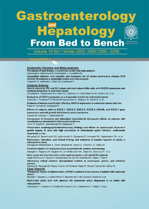
Gastroenterology and Hepatology From Bed to Bench Journal
Volume:15 Issue: 2, Spring 2022
- تاریخ انتشار: 1401/01/10
- تعداد عناوین: 14
-
-
Page 1
Anal fistula refers to a clinical condition with local pain and inflammation associated with purulent discharge that affects the quality of life. Due to the lack of studies, the presence of bias, and high heterogeneity in the studies, the present systematic review is the first to be performed on the population-based database in this field. The present systematic review and meta-analysis was performed according to MOOSE guidelines. After systematic searching in electronic databases, only four articles met the inclusion criteria. After preparing a checklist and extracting data from the relevant articles, a meta-analysis was performed. All studies on the prevalence of anal fistula are related to Europe, and so far, no study has been conducted on other continents. The overall prevalence of anal fistula in European countries was 18.37 (95% CI: 18.20-18.55%) per 100,000 individuals, and the highest prevalence was reported for Italy (23.20 (95% CI: 22.82 to 23.59) per 100,000 people). From the present population-based (224,097,362) study results, it can be concluded that there is a prominent knowledge gap in this context. Because all the studies included in the current study relate only to Europe, the need for further research in this field in other countries is inevitably sensible.
Keywords: Anal Fistulas, Prevalence, Systematic Review -
Page 9Aim
The purpose of the current study is to analyze the potential association between viral hepatitis and the severity of COVID-19.
BackgroundCoronavirus disease 2019 (COVID-19) is a worldwide concern that has created major issues with many aspects. It is important to identify the risk factors for severe outcomes of this disease. To date, no association between viral hepatitis and severe COVID-19 has not been established.
MethodsThrough November 5th, 2020, the databases of PubMed, Google Scholar, and medRxiv were systematically searched using specific keywords related to the focus of the study. All articles published on COVID-19 and viral hepatitis were retrieved. The Mantel- Haenszel formula with random-effects models was used to obtain the risk ratio (RR) along with its 95% confidence intervals (CIs) for dichotomous variables. The two-tailed p-value was set with a value ≤0.05 considered statistically significant. Restricted-maximum likelihood meta-regression was done for several variables, such as age, gender, hypertension, diabetes, and other liver disease.
Results:
Analysis results included a total of 16 studies with a total of 14,682 patients. Meta-analysis showed that viral hepatitis increases the risk of developing severe COVID-19 (RR 1.68 (95% CI 1.26 – 2.22), p = 0.0003, I2 = 21%, random-effect modeling). According to the meta-regression analysis, the association between viral hepatitis and severe COVID-19 was not influenced by age (p = 0.067), diabetes (p = 0.057), or other liver disease (p = 0.646).
ConclusionAn increase of severe COVID-19 risk is associated with viral hepatitis. To reduce the risk of COVID-19, patients with viral hepatitis should be monitored carefully.
Keywords: Hepatitis, Viral infection, Liver disease, Coronavirus, COVID-19 -
Page 15Aim
This study aimed to investigate the effects of natural adjuvants (G2 and PC) to activate natural killer cells in colorectal cancer.
BackgroundNatural killer (NK) cells are an element of the innate immune system that can recognize and kill cancer cells and provide hope for cancer therapy. One of the current methods in cancer immunotherapy is NK cell therapy. Immunotherapy with NK cells has been limited because of the low number and cytotoxicity level of NK cells. Natural adjuvants such as PC and G2 may stimulate the immune system. It seems that these adjuvants could increase cytotoxic NK cells.
MethodsTwelve patients with colorectal cancer and six healthy individuals qualified for inclusion in this study. Peripheral blood mononuclear cells (PBMCs) from each patient with two distinctive concentrations (105and 5×10⁴ cells/well) were treated with Interleukin2 (IL2), PC, and G2 adjuvant separately. The NK cell's surface markers, including CD16, CD56, and NKG2D, were evaluated by flow cytometry. The cytotoxicity effect of treated PBMCs as effector cells against NK sensitive cell line (K562) was assessed using the LDH assay method.
Results:
The results revealed a significant increase in the level of CD16+NKG2D+ NK cells in PBMCs treated with the G2 group compared with the control group in CRC PBMC (p<0.001) as well as the normal PBMC group (p < 0.01). In addition, the results indicated a significant increase in the level of CD56+NKG2D+ cells in the PBMC treated with PC (p < 0.05) and G2 (p < 0.001) groups compared with the PBMC group. The cytotoxicity result of PBMC from CRC patients in 10:1 ratio of the effector: target showed that the cells' cytotoxicity in the PBMCs treated with
PC (p<0.01) and G2 (p<0.05) was significantly higher than the untreated PBMC.ConclusionAccording to the result of this study, it can be stated that the PC and G2 adjuvants could be candidates for inducing cytotoxic natural killer cells.
Keywords: Colorectal cancer, Natural killer cells, PC adjuvant, G2 adjuvant, NKG2D, Cytotoxicity -
Page 24Aim
We aimed to determine the potential of CD10 as a marker for the early diagnosis of adenocarcinoma of the colon.
BackgroundAdenocarcinoma is diagnosed in one out of 20 individuals in the USA and western European countries. Its prognosis and treatment depend largely on the severity of the disease at the time of diagnosis. Additional new biological markers are being sought that can help diagnose colon cancer at an early stage. One such marker present in both serum and tumor tissue is CD10.
MethodsCD10 concentrations were tested by ELISA and immunohistochemistry in serum and tissue samples, respectively, from 113 patients diagnosed histopathologically and treated for adenocarcinoma of the colon. Additionally, the ROC curve with optimal cut-off point based on Youden’s criterion was calculated for CD10.
Results:
Serum concentrations of CD10 and its tissue expression in patients diagnosed with adenocarcinoma of the colon correlate with cancer staging based on the Astler-Coller-Dukes classification. To ascertain the optimal cut-off point for CD10 as a predictor of belonging to the study group, ROC curve was prepared for CD10. Optimal cut-off point for CD10 was 0.57, with prediction of belonging to the study group for CD10 ≥ 0.57.
ConclusionCD10 can be a useful marker in the early diagnosis of adenocarcinoma of the colon.
Keywords: CD10, Colorectal cancer, Immunohistochemistry, ELISA -
Page 32Aim
The current study aimed to focus on the role of histone deacetylation in reduced ARID1A expression in colorectal cancer cell lines.
BackgroundARID1A, a subunit of the switch/sucrose nonfermentable chromatin remodeling complex, has emerged as a bona fide tumor suppressor and is frequently downregulated and inactivated in multiple human cancers. Epigenetic modifications play an important role in dysregulation of gene expression in cancer. DNA methylation has been reported as an important regulator of ARID1A expression in colorectal cancer cell lines; however, the histone modification role in ARID1A suppression in colorectal cancer remains unclear.
MethodsThe expression levels of ARID1A mRNA were determined using real-time quantitative PCR in colorectal cancer cell lines including HCT116, SW48, HT29, SW742, LS180, and SW480. To evaluate the effect of histone deacetylation on ARID1A expression, all cell lines were treated with trichostatin A (TSA), a histone deacetylase inhibitor. SPSS software (Version 23) and GraphPad Prism (Version 6.01) were applied for data analysis using one-way ANOVA, followed by Tukey’s multiple comparison tests.
Results:
Treatment of colorectal cancer cell lines with TSA increased ARID1A expression in a cell line-dependent manner, suggesting that histone deacetylation is at least one factor contributing to ARID1A downregulation in colorectal cancer.
ConclusionHistone deacetylase inhibitors might provide a strategy to restore ARID1A expression and may bring benefits to the
colorectal cancer patients with a broader range of genetic backgrounds.Keywords: ARID1A, Histone acetylation, Colorectal cancer, Epigenetics -
Page 39Aim
The current study aimed to investigate the effect of valproic acid (VPA) on SOCS-1, SOCS-2, SOCS-3, SOCS-5, SOCS6, and SOCS-7 gene expression and cell growth inhibition in colon carcinoma IS1, IS2, and IS3 cell lines.
BackgroundCancer is a process induced by the accumulation of epigenetic alterations such as DNA methylation and histone deacetylation. The DNA methylation and histone deacetylation of tumor suppressor genes (TSGs) have been shown in various cancers. The methylation and deacetylation of suppressors of the cytokine signaling (SOCS) family, as TSGs, have been demonstrated in numerous cancers. Histone deacetylase inhibitors (HDACIs) have emerged as accessory therapeutic agents for human cancers.
MethodsIS1, IS2, and IS3 cells were cultured and treated with VPA. To determine cell viability, cell apoptosis, and the relative gene expression level, MTT assay, flow cytometry assay, and qRT-PCR, respectively, were performed. A database was established with the SPSS 16.0 software package (SPSS Inc., Chicago, Illinois, USA) for analysis. Data was acquired from three tests and is shown as means ± standard deviations. A significant difference was considered as p < 0.05.
Results:
VPA changed the expression levels of the SOCS-1, SOCS-2, SOCS-3, SOCS-5, SOCS6, and SOCS-7 genes, by which cell apoptosis was induced and cell growth inhibited in all three cell lines (p < 0.0001).
ConclusionVPA can induce apoptosis through reactivation of SOCS-1, SOCS-2, SOCS-3, SOCS-5, SOCS6, and SOCS-7 gene expression.
Keywords: Valproic acid, SOCS, Apoptosis, Colon carcinoma -
Page 45Aim
As few randomized clinical trials have verified the efficacy of selective and norepinephrine reuptake inhibitors in IBS, the current study made an inclusive comparison between them, and their effectiveness in IBS-C was proven.
BackgroundIrritable bowel syndrome with constipation (IBS-C) is a functional bowel disorder characterized by changes in bowel movements and abdominal pain in the absence of identifiable structural abnormalities. Despite much progress in the treatment of other types of IBS, limited treatments are available for IBS-C.
MethodsThe study population comprised 182 IBS-C patients who were randomly divided into 3 groups according to treatment type. One group was given 20 mg of dicyclomine and fluoxetine, the second group received dicyclomine along with duloxetine hydrochloride, and the third group received dicyclomine only for two months. The severity of symptoms was recorded by questionnaire at the beginning and end of the treatment.
Results:
The average age and BMI of the patients were 28.5 ± 5.2 years and 25.2 ± 2.4 kg/m2, respectively. Duloxetine was more effective than fluoxetine in reducing flatulence (p=0.043), abdominal pain intensity (p≤0.046), and duration (p≤0.003), in increasing the quality of life (p≤0.046), and the frequency of fecal excretion in patients (p≤0.004).
ConclusionBased on the study findings, fluoxetine and duloxetine had greater therapeutic effects on all symptoms of IBD than dicyclomine, with duloxetine, specifically, being more effective than fluoxetine. Further studies on larger groups are suggested to determine the best dosage and identify any potential side effects of these drugs.
Keywords: Fluoxetine, Duloxetine hydrochloride, Irritable bowel syndrome, Constipation -
Page 53Aim
The current study aimed to evaluate EGD findings effects on laparoscopic Roux-en-Y gastric bypass (RYGB) plan and time in areas with a high prevalence of Helicobacter pylori infection.
BackgroundEsophagogastroduodenoscopy (EGD) and Helicobacter pylori testing are routine parts of preoperative assessment of bariatric surgery at many centers
MethodsThis was a cross-sectional study of all patients underwent EGD and histopathological examination before laparoscopic RYGB in three gastroenterology centers in Iran between January 2018 and December 2020.
Results:
In total, 637 patients (52.4% female) were enrolled, of which 46.8% had no abnormal mucosal appearance. In 1.7%, surgery was canceled (gastric adenocarcinoma, gastric intestinal metaplasia, GIST, and esophageal varices). The prevalence of H. pylori was 61.5%, and there was no statistical difference between groups of normal and abnormal EGD; however, surgery was postponed after H. pylori eradication in both groups. Overall, 44.4% of patients with esophagitis (any grade), peptic ulcer disease, erosive and non-erosive gastritis/duodenitis, and short segment Barret’s esophagus needed medical management. Small- or medium-sized sliding hiatal hernias were seen in 18.7% of patients with no effect on surgery. Moreover, 88.8% of patients with normal mucosal appearance were asymptomatic, but 92.6% in the group with abnormal EGD were symptomatic (p=0.01). Changes in surgical plan and time occurred in 63.6%, but after eliminating H. pylori eradication, it was 15.4%.
ConclusionConsidering gastric cancer and the high prevalence of H. pylori in Iran, using EGD and histopathological examination as an investigation in the preoperative assessment would have a significant impact on patients undergoing RYGB surgery.
Keywords: Gastric bypass surgery, Esophagogastroduodenoscopy, Helicobacter py -
Page 59Aim
Compared to the prevalence and complications, there is still limited evidence in this regard.
BackgroundWith an incidence rate of 200,000 cases annually and the induction of numerous complications, caustic ingestion imposes a significant burden on the healthcare system. Apart from being fatal in some cases, this injury affects its victims’ quality of life as it is followed by many gastrointestinal problems. This injury mainly occurs accidentally among children, whereas in adults, it often occurs with suicidal intentions. Despite recent advances in internal medicine, gastroenterology, and toxicology, this type of injury remains a debilitating and, in some cases, fatal disorder for its victims.
MethodsThis study retrospectively evaluated the clinical, laboratory, and endoscopic findings of 150 patients admitted to a referral center of toxicology and forensic medicine and assessed factors associated with each type of injury.
Results:
The findings indicated a mortality rate as high as 7.3% in this population. Age, pH, and previous medical conditions were associated with more complications. Higher degrees of injury were also significantly associated with higher mortality. No significant difference was observed between types of corrosive substances.
ConclusionIt seems that the most effective intervention for controlling caustic ingestion injuries would be psychiatric support, primary healthcare, and household education.
Keywords: caustic, clinical, endoscopy, ingestion, outcome -
Page 66Aim
This study aimed to evaluate the effects of factors like demographic items, comorbidities, and drug history on the inadequacy of colonic preparation before colonoscopy.
BackgroundInadequate bowel preparation can lead to lower polyp detection rates, longer procedure times, and lower cecal intubation rates.
MethodsThis population-based study was conducted on 2476 Iranian adults who were referred to two tertiary centers for elective colonoscopy between 2017 and 2018. Bowel preparation quality was scored by the Boston bowel preparation scale (BBPS). Univariate and multivariate logistic regressions were used to find the independent predictors of bowel preparation inadequacy.
Results:
The results showed that 31.8% of patients had inadequate bowel preparation before their colonoscopy. Higher age, BMI>25, abdominal circumference>95 cm, low fruit consumption, and history of smoking were independently correlated with bowel preparation inadequacy. Additionally, using NSAIDs and SSRIs were correlated with bowel preparation adequacy in multivariate regression analysis. Finally, age, gender, ethnicity, BMI, abdominal circumference, fruit consumption, smoking, NSAIDs, SSRIs, education, constipation, physical activity, and diabetes entered the predictive model of this study. The area under the curve (AUC) reached 0.70 in the final step.
ConclusionThe independent risk factors associated with colonic preparation inadequacy were identified, and herein, a predictive model is suggested for identifying patients with a high risk of bowel preparation inadequacy before a colonoscopy so that alternative preparation techniques can be employed among high-risk groups to yield optimal preparation quality.
Keywords: Colonoscopy, Colon cleaning, Bowel preparation, Risk factors of bowel cleansing, Quality of colonoscopy -
Page 79Aim
This article aimed to evaluate nitric oxide (NO) and nitric oxide synthase (iNOS) markers in patients with erosive esophagitis (EE) and those with non-erosive reflux disease (NERD) and compare them with the control group.
BackgroundGastro-esophageal reflux disease (GERD) is one of the most common disturbances of the upper digestive tract. Inducible nitric oxide synthase (iNOS) is expressed in esophageal adenocarcinoma. NO, the product of this enzyme, has been implicated in the pathogenesis of this condition. Nevertheless, the data on whether iNOS and NO are expressed in the early stages of GERD is conflicting.
MethodsIn this study, tissue samples were obtained from fifty-four patients (27 with erosive esophagitis and 27 with non-erosive reflux disease) and 27 controls. Tissue concentrations of nitrite, nitrate, and iNOS were measured using Enzyme-Linked Immunesorbent Assay (ELISA). The Bradford method was used to determine the protein concentration of samples. The results were analyzed by SPSS software (version 22.0). In multiple comparisons, the Tukey test was performed, and p < 0.05 was considered as the level of significance.
Results:
Tissue amounts of iNOS were significantly higher (p= 0.001) in EE patients compared with the control group. There was a significant difference (p= 0.01) in this factor between EE patients and patients with NERD. Moreover, tissue levels of nitrite and nitrate were significantly higher (p = 0.001) in patient groups compared with the control group.
ConclusionIt was observed that NO and iNOS protein were increased in human esophagitis tissue. The results indicated that nitric oxide and iNOS levels are useful and effective markers in the pathogenesis of GERD. While the results are not certain, it is thought that a link exists between the expressions of iNOS and disease progression.
Keywords: Gastro-esophageal reflux disease, iNOS, Nitrite, Nitrate, Non-erosive reflux disease, Erosive esophagitis -
Page 87Aim
The current study aimed to determine the common dysregulated proteins between esophageal, gastric, and intestinal cancers.
BackgroundThough there are several documents about the role of AKT1 in promoting of esophageal, gastric, and intestinal cancers, there is not enough evidence about the dominant role of AKT1 relative to the other oncogene genes in the promotion of the three studied cancer types.
MethodsOne hundred proteins related to each of esophageal, gastric, or intestinal cancer were retrieved from the STRING database and interacted by Cytoscape software v 3.2.7. 2 to create the correlated interactomes. The network was analyzed by the “NetworkAnalyzer” application of Cytoscape to find the centrality parameters of the nodes. Results of network analysis and action map assessment were used to determine the common critical proteins between the three studied cancers.
Results:
One hundred proteins were extracted for each of the studied cancers. Among 42 common dysregulated proteins, 36 individuals were selected through network analysis and were screened through action map assessment. Eighteen proteins were introduced as the important common proteins. Finally, AKT1 was a candidate for the crucial dysregulated proteins common in the three analyzed diseases.
ConclusionThe findings indicate that AKT1, relative to the other oncogene genes, is a suitable candidate to be evaluated in patients as a prediagnostic tool to reduce endoscopy and colonoscopy rates.
-
Therapeutic impact of determination of RAS mutations in the plasma of patient with colorectal cancerPage 93
Stage IV colorectal cancer treatment includes targeted therapy depending on RAS status. During disease progression, loss or gain of RAS mutations could happen, supporting the hypothesis of the evolutionary pressure of therapy. Circulating tumor DNA (ctDNA) are nucleic acids released to the bloodstream by the tumor during its development and may be detected by liquid biopsy. The Idylla© Biocartis, a fully automated real-time-PCR-based molecular diagnostic system, was used in a patient with metastatic colorectal cancer with a NRAS mutation in progression after several therapeutic lines. The ctDNA mutational analysis was performed and revealed the absence of mutations in the KRAS, NRAS, and BRAF genes. The patient started the third line of palliative chemotherapy with irinotecan + cetuximab and achieved a partial response for the first time. The authors describe a case in which liquid biopsy determined the higher progression-free survival achieved.
Keywords: colorectal cancer, RAS gene, real-time PCR, circulating tumor cell, molecular targeted therapies -
Page 99
Right-sided aortic arch with aberrant left subclavian artery is a rare congenital anomaly of the aorta that occurs in less than 0.1% of the population. Patients are asymptomatic in most cases, and the anomaly is found incidentally; however, symptoms can occur due to the compression of other structures, mostly the trachea and esophagus. In this report, we present a case of esophageal compression by a right-sided aortic arch with aberrant left subclavian artery that mimicked gastro-esophageal reflux in a 3-month-old (87-day-old) infant with complaint of regurgitation, vomiting, and failure to gain weight who was diagnosed through a barium meal study.
Keywords: Aortic Arch, Regurgitation, Infant


