فهرست مطالب

Cell Journal (Yakhteh)
Volume:25 Issue: 1, Jan 2023
- تاریخ انتشار: 1401/10/12
- تعداد عناوین: 9
-
-
Pages 1-10Objective
Long non-coding RNA (lncRNA) H19 has essential roles in growth, migration, invasion, and metastasis of most cancers. H19 dysregulation is present in a large number of solid tumors and leukemia. However, the expression level of H19 in acute lymphoblastic leukemia (ALL) has not been elucidated yet. The current study aimed to explore H19 expression in ALL patients and cell lines.
Materials and MethodsThis experimental study was conducted in bone marrow (BM) samples collected from 25 patients with newly diagnosed ALL. In addition, we cultured the RPMI-8402, Jurkat, Ramos, and Daudi cell lines and assessed the effects of internal (hypoxia) and external (chemotherapy medications L-asparaginase [ASP] and vincristine [VCR]) factors on h19 expression. The expressions of H19, P53, c-Myc, HIF-1α and β-actin were performed using quantitative real-time polymerase chain reaction (qRT-PCR) method.
ResultsThere was significantly increased H19 expression in the B-cell ALL (B-ALL, P<0.05), T-cell ALL (T-ALL, P<0.01) patients and the cell lines. This upregulation was governed by the P53, HIF-1α, and c-Myc transcription factors. We observed that increased c-Myc expression induced H19 expression; however, P53 adversely affected H19 expression. In addition, the results indicated that chemotherapy changed the gene expression pattern. There was a considerable decrease in H19 expression after exposure to chemotherapy medications; nonetheless, hypoxia induced H19 expression through P53 downregulation.
ConclusionOur findings suggest that H19 may have an important role in pathogenesis in ALL and may act as a promising and potential therapeutic target.
Keywords: Acute Lymphoblastic Leukemia, lncRNA, H19, Hypoxia -
Pages 11-16Objective
Exercise can attenuate mitochondrial dysfunction caused by aging. Our study aimed to compare 12 weeks of high-intensity interval training (HIIT) and moderate-intensity continuous training (MICT) on the expression of mitochondria proteins [e.g., AMP-activated protein kinase (AMPK), Estrogen-related receptor alpha (ERRα), p38 mitogen-activated protein kinase (P38MAPK), and Peroxisome proliferator-activated receptor gamma coactivator 1-alpha (PGC1-α)] in gastrocnemius muscle of old female rats.
Materials and MethodsIn this experimental study, thirty six old female Wistar rats (18-month-old and 270-310 g) were divided into three groups: i. HIIT, ii. MICT, and iii. Control group (C). The HIIT protocol was performed for 12 weeks with 16-28 minutes (2 minutes training with 85-90% VO2max in high intensity and 2 minutes training with 45-75% VO2max low intensity). The MICT was performed for 30-60 minutes with the intensity of 65-70% VO2max. The gastrocnemius muscle expression of AMPK, ERRα, P38MAPK, and PGC1α proteins were determined by Western blotting.
ResultsThe expression of AMPK (P=0.004), P38MAPK (P=0.003), PGC-1α (P=0.028), and ERRα (P=0.006) in HIIT was higher than C group. AMPK (P=0.03), P38MAPK (P=0.032), PGC-1α (P=0.015), and ERRα (P=0.028) in MICT was higher than the C group. Also expression of AMPK (P=0.008), P38MAPK (P=0.009), PGC-1α (P=0.020) and ERRα (P=0.014) in MICT was higher than MICT group.
ConclusionIt seems that exercise training has beneficial effects on mitochondrial biogenesis, but the HIIT training method is more effective than MICT in improving mitochondrial function in aging.
Keywords: Aging, Exercise, Mitochondrial, Muscle -
Pages 17-24Objective
Although the role of obesity and diabetes mellitus (DM) in male infertility is well established, little information about the underlying cellular mechanisms in infertility is available. In this sense, nuclear factor kappa-B (NF-kB) has been recognized as an important regulator in obesity and DM; However, its function in the pathogenesis of male infertility has never been studied in obese or men who suffer from diabetes. Therefore, the main goal of current research is assessing NF-kB existence and activity in ejaculated human spermatozoa considering the obesity and diabetics condition of males.
Materials and MethodsIn an experimental study, the ELISA technique was applied to analyze NF-kB levels in sperm of four experimental groups: non-obese none-diabetic men (body mass index (BMI) <25 kg/m2 ; control group; n=30), obese non-diabetic men (BMI >30 kg/m2 ; OB group; n=30), non-obese diabetic men (BMI <25 kg/m2 ; DM group; n=30), and obese diabetic men (BMI >30 kg/m2 ; OB-DM group; n=30) who were presented to Royan Institute Infertility Center. In addition, protein localization was shown by Immunocytofluorescent assay. Sperm features were also evaluated using CASA.
ResultsThe diabetic men were older than non-diabetic men regardless of obesity status (P=0.0002). Sperm progressive motility was affected by obesity (P=0.035) and type A sperm progressive motility was affected by DM (P=0.034). The concentration of sperm (P=0.013), motility (P=0.025) and morphology (P<0.0001) were altered by obesity × diabetes interaction effects. The NF-kB activity was negatively influenced by the main impact of diabetics (P=0.019). Obesity did not affect (P=0.248) NF-kB activity. Uniquely, NF-kB localized to the midpiece of sperm and post-acrosomal areas.
ConclusionThe current study indicated a lower concentration of NF-kB in diabetic men, no effect of obesity on NF-kB was observed yet. Additionally, we revealed the main obesity and diabetes effects, and their interaction effect adversely influenced sperm characteristics.
Keywords: Diabetes Mellitus, Nuclear factor kappa-B, Obesity Morbid, Spermatozoa, Type II -
Pages 25-34Objective
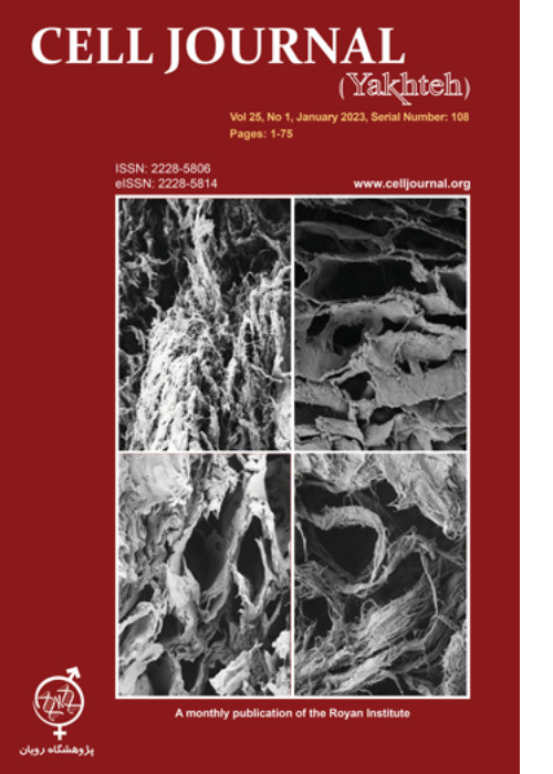
Decellularized uterine scaffold, as a new achievement in tissue engineering, enables recellularization and regeneration of uterine tissues and supports pregnancy in a fashion comparable to the intact uterus. The acellular methods are methods preferred in many respects due to their similarity to normal tissue, so it is necessary to try to introduce an acellularization protocol with minimum disadvantages and maximum advantages. Therefore, this study aimed to compare different protocols to achieve the optimal uterus decellularization method for future in vitro and in vivo bioengineering experiments.
Materials and MethodsIn this experimental study, rat uteri were decellularized by four different protocols (P) using sodium dodecyl sulfate (SDS), with different doses and time incubations (P1 and P2), SDS/Triton-X100 sequentially (P3), and a combination of physical (freeze/thaw) and chemical reagents (SDS/Triton X-100). The scaffolds were examined by histopathological staining, DNA quantification, MTT assay, blood compatibility assay, FESEM, and mechanical studies.
ResultsHistology assessment showed that only in P4, cell residues were completely removed. Masson’s trichrome staining demonstrated that in P3, collagen fibers were decreased; however, no damage was observed in the collagen bundles using other protocols. In indirect MTT assays, cell viabilities achieved by all used protocols were significantly higher than the native samples. The percentage of red blood cell (RBC) hemolysis in the presence of prepared scaffolds from all 4 protocols was less than 2%. The mechanical properties of none of the obtained scaffolds were significantly different from the native sample except for P3.
ConclusionUteri decellularized with a combination of physical and chemical treatments (P4) was the most favorable treatment in our study with the complete removal of cell residue, preservation of the three-dimensional structure, complete removal of detergents, and preservation of the mechanical property of the scaffolds.
Keywords: Acellularization, Female Infertility, Rat Uterus, Sodium Dodecyl Sulfate, Tissue Engineering -
Pages 35-44Objective
Organ transplantation is the last therapeutic choice for end-stage liver failure, which is limited by the lack of sufficient donors. Decellularized liver can be used as a suitable matrix for liver tissue engineering with clinical application potential. Optimizing the decellularization procedure would obtain a biological matrix with completely removed cellular components and preserved 3-dimensional structure. This study aimed to evaluate the decellularization efficacy through three anatomical routes.
Materials and MethodsIn this experimental study, rat liver decellularization was performed through biliary duct (BD), portal vein (PV), and hepatic vein (HV); using chemical detergents and enzymes. The decellularization efficacy was evaluated by measurement of DNA content, extracellular matrix (ECM) total proteins, and glycosaminoglycans (GAGs). ECM preservation was examined by histological and immunohistochemical (IHC) staining and scanning electron microscopy (SEM). Scaffold biocompatibility was tested by the MTT assay for HepG2 and HUVEC cell lines.
ResultsDecellularization through HV and PV resulted in a transparent scaffold by complete cell removal, while the BD route produced an opaque scaffold with incomplete decellularization. H&E staining confirmed these results. Maximum DNA loss was obtained using 1% and 0.5% sodium dodecyl sulfate (SDS) in the PV and HV groups and the DNA content decreased faster in the HV group. At the final stages, the proteins excreted in the HV and PV groups were significantly less than the BD group. The GAGs level was diminished after decellularization, especially in the PV and HV groups. In the HV and PV groups the collagen amount was significantly more than the BD group. The IHC and SEM images showed that the ECM structure was preserved and cellular components were entirely removed. MTT assay showed the biocompatibility of the decellularized scaffold.
ConclusionThe results revealed that the HV is a more suitable route for liver decellularization than the PV and BD.
Keywords: Biliary Duct, Decellularization, Hepatic Vein, Portal Vein, Tissue Engineering -
Pages 45-50Objective
Preeclampsia (PE) is a pregnancy related disorder with prevalence of 6-7%. Insufficient trophoblastic invasion leads to incomplete remodeling of spiral arteries and consequent decrease in feto-placental perfusion. Altered placental expression of tissue inhibitors of matrix metalloproteinase (TIMPs) is considered to be involved in this process while the balance between matrix metalloproteinases (MMPs) and TIMPs contributes to remodeling of the placenta and uterine arteries by degradation and refurbishing of extracellular matrix (ECM). Therefore, TIMPs, fetal expression pattern was evaluated with the aim of its potential to be used as a determinant for the (early) detection of PE.
Materials and MethodsIn this case-control study, cell free fetal RNA (cffRNA) released by placenta into the maternal blood was used to determine expression patterns of TIMP1, 2, 3 and 4 in the severe preeclamptic women in comparison with the normal pregnant women. Whole blood from 20 preeclamptic and 20 normal pregnant women in their 28-32 weeks of gestational age was collected. The second control group consisted of 20 normal pregnant women in either 14 or 28 weeks of gestation (each 10). cffRNA was extracted from plasma and real-time polymerase chain reaction (PCR) was done to determine the expression levels of TIMP1, 2, 3 and 4 genes.
ResultsStatistical analysis of the results showed significant higher expression of TIMP1-4 in the preeclamptic women in comparison with the control group (P=0.029, 0.037, 0.037 and 0.049, respectively). Also, an increased level of TIMPs expression was observed by comparing 14 to 28 weeks of gestational age in the normal pregnant women in the second control group.
ConclusionAn increased cffRNA expression level of TIMPs may be correlated with the intensity of placental vascular defect and may be used as a determinant of complicated pregnancies with severe preeclampsia.
Keywords: Cell Free Fetal RNA, Gene Expression, Preeclampsia, Tissue Inhibitors of Matrix Metalloproteinase -
Pages 51-61Objective
The multimodality treatment of cancer provides a secure and effective approach to improve the outcome of treatments. Cold atmospheric plasma (CAP) has got attention because of selectively target and kills cancer cells. Likewise, gold nanoparticles (GNP) have been introduced as a radiosensitizer and drug delivery with high efficacy and low toxicity in cancer treatment. Conjugating GNP with indocyanine green (ICG) can develop a multifunctional drug to enhance radio and photosensitivity. The purpose of this study is to evaluate the anticancer effects of GNP@ICG in radiotherapy (RT) and CAP on DFW melanoma cancer and HFF fibroblast normal cell lines.
Materials and MethodsIn this experimental study, the cells were irradiated to RT and CAP, alone and in combination with or without GNP@ICG at various time sequences between RT and CAP. Apoptosis Annexin V/PI, MTT, and colony formation assays evaluated the therapeutic effect. Finally, the index of synergism was calculated to compare the results.
ResultsMost crucially, the cell viability assay showed that RT was less toxic to tumors and normal cells, but CAP showed a significant anti-tumor effect on melanoma cells with selective toxicity. In addition, cold plasma sensitized melanoma cells to radiotherapy so increasing treatment efficiency. This effect is enhanced with GNP@ICG. In comparison to RT alone, the data showed that combination treatment greatly decreased monolayer cell colonization and boosted apoptotic induction.
ConclusionThe results provide new insights into the development of better approaches in radiotherapy of melanoma cells assisted plasma and nanomedicine.
Keywords: Apoptosis, Cold Plasma, Indocyanine Green, Melanoma, Radiation Therapy -
Pages 62-72Objective
Despite of antiviral drugs and successful treatment, an effective vaccine against hepatitis C virus (HCV) infection is still required. Recently, bioinformatic methods same as prediction algorithms, have greatly contributed to the use of peptides in the design of immunogenic vaccines. Therefore, finding more conserved sites on the surface glycoproteins (E1 and E2) of HCV, as major targets to design an effective vaccine against genetically different viruses in each genotype was the goal of the study.
Materials and MethodsIn this experimental study, 100 entire sequences of E1 and E2 were retrieved from the NCBI website and analyzed in terms of mutations and critical sites by Bioedit 7.7.9, MEGA X software. Furthermore, HCV-1a samples were obtained from some infected people in Iran, and reverse transcriptase-polymerase chain reaction (RTPCR) assay was optimized to amplify their E1 and E2 genes. Moreover, all three-dimensional structures of E1 and E2 downloaded from the PDB database were analyzed by YASARA. In the next step, three interest areas of humoral immunity in the E2 glycoprotein were evaluated. OSPREY3.0 protein design software was performed to increase the affinity to neutralizing antibodies in these areas.
ResultsWe found the effective in silico binding affinity of residues in three broadly neutralizing epitopes of E2 glycoprotein. First, positions that have substitution capacity were detected in these epitopes. Furthermore, residues that have high stability for substitution in these situations were indicated. Then, the mutants with the strongest affinity to neutralize antibodies were predicted. I414M, T416S, I422V, I414M-T416S, and Q412N-I414M-T416S substitutions theoretically were exhibited as mutants with the best affinity binding.
ConclusionUsing an innovative filtration strategy, the residues of E2 epitopes which have the best in silico binding affinity to neutralizing antibodies were exhibited and a distinct peptide library platform was designed.
Keywords: Broadly Neutralizing Antibody, Mutation, Peptide Library, Sequence Analysis -
Pages 73-75
Considering HER2 as one of the well-known biomarkers in the cancer field, and published articles regarding serum levels of HER2, in this paper we tried to highlight the issue that most studies don’t stratify the HER-2 concentration of individuals in terms of gender. In this brief survey, healthy individuals with no prior non-communicable diseases were categorized as males (n=34) and females (n=43), and all samples were evaluated for plasma HER-2 levels at once. Surprisingly, the plasma level of HER-2 of healthy male individuals (mean= 2.28 ± 0.21 ng/mL) was significantly (P<0.0001) higher than the plasma level of HER-2 of healthy females (mean: 0.06 ± 0.09 ng/mL), with no overlap. Therefore, we suggest that more studies are required to re-check the cutoff values for HER-2 plasma levels based on gender since the clinical implications of a unique HER-2 cutoff for both genders may be seriously concerning.


