فهرست مطالب

Cell Journal (Yakhteh)
Volume:25 Issue: 4, Apr 2023
- تاریخ انتشار: 1402/02/31
- تعداد عناوین: 8
-
-
Pages 217-221Objective
Recent studies imply extensive applications for the human amniotic membrane (hAM) and its extract in medicine and ophthalmology. The content of hAM meets many requirements in eye surgeries, such as refractive surgery as the most important and commonly used method for treating the dramatically increasing refractive errors. However, they are associated with complications such as corneal haziness and corneal ulcer. This study was designed to evaluate the impact of amniotic membrane extracted eye drop (AMEED) on Trans-PRK surgery complications.
Materials and MethodsThis interventional and historical study was performed during two years (July 1, 2019-September 1, 2020). Thirty-two patients (64 eyes), including 17 females and 15 males, aged 20 to 50 years (mean age of 29.59 ± 6.51) with spherical equivalent between -5 to -1.5 underwent Trans Epithelial Photorefractive Keratectomy (Trans-PRK) surgery. One eye was selected per case (case group) and the other eye was considered as control. Randomization was done using the random allocation rule. The case group was treated with AMEED, and the artificial tear drop every 4 hours. The control eyes received artificial tear drops instilled every 4 hours. The evaluation continued for three days after the Trans-PRK surgery.
ResultsA significant decrease in CED size was found in the AMEED group on the second day after surgery (P=0.046). Also, this group had a substantial reduction in pain, hyperemia, and haziness.
ConclusionThis study showed that AMEED drop can increase the healing rate of corneal epithelial lesions after Trans- PRK surgery and reduce the early and late complications of Trans-PRK surgery. Researchers and Ophthalmologists should consider AMEED as a selection in patients with persistent corneal epithelial defects and patients who have difficulty in corneal epithelial healing. We understood AMEED has a different effect on the cornea after surgery; therefore, the researcher must know AMEED’s exact ingredients and help expand AMEED uses.
Keywords: Amnion, Photorefractive Keratectomy, Refractive Surgical Procedures, Wound Healing -
Pages 222-228Objective
Gastric cancer is the fifth most common neoplasm and the fourth reason for mortality globally. Incidence rates are highly variable and dependent on risk factors, epidemiologic and carcinogenesis patterns. Previous studies reported that Helicobacter pylori (H. pylori) infection is one the strongest known risk factor for gastric cancer. USP32 is a deubiquitinating enzyme identified as a potential factor associated with tumor progression and a key player in cancer development. On the other hand, SHMT2 is involved in serine-glycine metabolism to support cancer cell proliferation. Both USP32 and SHMT2 are reported to be upregulated in many cancer types, including gastric cancer, but its complete mechanism is not fully explored yet. The present study explored possible mechanism of action of USP32 and SHMT2 in the progression of gastric cancer.
Materials and MethodsIn this experimental study, Capsaicin (0.3 g/kg/day) and H. pylori infection combination was used to successfully initiate gastric cancer conditions in mice. It was followed by 40 and 70 days of treatment to establish initial and advanced conditions of gastric cancer.
ResultsHistopathology confirmed formation of signet ring cell and initiation of cellular proliferation in the initial gastric cancer. More proliferative cells were also observed. In addition, tissue hardening was confirmed in the advanced stage of gastric cancer. USP32 and SHMT2 showed progressive upregulated expression, as gastric cancer progress. Immunohistologically, it showed signals in abnormal cells and high-intensity signals in the advanced stage of cancer. In USP32 silenced tissue, expression of SHMT2 was completely blocked and reverted cancer development as evident with less abnormal cell in initial gastric cancer. Reduction of SHMT2 level to one-fourth was observed in the advanced gastric cancer stages of USP32 silenced tissue.
ConclusionUSP32 had a direct role in regulating SHMT2 expression, which attracted therapeutic target for future treatment.
Keywords: Cancer, Gastric Cancer, H. pylori, SHMT2, USP32 -
Pages 229-237Objective
The study of pathophysiology as well as cellular and molecular aspects of diseases, especially cancer, requires appropriate disease models. In vitro three-dimensional (3D) structures attracted more attention to recapitulate diseases rather than in vitro two-dimensional (2D) cell culture conditions because they generated more similar physiological and structural properties. Accordingly, in the case of multiple myeloma (MM), the generation of 3D structures has attracted a lot of attention. However, the availability and cost of most of these structures can restrict their use. Therefore, in this study, we aimed to generate an affordable and suitable 3D culture condition for the U266 MM cell line.
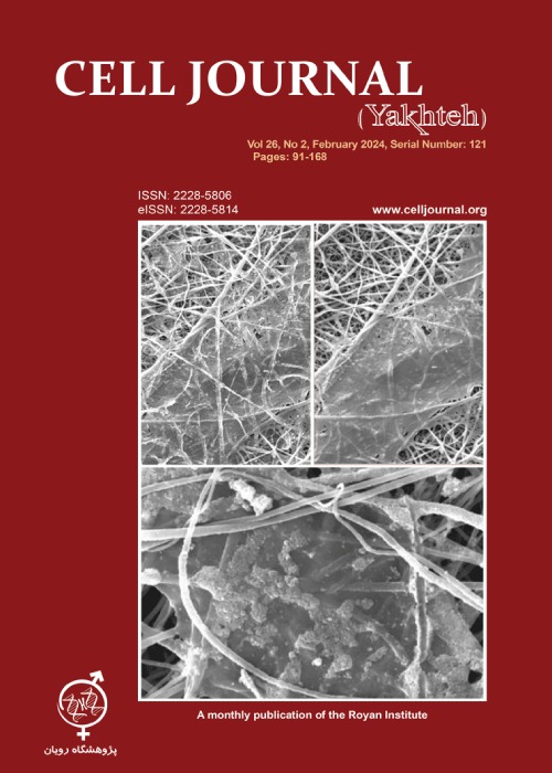
Materials and MethodsIn this experimental study, peripheral blood-derived plasma was used to generate fibrin gels for the culture of U266 cells. Moreover, different factors affecting the formation and stability of gels were evaluated. Furthermore, the proliferation rate and cell distribution of cultured U266 cells in fibrin gels were assessed.
ResultsThe optimal calcium chloride and tranexamic acid concentrations were 1 mg/ml and 5 mg/ml for gel formation and stability, respectively. Moreover, the usage of frozen plasma samples did not significantly affect gel formation and stability, which makes it possible to generate reproducible and available culture conditions. Furthermore, U266 cells could distribute and proliferate inside the gel.
ConclusionThis available and simple fibrin gel-based 3D structure can be used for the culture of U266 MM cells in a condition similar to the disease microenvironment.
Keywords: Blood Plasma, Fibrin, Multiple Myeloma, Three-Dimensional Culture -
Pages 238-246Objective
Choosing the optimal method for human sperm cryopreservation seems necessary to reduce cryoinjury. The aim of this study is to compare two cryopreservation methods including rapid-freezing and vitrification, in terms of cellular parameters, epigenetic patterns and expression of paternally imprinted genes (PAX8, PEG3 and RTL1) in human sperm which play a role in male fertility.
Materials and MethodsIn this experimental study, semen samples were collected from 20 normozoospermic men. After washing the sperms, cellular parameters were investigated. DNA methylation and expression of genes were investigated using methylation-specific polymerase chain reaction (PCR) and real-time PCR methods, respectively.
ResultsThe results showed a significant decrease in sperm motility and viability, while a significant increase was observed in DNA fragmentation index of cryopreserved groups in comparison with the fresh group. Moreover, a significant reduction in sperm total motility (TM, P<0.01) and viability (P<0.01) was determined, whereas a significant increase was observed in DNA fragmentation index (P<0.05) of the vitrification group compared to the rapid-freezing group. Our results also showed a significant decrease in expression of PAX8, PEG3 and RTL1 genes in the cryopreserved groups compared to the fresh group. However, expression of PEG3 (P<0.01) and RTL1 (P<0.05) genes were reduced in the vitrification compared to the rapid-freezing group. Moreover, a significant increase in the percentage of PAX8, PEG3 and RTL1 methylation was detected in the rapid-freezing group (P<0.01, P<0.0001 and P<0.001, respectively) and vitrification group (P<0.01, P<0.0001 and P<0.0001, respectively) compared to the fresh group. Additionally, percentage of PEG3 and RTL1 methylation in the vitrification group was significantly increased (P<0.05 and P<0.05, respectively) compared to the rapid-freezing group.
ConclusionOur findings showed that rapid-freezing is more suitable method for maintaining sperm cell quality. In addition, due to the role of these genes in fertility, changes in their expression and epigenetic modification may affect fertility.
Keywords: Epigenetics, Male Fertility, Rapid-Freezing, Vitrification -
Pages 247-254Objective
Thyroid hormones are involved in the pathogenesis of various neurological disorders. Ischemia/hypoxia that induces rigidity of the actin filaments, which initiates neurodegeneration and reduces synaptic plasticity. We hypothesized that thyroid hormones via alpha-v-beta-3 (αvβ3) integrin could regulate the actin filament rearrangement during hypoxia and increase neuronal cell viability.
Materials and MethodsIn this experimental study, we analysed the dynamics of actin cytoskeleton according to the G/F actin ratio, cofilin-1/p-cofilin-1 ratio, and p-Fyn/Fyn ratio in differentiated PC-12 cells with/without T3 hormone (3,5,3'-triiodo-L-thyronine) treatment and blocking αvβ3-integrin-antibody under hypoxic conditions using electrophoresis and western blotting methods. We assessed NADPH oxidase activity under the hypoxic condition by the luminometric method and Rac1 activity using the ELISA-based (G-LISA) activation assay kit.
ResultsThe T3 hormone induces the αvβ3 integrin-dependent dephosphorylation of the Fyn kinase (P=0.0010), modulates the G/F actin ratio (P=0.0010) and activates the Rac1/NADPH oxidase/cofilin-1 (P=0.0069, P=0.0010, P=0.0045) pathway. T3 increases PC-12 cell viability (P=0.0050) during hypoxia via αvβ3 integrin-dependent downstream regulation systems.
ConclusionThe T3 thyroid hormone may modulate the G/F actin ratio via the Rac1 GTPase/NADPH oxidase/ cofilin1signaling pathway and αvβ3-integrin-dependent suppression of Fyn kinase phosphorylation.
Keywords: Actin Filament, Hypoxia, Integrin, PC-12, Thyroid Hormone -
Pages 255-263Objective
The biological factors secreted from cells and cell-based products stimulate growth, proliferation, and migration of the cells in their microenvironment, and play vital roles in promoting wound healing. The amniotic membrane extract (AME), which is rich in growth factors (GFs), can be loaded into a cell-laden hydrogel and released to a wound site to promote the healing of the wound. The present study was conducted to optimize the concentration of the loaded AME that induces secretion of GFs and structural collagen protein from cell-laden AME-loaded collagen-based hydrogels, to promote wound healing in vitro.
Materials and MethodsIn this experimental study, fibroblast-laden collagen-based hydrogel loaded with different concentrations of AME (0.1, 0.5, 1, and 1.5 mg/mL, as test groups) and without AME (as control group), were incubated for 7 days. The total proteins secreted by the cells from the cell-laden hydrogel loaded with different concentrations of AME were collected and the levels of GFs and type I collagen were assessed using ELISA method. Cell proliferation and scratch assay were done to evaluate the function of the construct.
ResultsThe results of ELISA showed that the concentrations of GFs in the conditioned medium (CM) secreted from the cell-laden AME-loaded hydrogel were significantly higher than those secreted by only the fibroblast group. Interestingly, the metabolic activity of fibroblasts and the ability of the cells to migrate in scratch assay significantly increased in the CM3-treated fibroblast culture compared to other groups. The concentrations of the cells and the AME for preparation of CM3 group were 106 cell/mL and 1 mg/mL, respectively.
ConclusionWe showed that 1 mg/ml of AME loaded in fibroblast-laden collagen hydrogel significantly enhanced the secretion of EGF, KGF, VEGF, HGF, and type I collagen. The CM3 secreted from the cell-laden AME-loaded hydrogel promoted proliferation and scratch area reduction in vitro.
Keywords: Amniotic Membrane Extract, Fibroblast, Growth Factor, Hydrogel, Wound Healing -
Pages 264-272Objective
This study was conducted to clarify the expression characteristics of cell cycle exit and neuronal differentiation 1 (CEND1) in glioma and its effects on the proliferation, migration, invasion, and resistance to temozolomide (TMZ) of glioma cells.
Materials and MethodsIn this experimental study, CEND1 expression in glioma tissues and its relationship with patients’ survival were analyzed through bioinformatics. Quantitative real-time polymerase chain reaction (qRT-PCR) and immunohistochemistry were performed to detect CEND1 expression in glioma tissues. The cell counting kit-8 (CCK-8) method was adopted to detect cell viability and the effects of different concentrations of TMZ on the inhibition rate of glioma cell proliferation, and the median inhibitory concentration of TMZ (IC50 value) was calculated. 5-Bromo- 2'-deoxyuridine (BrdU), wound healing and Transwell assays were performed to evaluate the impacts of CEND1 on glioma cell proliferation, migration, and invasion. Besides, the Kyoto Encyclopedia of Genes and Genomes (KEGG) analysis, Gene Ontology (GO) analysis, and Gene Set Enrichment Analysis (GSEA) were applied to predict the pathways regulated by CEND1. Nuclear factor-kappa B p65 (NF-κB p65) and phospho-p65 (p-p65) expression were detected by Western blot.
ResultsCEND1 expression was reduced in glioma tissues and cells, and its low expression was significantly associated with the shorter survival of glioma patients. CEND1 knockdown promoted glioma cell growth, migration, and invasion, and increased the IC50 value of TMZ, whereas up-regulating CEND1 expression worked oppositely. Genes co-expressed with CEND1 were enriched in the NF-κB pathway, and knocking down CEND1 facilitated p-p65 expression, while CEND1 overexpression suppressed p-p65 expression.
ConclusionCEND1 inhibits glioma cell proliferation, migration, invasion, and resistance to TMZ by inhibiting the NF- κB pathway.
Keywords: CEND1, Glioma, Proliferation, Temozolomide -
Pages 273-286Objective
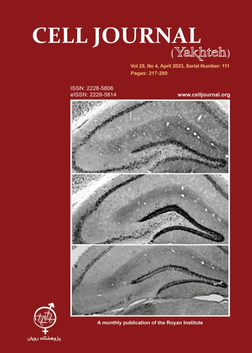
The mechanisms behind seizure suppression by deep brain stimulation (DBS) are not fully revealed, and the most optimal stimulus regimens and anatomical targets are yet to be determined. We investigated the modulatory effect of low-frequency DBS (L-DBS) in the ventral tegmental area (VTA) on neuronal activity in downstream and upstream brain areas in chemically kindled mice by assessing c-Fos immunoreactivity.
Materials and MethodsIn this experimental study, 4-6 weeks old BL/6 male mice underwent stereotaxic implantation of a unilateral stimulating electrode in the VTA followed by pentylenetetrazole (PTZ) administration every other day until they showed stage 4 or 5 seizures following 3 consecutive PTZ injections. Animals were divided into control, sham-implanted, kindled, kindled-implanted, L-DBS, and kindled+L-DBS groups. In the L-DBS and kindled+L-DBS groups, four trains of L-DBS were delivered 5 min after the last PTZ injection. 48 hours after the last L-DBS, mice were transcardially perfused, and the brain was processed to evaluate c-Fos expression by immunohistochemistry.
ResultsL-DBS in the VTA significantly decreased the c-Fos expressing cell numbers in several brain areas including the hippocampus, entorhinal cortex, VTA, substantia nigra pars compacta, and dorsal raphe nucleus but not in the amygdala and CA3 area of the ventral hippocampus compared to the sham group.
ConclusionThese data suggest that the possible anticonvulsant mechanism of DBS in VTA can be through restoring the seizure-induced cellular hyperactivity to normal.
Keywords: Deep Brain Stimulation, Epilepsy, Pentylenetetrazole, Ventral Tegmental Area


