فهرست مطالب

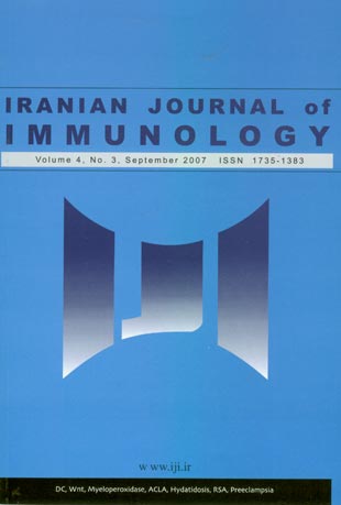
Iranian journal of immunology
Volume:4 Issue: 3, Summer 2007
- 60 صفحه،
- تاریخ انتشار: 1386/07/05
- تعداد عناوین: 7
-
-
Pages 127-144Dendritic cells (DCs) are antigen presenting cells with unique capability to take up andprocess antigens in the peripheral blood and tissues. They subsequently migrate to draining lymph nodes where they present these antigens and stimulate naive T lymphocytes. During their life cycle, DCs go through two maturation stages and are referred to as immature and mature cells, respectively. While immature DCs are very good at capturing antigens, mature DCs are suitably equipped to present antigens to T cells and to initiate an immune response. DCs with different phenotypes serve as sentinels in nearly all tissues including the peripheral blood, where they are continuously exposed to antigens. Very small numbers of activated DCs are extremely efficient at generating immune response against viruses, other pathogens and in experimental models of tumors.Protection against infectious microorganisms and probably against tumors is provided by complex interactions of the innate and adaptive immune systems. For the initiation to occur, pathogens must first be recognized as a “danger”. DC possesses specific receptorsto detect such danger signals. The unique immune-stimulating properties of DC and the feasibility of manipulating their function arouse much enthusiasm and hold great promise for the treatment of cancer. Early clinical trials showed that DC can induce immune responses in cancer patients. Nonetheless, cancer treatments based on DC administrationrequire further studies that will optimize this promising treatment modality.
-
Pages 145-154BackgroundWnt molecules play a key role in growth, proliferation and development of some embryonic and adult organs as well as hematopoietic stem cells. Wnt signaling pathways are aberrantly activated in many tumor types, including solid tumors and hematologic malignancies.ObjectiveTo investigate the expression profile of a large number of Wnt genes in leukemic cells from Iranian patients with acute myeloblastic leukemia.MethodsRT-PCR method was used to determine the Wnt genes expressionin bone marrow (BM) and/or peripheral blood (PB) samples from 16 patients with AMLand PB samples of 36 normal subjects.ResultsAmong 14 Wnt molecules included inthis study, Wnt-7A and Wnt-10A were significantly down-regulated (p = 0.002 and p <0.0001, respectively) and Wnt-3 was significantly over-expressed (p < 0.02) in AML patients compared to normal subjects. No significant association was found between Wnt expression and FAB classification of the patients.ConclusionOur results demonstrated for the first time aberrant expression of Wnt-7A, Wnt-10A and Wnt-3 genes in Iranian AML patients. This may be of relevance to the tumorigenesis process in this malignancy.
-
Pages 155-160BackgroundPolymorphisms in the immune related genes are important in the clinical outcome of Helicobacter pylori infection. Myeloperoxidase -463 G/A polymorphismhas been shown to reduce enzyme expression and activity.Objectivethe aim of the present study is to investigate the association of myeloperoxidase G-463A polymorphism with clinical outcome of Helicobacter pylori infection.Methodstwo hundred and eighty five patients with positive culture of Helicobacter pylori from their gastric biopsies are included in this study. Human leukocyte DNA was extracted using salting out method and myeloperoxidase G-463A polymorphism was investigated by PCRRFLP. All clinicopathological data were collected from individual records.ResultsWhen the patients were categorized according to the high (GG) and low + intermediate (AG+AA) genotypes of myeloperoxidase producers, there was a significant association between myeloperoxidase G-463A genotypes and clinical outcome of Helicobacter pylori infection (p=0.006). In search for combined effect of cagA status and myeloperoxidase genotypes on clinical presentations, only in cagA- Helicobacter pylori infected patients a significant association between myeloperoxidase genotypes and clinical outcome was found (p=0.0001). Also this association was found only in patients infectedwith vacA s1m1 genotype (p=0.008).ConclusionsOur findings suggest that the myeloperoxidase G-463A polymorphism is a host genetic factor which determines the clinical outcome of Helicobacter pylori infection. Moreover, the combination of host and bacterial genetics could provide a better understanding of clinical outcome after infection with Helicobacter pylori.
-
Pages 161-166BackgroundThe clinical significance of antiphospholipid antibodies in patients with chronic hepatitis C virus (HCV) and some other viral infections is controversial.ObjectiveTo study the prevalence of anticardiolipin antibody (ACLA) and antibeta2glycoproteinI antibody (antibeta2GPI antibody) in HCV and hepatitis B virus(HBV) infected patients and its association with liver clinical parameters.MethodsSerum levels of ACLA, antibeta2GPI antibody as well as platelet count, ALT (alanine transaminase), PT (prothrombine time), disease duration and liver histologic findings of 38 patients with HBV and 15 patients with HCV infections were compared with those of 58 healthy controls.ResultsSerum titres of ACLA in HCV and HBV patients (13.4 ±7.1 GPL units/ml), and in each of the HCV (15.18±9.91 GPL units/ml) and HBV (12.7 ± 5.7 GPL units/ml) patients were significantly higher than that of the control group (3.4±2.3GPL units/ml). However, there was no significant difference in serum levels of antibeta2GPI antibody from patients with HCV and HBV (3.3 ± 1.3 GPL units/ml) or HCV alone (2.79 ± 1.01 GPL units/ml) or HBV alone (3.4±1.3GPL units/ml) and that of the control group (3.3±1.1GPL units/ml).ConclusionThe findings suggest that the presence of ACLA has no pathologic significance in patients with HBV and HCV infections.
-
Pages 167-172BackgroundHydatidosis is one of the cosmopolitan parasitic zoonoses caused by the larval stage of Echinococcus granulosus. Diagnosis of hydatidosis is still an unresolved problem. Serological tests using crude antigens for diagnosis of E. granulosus are sensitive, however their specificity are not satisfactory. Therefore, WHO recommended specific serological methods using specific antigens, specially native AgB for proper diagnosis.ObjectivesThis study was designed to evaluate the ELISA and counter current immunoelectrophresis (CCIEP) method using native antigen B (Ag B) for serodiagnosis ofhuman hydatidosis in Fars Province, Iran, an endemic area for this parasitic disease.MethodsNative AgB was purified from sheep hydatid fluid. Serum samples obtained from 40 pathologically confirmed cases of hydatidosis along with samples from patients with fascioliasis, toxocariasis, taeniasis and cancer patients and sera from healthy individuals were tested by ELISA using native antigen B or tested by countercurrent immunoelectrophresis (CCIEP) using crude sheep hydatid cyst fluid.ResultsSensitivity of the ELISA system was determined to be 92.5% and the specificity was found to be 97.3%. Positive and negative predictive values of the system were 92.5% and 97.3%, respectively. For countercurrent immunoelectrophresis the sensitivity of the assay was 97.5% and its specificity was 58.18%. This ELISA system is much more specific in detecting anti hydatid cyst antibody than CCIEP, while CCIEP is more sensitive in detecting anti hydatid cyst antibody.ConclusionThe new ELISA system using native antigen B is a suitable method and preferable to CCIEP for immunodiagnosis of human hydatidosis.
-
Pages 173-178BackgroundRecurrent spontaneous abortion (RSA) is defined as three or more sequential abortions before the twentieth week of gestation. There are evidences to support an allo-immunologic mechanism for RSA. One of the methods for treatment of RSA is leukocyte therapy; however there is still controversy about effectiveness of this method.ObjectivesTo evaluate the effectiveness of leukocyte therapy for treatment of RSA.MethodsNinety two non-pregnant women with at least three sequential abortions (60 primary & 32 secondary aborters) recognized as RSA were referred to ourLaboratory for immunotherapy. All the cases were immunized by isolated lymphocytes from their husbands. Fifty to 100 million washed and resuspended mononuclear cells were injected by I.V., S.C., and I.D. route. The result of each injection was checked by WBC cross matching between couples after four weeks of injections. Immunization was repeated in fifth week to a maximum of 3 times if needed. Eighty one age-matched nonpregnant RSA women (52 primary and 29 secondary aborters) with at least three sequential abortions were also included in this study as controls. The control group wasnot immunized.Results67 out of 92 (72.8%) immunized cases and 44 out of 81 controls(54.3%) showed a successful outcome of pregnancy (p<0.02). Comparison of primaryand secondary aborters indicated a significantly better outcome only in primary (75% vs. 42.3%. p<0.001) but not in secondary aborters (68.8% vs. 75.9%, p = 0.7).ConclusionThe present investigation showed the effectiveness of leukocyte therapy in primary but not in secondary RSA patients. Despite the current controversy and limitation of leukocyte therapy in RSA, the results of our investigation provide evidence supporting the use of allo-immunization in improving the outcome of pregnancy in primary RSA patients.
-
Pages 179-185BackgroundPreeclampsia is a major cause of mortality and morbidity and is also a leading cause of preterm birth and intrauterine growth retardation. Several studies havereported abnormal levels of cytokines in women with preeclampsia.ObjectivesTo detect serum levels of various cytokines in pregnant women with and without preeclampsia in the third trimester of pregnancy.MethodsThirty patients with preeclampsia and thirty normal pregnant women were enrolled in the study. Blood samples were taken and serum levels of IFN γ, IL-12p70, IL-18, IL-15, IL-4 and IL-10 were measured by enzyme-linked immunosorbent assay (ELISA).ResultsPreeclamptic women had significantly increased levels of circulating IL-12p70 (p < 0.05), IL-18 (p < 0.001), IL-4 (p < 0.001), IL-15 (p < 0.05) and IFN γ (p < 0.001). By contrast, circulating levels of IL-10 were not significantly different between the two groups.ConclusionsThe present study supports the hypothesis of altered immune response in preeclampsia and suggests that dysregulation of cytokine expression occurs in preeclampsia with increased levels of IFN γ, IL-12p70, IL-15, IL-18 and IL-4.


