فهرست مطالب

Urology Journal
Volume:4 Issue: 4, Autumn 2007
- 92 صفحه،
- تاریخ انتشار: 1386/12/01
- تعداد عناوین: 18
-
-
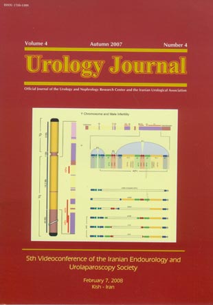
Page 191IntroductionWe reviewed the most recent advances in the genetics of male infertility focusing on Y chromosome microdeletions.Materials And MethodsWe searched the literature using the PubMed and skimmed articles published from January 1998 to October 2007. The keywords were the Y chromosome, microdeletions, male infertility, and azoospermia factor (AZF). The full texts of the relevant articles and their bibliographic information were reviewed and a total of 78 articles were used.ResultsThree regions in the long arm of the Y chromosome, known as AZFa, AZFb, and AZFc, are involved in the most frequent patterns of Y chromosome microdeletions. These regions contain a high density of genes that are thought to be responsible for impaired spermatogenesis. In 2003, the Y chromosome sequence was mapped and microdeletions are now classified according to the palindromic structure of the euchromatin that is composed of a series of repeat units called amplicons. Although it has been shown that the AZFb and AZFc are overlapping regions, the classical AZF regions are still used to describe the deletions in clinical practice.ConclusionY chromosome microdeletions are the most common genetic cause of male infertility and screening for these microdeletions in azoospermic or severely oligospermic men should be standard. Detection of various subtypes of these deletions has a prognostic value in predicting potential success of testicular sperm retrieval for assisted reproduction. Men with azoospermia and AZFc deletions may have retrievable sperm in their testes. However, they will transmit the deletions to their male offspring by intracytoplasmic sperm injection.
-
Page 192IntroductionWe reviewed the most recent advances in the genetics of male infertility focusing on Y chromosome microdeletions.Materials And MethodsWe searched the literature using the PubMed and skimmed articles published from January 1998 to October 2007. The keywords were the Y chromosome, microdeletions, male infertility, and azoospermia factor (AZF). The full texts of the relevant articles and their bibliographic information were reviewed and a total of 78 articles were used.ResultsThree regions in the long arm of the Y chromosome, known as AZFa, AZFb, and AZFc, are involved in the most frequent patterns of Y chromosome microdeletions. These regions contain a high density of genes that are thought to be responsible for impaired spermatogenesis. In 2003, the Y chromosome sequence was mapped and microdeletions are now classified according to the palindromic structure of the euchromatin that is composed of a series of repeat units called amplicons. Although it has been shown that the AZFb and AZFc are overlapping regions, the classical AZF regions are still used to describe the deletions in clinical practice.ConclusionY chromosome microdeletions are the most common genetic cause of male infertility and screening for these microdeletions in azoospermic or severely oligospermic men should be standard. Detection of various subtypes of these deletions has a prognostic value in predicting potential success of testicular sperm retrieval for assisted reproduction. Men with azoospermia and AZFc deletions may have retrievable sperm in their testes. However, they will transmit the deletions to their male offspring by intracytoplasmic sperm injection.
-
Page 207IntroductionOur aim was to compare transureteral lithotripsy (TUL) and extracorporeal shock wave lithotripsy (SWL) in the management of upper ureteral calculi larger than 5 mm in diameter.Materials And MethodsPatients who had upper ureteral calculi greater than 5 mm in diameter were enrolled in this clinical trial. The calculi had not responded to conservative or symptomatic therapy. Semirigid ureteroscopy and pneumatic lithotripsy were used for TUL in 52 patients and SWL was performed in 48. Analysis of the calculi compositions was done and the patients were followed up by plain abdominal radiography and ultrasonography 3 month postoperatively.ResultsThe stone-free rates were 76.9% in the patients of the TUL group and 68.8% in the patients of the SWL group. These rates in the patients with mild or no hydronephrosis were 85.7% and 59.1% for the SWL and TUL groups, respectively. In the TUL group, half of the patients with no hydronephrosis developed upward calculus migration. The stone-free rates were 75.0% and 89.3% for the patients with moderate hydronephrosis and 70.0% and 100.0% for those with severe hydronephrosis in the SWL and TUL groups, respectively. All of the failed cases were treated by double-J stenting and TUL or SWL successfully. There were no serious complications. Upward calculus migration after TUL was more frequent in cases with no hydronephrosis or mild hydronephrosis (41.0%).ConclusionUpper ureteral calculi smaller than 1 cm can be safely and effectively managed using semirigid ureteroscopy and pneumatic lithotripsy. However, the SWL approach has still its role if an experienced endourologist is not available.
-
Page 212IntroductionThe aim of this study was to investigate low-dose intrathecal meperidine for prevention or alleviation of shivering after induction of spinal anesthesia for transurethral resection of the prostate (TURP).Materials And MethodsIn a randomized controlled trial, 80 patients scheduled for TURP under spinal anesthesia were assigned into two groups of case and control. Spinal anesthesia was performed using 75 mg of hyperbaric lidocaine 5% plus meperidine, 15 mg, in the patients of the case group and the same dose of lidocaine plus normal saline in the patients of the control group. Shivering episodes were recorded during the operation and in the recovery room. Data on systolic blood pressure, heart rate, arterial oxygen saturation, and body temperature were collected before the induction of anesthesia; 5, 15, and 30 minutes after the induction; and in the recovery room.ResultsMaximum level of sensory block was similar in the patients of the case and control groups. Shivering was not seen in the patients who received meperidine, while in the control group, 11 (27.5%) experienced some degrees of shivering (P =. 001). Blood pressure, body temperature, and arterial oxygen saturation did not have a clinically significant change and they were not different between the two groups. Side effects of opioids were unremarkable.ConclusionLow-dose intrathecal meperidine is effective and safe in reducing the incidence of shivering associated with spinal anesthesia for TURP.
-
Page 217IntroductionOur aim was to study the changes in resistive index (RI) of the ipsilateral and contralateral kidneys following electromagnetic extracorporeal shock wave lithotripsy (SWL) of the kidney calculi.Materials And MethodsUsing color Doppler ultrasonography, the RI was determined in 21 patients with unilateral caliceal and pelvic kidney calculi. The RI of the interlobar renal arteries were measured for the regions near and far from the calculi (distance, less and more than 2 cm), before, 30 minutes after, and 1 week after SWL. The same measurements were carried out for the contralateral kidney. Changes in the RI values and their relation with age were evaluated.ResultsThe RI near the calculi increased 30 minutes after SWL from 0.594 ± 0.062 to 0.620 ± 0.048 (P =. 003; 95% confidence interval, 0.020 to 0.073), but returned to the pre-SWL values 1 week later. The RI values of the region remote from the calculus and in the contralateral kidney did not change significantly. There was a weak correlation between age and the RI far from the calculus before and 1 week after SWL. There were no relationships between the RI and age, sex, weight, blood pressure, and smoking.ConclusionThe results suggest that SWL of the kidney calculi changes the RI only near the calculus which is immediate, transient, and not age-related.
-
Page 221IntroductionThe aim of this study was to compare the results and complications of extracorporeal shock wave lithotripsy (SWL) plus retrograde ureteroscopic lithotripsy using laser and pneumatic lithotriptors with SWL monotherapy for renal pelvic calculi between 2 cm and 3 cm.Materials And MethodsA total of 55 patients with 2- to 3-cm pelvic calculi were assigned into groups 1 and 2, including 22 and 33 patients, respectively. Patients in group 1 first underwent laser pneumatic lithotripsy and insertion of a double-J ureteral catheter and then underwent SWL 2 to 4 weeks thereafter. In group 2, the patients underwent SWL after double-J ureteral catheter insertion. The stone-free rate, complications, and cost effectiveness were evaluated 3 months postoperatively.ResultsFive patients (22.7%) in group 1, had their calculi completely fragmented after ureteroscopy and retrograde lithotripsy without any need for further SWL. In 9 patients (40.9%), after a single session of SWL, and in 3 (13.6%), after 2 sessions, fragmentation was completed. In group 2, successful treatment was achieved after 1 and 2 SWL sessions in 6 (18.2%) and 8 (24.2%) patients, respectively. The stone-free rate was significantly higher in the patients of group1 than those in group 2 (77.3% versus 42.4%, respectively; P =. 01). The period of anesthesia was 23.1 minutes (during ureteroscopy) in group 1 and 13.2 minutes in group 2 (during cystoscopy or ureteroscopy and insertion of ureteral catheter). No significant complication was reported in neither of the groups. The mean costs of the treatment were US $ 400 and US $ 370 in groups 1 and 2, respectively.ConclusionUreteroscopic lithotripsy before SWL is a rational method for the treatment of the rather large renal pelvic calculi with fairly acceptable costs.
-
Page 226IntroductionA simple technique to dilate urethral stricture using guide wire and sheath dilator has been described in pediatric urology. The aim of this study was to report the long-term outcome of the children who underwent dilation of the urethral stricture using guide wire and sheath dilator.Materials And MethodsFrom 1999 to 2004, a total of 52 children with documented urethral stricture were managed by urethral dilation using guide wire. Data on the cause of urethral stricture, operation, postoperative recovery, follow-up cystoscopic appearance, and patient’s outcome were audited and analyzed.ResultsThe mean age of the patients was 5.6 ± 2.3 years (range, 2 to 18 years). The mean period of the follow-up was 4.5 ± 2.4 years (range, 3.8 to 6.5 years). Twenty-two patients (42.3%) did not require any further surgical treatments. However, urethral stricture in 13 patients (25.0%) progressed significantly, and therefore, they needed further surgical interventions. The complications included minor urinary tract infections in 3 and bladder spasm in 2 patients. No case of false passage or sepsis was encountered.ConclusionGuide wire-assisted urethral dilation avoids the risks associated with blind dilation techniques and continues to be a safe alternative for urethral strictures in selected cases. However, in our experience, less than half of the patients became “recurrence free” after two dilation attempts. We recommend that urethral dilation be considered only in selected cases and emergency settings.
-
Page 230IntroductionThe aim of this study was to investigate the probable differences in P53 expression between papillary urothelial neoplasm of low malignant potential (PUNLMP) and varying grades of transitional cell carcinoma (TCC) of the bladder.Materials And MethodsTen biopsy specimens of the patients with PUNLMP, 20 of the patients with papillary low-grade TCC, 20 of those with invasive high-grade TCC, and 10 of healthy individuals were stained for P53 protein by immunohitochemical methods. Histological grading was performed according to the World Health Organization/International Society of Urological Pathology consensus classification of urothelial neoplasms of the urinary bladder.ResultsNuclear P53 protein in invasive high-grade TCC was slightly more frequent than that in noninvasive low-grade papillary TCC (P =. 35). Ten percent of specimens with PUNLMP had nuclear P53 accumulation, while in low-grade and high-grade TCCs, 75% and 85% of the specimens were positive for P53 protein accumulation (P <. 001). Expression of P53 was nil in all normal transitional epithelium specimens.ConclusionOverexpression of P53 in papillary low-grade TCC and invasive high-grade TCC, while lacking of expression in PUNLMP indicates that mutations of P53 gene are not usually associated with the development of urothelial neoplasms and they may play a crucial role only in progression of PUNLMP to low-grade TCC.
-
Page 234IntroductionThe aim of this study was to evaluate the results of kidney transplantation in patients with Alport syndrome.Materials And MethodsA total of 15 patients with Alport syndrome underwent kidney transplantation and the result of their transplantation was compared with the results in patients without Alport Syndrome. Rejection episodes and the presence of antiglomerular basement membrane (anti-GBM) nephritis were assessed in these patients.ResultsFifteen patients with Alport syndrome were compared with a control group including 212 kidney allograft recipients. One patient with Alport syndrome (6.7%) and 30 controls (14.2%) experienced delayed graft function. Renal artery thrombosis was reported in 1 patient (6.7%) with Alport syndrome and 10 (4.7%) in the control group, which led to nephrectomy in all cases. Acute rejection was confirmed in 2 patients (13.3%) by kidney biopsy and classic treatment yielded relative response. However, they lost their grafts 35 and 44 months after the transplantation. On pathologic examination, no specific finding of anti-GBM nephritis was found. In the control group, 43 cases of acute rejection (20.3%) were reported and 12 patients (5.7%) returned to dialysis. The 1-, 3-, and 5-year graft survival rates were 100%, 92%, and 84% in the patients with Alport syndrome, which was not different from those in the control group (P =. 53).ConclusionIn spite of the risk of anti-GBM nephritis in the patients with Alport Syndrome, it seems that kidney transplantation can yield favorable results and anti-GBM nephritis is not a common etiology of rejection.
-
Page 238IntroductionWe evaluated the ratio of free to total prostate-specific antigen (PSA) and PSA to protein concentrations in saliva and serum of healthy men.Materials And MethodsConcentrations of protein, free PSA, and total PSA in serum and saliva were measured in 30 healthy men aged 42 to 73 years, and their ratios were compared between the two fluids.ResultsThere was a significant direct correlation between serum free-total PSA ratios of serum and saliva (P =. 04) and between total PSA-protein ratios of serum and saliva (P =. 02). Also, there were significant correlations between total and free PSA levels in saliva (P =. 05) and between those in serum (P <. 001). Significant inverse and direct correlations were detected between the body mass index and serum values of total PSA-protein (P =. 04) and free-total PSA (P =. 01), respectively.ConclusionWe can use saliva sample instead of serum sample for estimation of free-total PSA and total PSA-protein levels in men without prostate diseases. There is, however, a pressing need for much additional research in this area before the true clinical value of saliva as a diagnostic fluid can be determined.
-
Page 254
-
Acknowledgment: Reviewers in Volume 4Page 255
-
Subject Index to Volume 4Page 256
-
Author Index to Volume 4Page 259


