فهرست مطالب

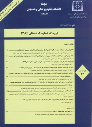
مجله دانشگاه علوم پزشکی رفسنجان
سال ششم شماره 2 (پیاپی 23، تابستان 1386)
- تاریخ انتشار: 1386/04/11
- تعداد عناوین: 9
-
-
صفحه 91زمینه و هدفدرمان اساسی لیشمانیوز جلدی بیشتر بر اساس ترکیبات پنج ظرفیتی آنتیوان هم چون گلو کانتیم و پنتوستام بوده است اما به دلایلی هم چون مقاومت دارویی و کم اثر شدن دارویی ک در حال حاضر به کار می روند به ترکیبات یا داروهای جدید نیاز مبرم می باشد. در این مطالعه تاثیر ضد انگلی مشتقات جدید 1و3و4 تیادی آزول علیه انگل لیشمانیا ماژور به دو صورت تاثیر بر آماستیگوت ها و پروماستیگوت ها در محیط کشت در مقایسه با تارتارامتیک بررسی شده است.مواد و روش هادراین مطالعه آزمایشگاهی پس از این که پروماستیگوت های انگل لیشمانیا ماژور در محیط کشت به فاز ثابت رشد رسیدند آنها را به نسبت 5 به ا، به ماکروفاژهای صفاق موش اضافه نموده و سپس رقت های مختلف مشتقات جدید 1و3و4 تیادی آزول و داروی کنترل (تارتارامتیک) را اضافه نمودیم و پس از 5 روز میزان تاثیر با محاسبه psi مورد بررسی قرارگرفت. هم چنین جهت برسی اثر این مشتقات بر فوم خارج سلولی انگل، از روش MTT استفاده گردید که میزان OD رنگ حاصله از احیا MTT به فورمازان پس از 72 ساعت خوانده شده و IC50 محاسبه گردید.یافته هابعضی از این مشتقات در غلظت 6/4 میکروگرم بر میلی لیتر تاثیراتی بین67%-6% علیه فرم های داخل سلولی انگل داشته و در روش MTT تاثیر ترکیبات بر فرم پروماستیگوتی انگل IC50 بین 6/7-6/3 میکروگرم بر میلی لیتر بوده است.نتیجه گیرینتایج به دست آمده از پژوهش نشان میدهد که دو ترکیب از مشتقات به کار رفته تاثیر قابل ملاحظه ای بر لیشمانیا ماژور داشته است و به نظر میرسد که میتوان از آن ها به عنوان ترکیبات جایگزین مناسب در مطالعات آینده استفاده نمود.کلیدواژگان: لیشمانیا ماژور، 1و3و4 تیادی آزول، تارتارامتیک
-
صفحه 101زمینه و هدفشواهد متعددی نشان داده است که هسته میخی شکل در بی دردی مشارکت دارد. در مطالعه حاضر اثر تزریق آگونیست (باکلوفن) و آنتاگونیست (35348CGP) گابا B به درون هسته میخی شکل بر روی اثرات ضد دردی مرفین با آزمون فرمالین در موش های صحرایی نر مورد مطالعه قرار می گیرد.مواد و روش هادر این مطالعه تجربی پس از کانول گذاری هسته میخی شکل موش های صحرایی نر اثرات تزریق درون هسته ای آگونیست و آنتاگونیست گیرنده گابا B (به ترتیب باکلوفن و 35348CGP) بر اثرات ضد دردی مرفین با آزمون فرمالین مورد مطالعه قرار گرفت.یافته هاتزریق مرفین (5/0 میکرولیتر سالین حاوی 10 میکروگرم مرفین) یا مقادیر مختلف باکلوفن (25/0، 5/0 و 1 میکروگرم به ازای هر موش) دارای اثرات ضد دردی در هر دو مرحله حاد و مزمن آزمون فرمالین بودند. پاسخ های ضد دردی ناشی از تزریق مرفین و باکلوفن هر دو با تزریق 35348CGP کاهش می یافت. تزریق 1 میکروگرم باکلوفن همراه با تزریق درون صفاقی نالوکسان اثرات ضد دردی کمتری را نشان داد. تزریق 35348CGP به تنهایی نیز در مرحله اول (حاد) آزمون فرمالین دارای اثرات ضد دردی بود. تزریق توامان مقادیر مختلف باکلوفن با مرفین اثرات ضد دردی آن را تشدید نکرد در حالی که تزریق 35348CGP توانست در مرحله حاد آزمون فرمالین به طور معنی داری اثرات ضد دردی مرفین را تقویت نماید.نتیجه گیریاحتمالا بخشی از اثرات ضد دردی اعمال شده از طریق گیرنده های گابا B در هسته میخی شکل با دخالت و تعامل گیرنده های اوپیوییدی این هسته در آزمون فرمالین بروز می کند.
کلیدواژگان: هسته میخی شکل، 35348CGP، باکلوفن، گابا B، مرفین -
صفحه 109زمینه و هدفکم خونی فقر آهن شایع ترین علت کم خونی در جهان و شایع ترین کمبود غذایی کودکان است. این کم خونی بیشتر در شیرخواران 9 تا 24 ماهه، به دلیل رشد سریع و عدم مصرف قطره آهن خوراکی شایع است. به دلیل تشخیص های اتفاقی در کودکان قبل از سن مدرسه و این که کم خونی آهن باعث اختلالات رشدی، خلقی، ایمنی و کاهش یادگیری می شود، بر آن شدیم که در محدوده سنی 6-4 ساله، کم خونی آهن را مورد بررسی قرار دهیم.مواد و روش هااین مطالعه مقطعی بر روی 560 کودک 6-4 ساله مهدهای کودک شهرستان رفسنجان انجام شد. نمونه گیری به صورت تصادفی خوشه ایو طبقه بندی شده بود. بعد از اخذ رضایت نامه کتبی و تکمیل پرسشنامه حاوی سوالاتی در مورد علایم کم خونی و عادات غذایی، جهت بررسی هموگلوبین، هماتوکریت، آهن خون و TIBC از کودکان نمونه خون گرفته شد. اطلاعات وارد نرم افزار SPSS شد و مورد تجزیه و تحلیل آماری قرار گرفت.نتایج2/48 % کودکان پسر و 8/51% نفر دختر بودند. شیوع کم خونی در جمعیت مورد بررسی 1/11% بود که با جنس و سن کودک ارتباطی نداشت. شیوع فقر آهن 18/5% بود که به جنس و سن کودک ارتباطی نداشت. شیوع فقر آهن در کودکانی که آهن تکمیلی دریافت نکرده بودند و یا مادرشان در طول بارداری آهن مصرف نمی کردند بیشتر بود.نتیجه گیریکم خونی فقر آهن در کودکان 6-4 ساله مهد کودک های شهرستان رفسنجان نسبت به سایر مناطق در حال توسعه کمتر است. برنامه ریزی برای پیشگیری از این نوع کم خونی با آموزش مادران جهت تغذیه مناسب و تجویز مکمل های آهن توصیه می شود.
کلیدواژگان: کم خونی، فقر آهن، کودکان، مهد کودک -
صفحه 115زمینه و هدفبافت استخوان مرکز عمده تجمع سرب محسوب شده و به عنوان یکی از اهداف اصلی سمیت این فلز سنگین مطرح است. در مطالعه حاضر ما اثر سرب را بر رشد سلول های کشت اولیه ستون مهره جنین انسان و بیان پروتئین Bax بررسی کردیم.مواد و روش هانوع مطالعه حاضر آزمایشگاهی است. به این منظور ابتدا کشت اولیه از سلول های ستون مهره جنین انسان پس از هضم آنزیمی بافت تهیه شد و میزان حضور سلول های استئوبلاست در کشت با بررسی آنزیم آلکالین فسفاتاز تعیین گردید. سپس اثر مجاورت با رقت های ده میکرومول تا 5/1 میلی مول سرب بر تکثیر سلول در محیط کشت حاوی 5 و 10% سرم جنین گاوی (Fetal Bovine Serum، FBS)با روش سنجش MTT (Methyl Thiazolyl Blue Tetrazolium Bromide) بررسی شد. در ادامه، اثر 1/0 میکرومول سرب، بر بیان ژن bax، با روش ایمونوسیتوشیمی بررسی شد.یافته هادرصد سلول های استئوبلاست در کشت اولیه با بررسی آنزیم آلکالین فسفاتاز، 80 تا 85% تعیین شد. رقت های 100 تا 1500 میکرومول سرب تکثیر سلول را در محیط کشت حاوی 10%، 40 تا 81% افزایش داد و بیشترین تحریک رشد در رقت یک میلی مول مشاهده شد (001/0p<). هم زمان با کاهشFBS، فعالیت های مهاری و تکثیری سرب افزایش یافت، به ترتیبی که در رقت های بین 10 تا 1000 میکرومول رشد سلول بین 15 تا 103% افزایش یافت، و در رقت 5/1 میلی مول، 72% کاهش رشد مشاهده شد (01/0p<). بیشترین تحریک تکثیر سلول در رقت 500 میکرومول اعمال شد (001/0p<).نتیجه گیریمجاورت سلول ها با یک دهم میکرومول سرب، افزایش پروتئین Bax را در سیتوپلاسم سلول نسبت به کشت کنترل به دنبال داشت. نتایج این مطالعه، احتمال اختلال در عملکرد فیزیولوژیک طبیعی بافت استخوان به وسیله سرب را مطرح می کند.
کلیدواژگان: استات سرب، استئو بلاست، Bax، MTT assay -
صفحه 123زمینه و هدفکسب موفقیت در درمان ریشه دندان به اجرای صحیح مرحله پرکردن آن بستگی دارد. اخیرا مواد و روش های متعددی جهت پرکردن ریشه دندان ارایه شده است. هدف از این مطالعه، مقایسه آزمایشگاهی ریز نشت آپیکالی روش تراکم جانبی با روش عمودی گرم مخروط های منفرد با تقارب 4% در دندان های آماده سازی شده با فایل چرخشی مستر FlexMaster به شیوه کراون- داون می باشد.مواد و روش هادر این مطالعه آزمایشگاهی ریشه مزیال 36 دندان مولر پایین تازه کشیده شده انسان به طور تصادفی در دو گروه آزمایشی پانزده دندانی و دو گروه شاهد منفی و مثبت قرار گرفتند. پس از پاکسازی و شکل دهی با سیستم چرخشی Flexmaster درگروه اول، کانال های ریشه با کن گوتای پرکا تقارب 2% و به روش تراکم جانبی سرد و در گروه دوم، با کن گوتا پرکا تقارب 4% و ENDOTWIN به روش تراکم عمودی گرم و با استفاده از سیلر AH Plus پر شدند. سطح ریشه هر دندان به وسیله دو لایه لاک ناخن و یک لایه موم چسب (به جز 2 میلی متری آپکس) پوشیده شد. نمونه ها به مدت 2 روز در رنگ گذاشته شده با دیسک برش داده شد. میزان نفوذ رنگ توسط آزمایشگر ارزیابی گردید.یافته هااختلاف آماری بین گروه جانبی سرد و گروه کن منفرد با روش عمودی گرم مشاهده نگردید (83/0=p). میانگین نفوذ رنگ در گروه تراکم حانبی سرد 65/0 و برای گروه تراکم عمودی گرم 61/0 میلی متر می باشد.نتیجه گیریاین مطالعه نشان داد که کانال های آماده سازی شده با سیستم چرخشی FlexMaster را می توان با مخروط گوتا پرکا با تقارب 2% و به روش جانبی پر نمود. لذا روش تراکم جانبی جایگزین مناسبی برای پرکردن ریشه دندان و برآوردن نیازها می باشد.
کلیدواژگان: آماده سازی کانال ریشه، فایل چرخشی FlexMaster، روش تراکم جانبی سرد، Endotwin، ریزنشت آپیکالی -
صفحه 129زمینه و هدفکمبود آهن و کم خونی ناشی از آن، یکی از مشکلات عمده بهداشت عمومی در دنیاست و کودکان زیر 2 سال از مهم ترین گروه های آسیب پذیر می باشند. تجویز آهن کمکی به شیرخوار یکی از روش های پیشگیری از فقر آهن است که در حال حاضر توسط مراکز بهداشتی کشور اجرا می شود. ولی با وجود ضرورت مصرف آهن، تعدادی از مادران علی رغم در دسترس بودن قطره آهن در تمام مراکز بهداشتی، به دلایلی منطقی یا غیرمنطقی از خوراندن قطره آهن به شیرخوار خود امتناع می ورزند. هدف از انجام این طرح، تعیین فراوانی مصرف قطره آهن کمکی و شناخت موانع مصرف آن می باشد.مواد و روش هادر این مطالعه مقطعی، 1200 شیرخوار 24-6 ماهه تحت پوشش هفت مرکز بهداشتی شهر رفسنجان در سال 1382 به طور تصادفی طبقه بندی شده انتخاب شدند. بعد از جلب رضایت مادر، پرسش نامه ای که شامل اطلاعات دموگرافیک کودک، مادر و چگونگی مصرف قطره آهن بود تکمیل شد و در صورت مصرف نامنظم، موانع مهم آن توسط مادران ارایه گردید.یافته هااز کل 1200 شیرخوار مورد مطالعه، 7/61% به طور منظم قطره آهن دریافت می کردند (64-58 CI=95%). شایع ترین دلیل مصرف نامنظم آهن، سیاه شدن دندان ها (1/25%) و کم ترین علت، سیاه شدن مدفوع (5/1%) بوده است. ارتباط معنی داری بین مصرف نامنظم قطره آهن با سن شیرخوار، رتبه تولد، سطح تحصیلات مادر، شغل و سن مادر مشاهده شد ولی با جنس شیرخوار ارتباط معنی دار دیده نشد.نتیجه گیریبا توجه به میزان بالای مصرف نامنظم قطره آهن و دلایل غیرمنطقی آن، آموزش بیشتر مادران و یا غنی سازی غذاهای شیرخوار با آهن پیشنهاد می شود. شیرخوارانی که قطره آهن را به صورت نامنظم دریافت می کنند بایستی از نظر کم خونی فقر آهن ارزیابی و درمان شوند.
کلیدواژگان: آهن تکمیلی، کم خونی فقر آهن، کودکان -
صفحه 135زمینه و هدفیکی از فاکتورهای مهم در روش های کمک باروری، تشکیل و رشد جنین در محیط آزمایشگاه و استفاده از محیط کشت مناسبی می باشد که تمام شرایط لازم را برای رشد آن داشته باشد. در این مطالعه، شرایط دو محیط کشت MS10%+ 10Ham’s F - و GIIII با هم مقایسه شدند.مواد و روش هااین مطالعه آزمایشگاهی بر روی 100 زوج نابارور مراجعه کننده به مرکز ناباروری، که در سیکل درمانی IVF-ICSI قرار داشتند، در دو گروه یکسان انجام گرفت. تخمک های به دست آمده از گروه اول در محیط کشت MS10%+ 10Ham’s F - و گروه دوم در GIIII، کشت داده شدند. سپس پارامترهای تخمک و جنین های تشکیل یافته ارزیابی شده و داده ها با نرم افزار SPSS تجزیه و تحلیل گردیدند.نتایجمیانگین سن بیماران، 01/28 سال و مدت زمان ناباروری، 87/6 سال بود. تعداد پرونوکلئوس های Moderate، در دو گروه اول و دوم به ترتیب 14/2 و 22/3 عدد، جنین های سه سلولی 26/1 و 54/0 عدد، جنین های Grade B، 18/1 و 78/1 عدد و جنین های Grade C نیز 08/1 و 56/0 عدد به دست آمد. میانگین تعداد بلاستومرهای Grade B در محیط کشت MS10%+10-Ham’s F کمتر از GIIIIبود.نتیجه گیریدر این بررسی مشخص گردید که محیط GIIII در بسیاری از فاکتورهای مربوط به کشت جنین، برتری خود را حفظ نموده و سرم مادری به عنوان نگهدارنده تاثیر چندانی نداشته است.
کلیدواژگان: لقاح آزمایشگاهی، محیط کشت، GIIII، سرم مادری -
صفحه 143زمینه و هدفدر حال حاضر درمان انتخابی شقاق مزمن مقعد عمل جراحی است. با شناخت بهتر پاتوژنز بیماری و با هدف درمان سالم تر و اقتصادی تر گرایش زیادی به درمان غیر جراحی معطوف شده است. نیتروگلیسیرین، ایزوسورباید، بتانکول، ال - آرژنین، نیفیدیپین و دیلتیازم، آنتاگونیست های آدرنرژیک بصورت خوراکی یا موضعی و تزریق توکسین بوتولینوم از درمان هستند که در شقاق مزمن مقعد بکار رفته اند. در این مطالعه اسفنکتروتومی جراحی با ژل دیلتیازم موضعی در درمان شقاق مقعد مورد مقایسه قرار گرفت.مواد و روش هادر این مطالعه کارآزمایی بالینی دو گروه 35 نفری مورد بررسی قرار گرفتند، که گروه اول با روش اسفنکتروتومی جراحی و گروه دوم با ژل دیلتیازم موضعی تحت درمان قرار گرفتند. پس از شروع درمان در هفته های 2، 4 و 6، معاینه و ثبت یافته ها انجام شد.یافته هادو هفته پس از شروع درمان، تسکین درد در گروه اسفنکتروتومی شده 75% و در گروه دیلتیازم 45% بود که اختلاف معنی داری (005/0p<) را نشان داد اما پس از چهار هفته، تسکین درد در دو گروه مشابه بود. در چهار هفته اول اسفنکترتومی باعث 85% التیام زخم در مقابل 40% در بیماران دریافت کننده دیلتیازم گردید (0001/0 < p) ولی در هفته ششم التیام در هر دو گروه مشابه بود (351/0= p).نتیجه گیریاسفنکتروتومی جراحی و ژل دیلتیازم به طور مساوی در درمان شقاق مزمن مقعد موثرند ولی حسن ژل دیلتیازم در کم عارضه بودن، خوب تحمل شدن و عدم نیاز به بستری، می باشد و باید به عنوان اولین قدم درمانی در شقاق مزمن مقعد مورد نظر باشد و جراحی برای موارد خاص مد نظر قرار گیرد.
کلیدواژگان: شقاق مزمن مقعد، اسفنکتروتومی، ژل دیلتیازم - گزارش مورد
-
صفحه 151A Rare Anatomical Innervation of the Musculocutaneous Nerve M.M. Taghavi MSc, M. Shariati Kohbanani MSc, S.M. Seyedmirzaee MD Received: 24/09/06 Sent for Revision: 20/02/07 Received Revised Manuscript: 07/04/07 Accepted: 27/05/07 Background and Objectives: Anatomically the musculocutaneous nerve (C5,6) is a branch of lateral cord of the brachial plexus and its motor nerve fibers innervates the muscles of anterior compartment of the arm. This nerve penetrates into the coracobrachialis of arm muscle and lies between biceps and brachialis muscles. At the lateral bicipital groove becomes superficial and the finally converts to lateral cutaneous nerve of the foream. Here we report a rare case of musculocutaneous nerve variation. Case Report: We found a rare anatomical form of musculocutaneous nerve during upper left limb dissection of a male cropse who was in dissecting room of Rafsanjan Medical School. His body was tall with muscular limbs, weighed 65-75Kg, 175 cm height, and fifty years old. The following variations were observed after dissecting of the axillary and arm regions. 1) The Musculocutaneous nerve arised from the lateral root of the median nerve. 2) The coracobrachialis muscle was innervated by a branch of the lateral cord of the brachial plexus. 3) The Musculocutaneous nerve did not penetrate into the coracobrachialis muscle but rather passed between the brachialis and biceps muscles. At the level of lateral bicipital groove, it then became a superficial nerve as the lateral cutaneous nerve of forearm. Hense, it was very close to the brachial artery and median nerve in the upper one-third of arm. Conclusion: This study describes a rare innervation of the musculocutanous nerve and requires further study to understand the nature of this unique structure. This atypical innervation is extremely important for surgical procedures performed on the arm muscles and adjecent vessels. Key words: Musculocutaneous nerve, Variation, Brachial plexus
-
Page 91Background And ObjectiveThe main therapeutic compounds available against Leishmaniasis disease is pentavalent antimonyfcg compounds i.e. Glucantime and Pentostam. New antileishmanial compound is needed due to the emerge of drug-resistant leishmania agents in recent years. In the present study the antileishmanial activity of new 1, 3, 4 thiadiazole derivatives were evaluated.Materials And MethodsPromastigote stages of the parasites were cultured in RPMI-1640 containing 10% FBS, 100 IU/ml penicillin and 100 µg/ml streptomycin. Mouse peritoneal exudate macrophages (MPEM) isolated from the peritoneal cavity of BALB/c mice were used and the macrophages were counted and the cell suspension was adjusted to 5×10⁵ cell/ml. Macrophage monolayers in 8-well chamber slides were infected with stationary phase promastigote, at a 5:1 parasite/cell proportion and incubated at 37oC and 5% CO2. Serial dilution of thiadiazole compounds and tartar emetic as the control was added to the slide chambers and parasite survival index (PSI) was measured after 5 days. The Thiazolyl blue reduction (MTT test) was used to determine the antileishmanial effect of the compounds on extra cellular forms of the parasite and after 72 h. The OD's were read by 96-well scanner and IC50 were calculated.ResultsTwo thiadiazole compounds showed 6-67% antileishmanial activity in 4.6µg/ml concentration against intracellular forms of the parasites and also in MTT assay IC50 of 3.6 -7.6 µg/ml was determined.ConclusionDue to high antileishmanial activity of some compounds, further studies on structure and activity of these compounds and new highly active derivatives is expected.Keywords: Leishmania major, 1, 3, 4 Thiadiazole, Tartar emetic
-
Page 101Background And ObjectiveThere are several evidences that show cuneiformis nucleus is involved in nociception. In the present study the effect of intra cuneiformis microinjection of GABAB agonist (baclofen) and antagonist (CGP35348) on morphine induced antinociception in rat were investigated.Materials And MethodsIn this expremental study, through canulation of cuneifoprmis nucleus in rat the effect of intra cuneiformis (CNF) microinjection of GABAB receptor agonist (baclofen) and antagonist (CGP35348) on morphine – iduced antinociception were investigated by formalin test.ResultsMicroinjection of morphine (10μg /0.5 μl/saline) or different doses of baclofen (0.25,0.5,1μg per rat) had antinociception in the both first and second phases of formalin test. The response induced by morphine or baclofen in both phases were reduced by CGP35348. The responses induced by combination of baclofen (1μg per rat) and intraperitoneal (ip) injection of naloxan were reduced in both phases of formalin test. Microinjection of CGP35348 alone has produced antinociception in first phase of the formalin test. Morphine with different doses of baclofen did not increase the antinociception effect whereas microinjection of CGP35348 administration significantly increased the antinociception in acute phase.ConclusionIt may be concluded that CNF GABAB receptor induced antinociception via opioid receptor in the formalin test.Keywords: Cuneiformis, CGP35348, Baclofen, GABAB, Morphine
-
Page 109Background And ObjectiveIron deficiency is the most common etiological factor of the anemia and also the most common nutritional deficiency in children. The iron deficiency anemia is prevalent in 9-24 months babies due to accelerated growth rate and lack of supplemental iron in their diet. This anomaly leads to growth, psychological, immunological and educational problems in children. Diagnosis of the anemia is quite accidental in children before the school age. This study evaluates the iron deficiency anemia in children aged 4-6 years old (y/o).Materials And MethodsA cross-sectional study on 560 childrens (aged 4-6 y/o) was done at kindergardens of Rafsanjan City. The sampling method was achieved in randomized, clustered and classified forms. After an informational session with parents and recieving their written consent, a questionnaire including anemic symptoms and nutritional habits was completed. The blood sample of each child was obtained to evaluate Hemoglobin, Hematocrit, serum Iron and TIBC. All data were then analyzed with SPSS Software.ResultsOf the total children samples 48.2% of were male and 51.8% of them were female. The prevalence of anemia in investigated population was 11.1% which was not related to the age and sex of the child. The prevalence of iron deficiency anemia was 5.18%, which was not also related to the age and sex of the child. There were higher incidence of anemia among children who didnt have an appropriate nutritional education for children.ConclusionIron deficiency anemia in 4-6 y/o children of kindergardens of Rafsanjan City is less than other developed regions. Good preventive measures for anemia along with parental education for appropriate diet and taking iron supplements are strongly recommended.Keywords: Iron deficiency anemia, Children, Kindergarden
-
Page 115Background And ObjectiveBone is the main source of lead concentration and is presumed as one of the main targets of the toxic effects of this heavy metal. This study have evaluated the toxic effects of lead on the primary culture of the vertebrate of the human fetus and the expression of the Bax protein in the cell.Materials And MethodsThe present investigation is a laboratory study which initially a primary culture of the vertebrate of the human fetus was prepared by the enzymatic digestion and quantity of the osteoblast cells were then determined by Alkaline phosphatase assay. The effects of lead exposure at serially made concentrations of 10µmol to 1.5 µmol on the cell proliferation, was evaluated in a culture containing 5 and 10 percentages of fetal bovine serum (FBS) by MTT assay (Methyl Thiazolyl Blue Tetrazolium Bromide). In addition the effects of 0.1 µmol of lead on Bax gene expression in osteoblast cells was analyzed by immunocytochemistry method.ResultsQuantitative analysis of osteoblast cells in the primary culture by the Alkaline phosphatase assay was determined as 80 to 85%. The lead concentrations of 100 to 1500 micromole caused 40 to 81% increase in the cell proliferation in culture containing 10% of FBS. The most growth stimulation was observed at the concentration of 1µmol (p<0.001). By decreasing the FBS, the inhibitory proliferative activities of lead increased as such that the cell growth showed an increase of 15 to 103 % with concentration of 10 to 1000µmol, and a decrease was observed in cell growth about 72% in a lead concentration of 1.5 µmol (p<0.001). The most cell proliferation stimulation was seen in a concentration of 500 µmol (p<0.001). Osteoblast cells exposure to 0.1 µmol of lead caused an increase in the amount of Bax protein in the cytoplasm in compare with the control culture.ConclusionThe result of this study shows that lead may disturb the natural physiologic function of the bone cells and this heavy metal may act as a mitogenic element.Keywords: Lead acetate, Osteobast, Box, MTT assay
-
Page 123Background And ObjectiveThe success rate of root canal treatment depends on the proper execution of the final phase. Recently, various materials and techniques have been presented on the market for root canal obturation. The purpose of this study was to compare in vitro lateral condensation technique of the microleakage seal of with warm vertically-obturated single 4% tapered gutta-percha cone in root canals prepared by rotary system FlexMaster in crown-down manner.Materials And MethodsIn this laboratory study the mesial root canals of 36 freshly extracted human mandibular molar teeth randomly were divided in to two groups of 15 teeth and two groups were designate as a negative and positive control respectively. After cleaning and shaping with FlexMaster rotary system, the teeth were obturated as follows: in group one, a lateral compaction technique with 2% tapered gutta-percha and in group two, obturated warm vertically (ENDOTWIN) with a single 4% tapered gutta-percha cone and AH plus endodontic sealer were applied. Nevertheless, the coronal portion and the root surface of each tooth was covered with two layers of nail varnish and a layer of sticky wax. The specimens were then immersed in blue Indian ink dye for two days, prior to sectioning by disc. Dye penetration was evaluated, and the data were analyzed with T-student test.ResultsThere was no significant difference between the two groups (p=0.83). The mean of dye penetration in lateral compaction group was 0/65 mm and for warm vertically compaction group was 0/61mm.ConclusionThis study showed that canals prepared with Flex Master rotary systems can be obturated with 2% taper gutta-percha plus AHPLUS root canal sealer. Thus lateral compaction could possibly be a suitable technique for root canal obturation and other requirements.Keywords: Root canal preparation, Flexmaster rotary system, 4% Tapered gutta, percha, Cold Lateral Compaction, Endotwin, Plugger, Apical microleakage
-
Page 129Background And ObjectivesIron deficiency and anemia are among the most problems encountered the general hygiene system in the world because infants under 2 years of age are at risk of having iron deficiency. Therefore it is necessary to supplement the infant with iron as an important prophylactic method for anemia. The supplementation of infant with iron is routinely done in health centers of iran. Although, iron drop supplement is available for children, some mothers don’t provide this necessary nutritional element for their infants. This study was conducted to evaluate the iron supplemental dose, and compliance of mothers in giving iron drop to their infants. In addition, we looked for possible demographic characters of mothers and their reasons that prevent their infants from accessing the iron drop.Materials And MethodsThis descriptive study was performed on 1200 infants (6-24 months who were referred to health care centers of Rafsanjan city in 2001. Systematic randomized sampling method was used for the study. A questionnaire containing demographic characters of mothers and infants, daily intake of iron drop, as well as lack of iron drop intake along with mother's reasons such as blackening of teeth, black stool, lack of Iron drop, unfavorable taste, mothers forgetfullness, lack of need for iron in infants, and lack of recommendation by physicians. The data were gathered and analyzed by using SPSS12 soft ware, T and X2 tests.ResultsAmong 1200 infants investigated 61% were taking the iron drop daily (95% CI, 58-64). The most common reason for not taking the supplement daily was blackening of teeth 25.1% (95% CI, 21-29) and the least frequent one was black stool 1.5% (95% CI, 0-3). The relationship between avoding daily intake of iron drop and infant age, growth level, level of mother's education, mother's job and here age were significant but it was not significant for sex.ConclusionConsidering the importance of infant daily intake of the iron drop and lack of logical reasons of mothers to give the supplement to their infants, a meticulous and intensive public campaign is needed to augment awareness of the mother to provide the iron for their infant either as a supplement or in the infant’s food. Nevertheless, infants who don’t have enogh of this necessary element in their diet should be examined for iron deficiency and subsequent treatment.Keywords: Iron supplementation, Iron deficiency anemia, Infant
-
Page 135Background And ObjectivesAppropriate culture media and environment for embryonic cell growth considered to be major factors for generation of an embryo in vitro. In this study two embryonic growth condition of GIIII and Ham’s F-10 + Maternal Serum (MS) %10 were compared.Materials And MethodsThis investigation was prospectively performed on 100 infertile couples that were treated by IVF-ICSI. The participants were divided into two equal groups. Ovules obtained from first group were treated with Ham’s F10 + Maternal Serum %10 culture media and the second group with GIIII. The variables affecting ovule and embryonic growth were measured, and collected data were analyzed by SPSS. Reasults: The mean of age for tested ladies was 28.01 and the mean for duration of infertility was 6.87 years. The number of pronucleous with moderate quality were 2.14 in-group I and 3.22 in-group II (P=0.023). The number of embryo with three cells were 1.26 and 0.54 and for grade B embryonic cell were 1.18 and 1.78 and grade C embryonic cell were 1.08 and 0.56 in group I and II respectively (p=0.29). The mean of grade B blastomer in GIIII media was more than Ham’s F10 + Maternal Serum 10%. Compairing the means the mean of grade B blastomers in GIIII media was shown to be more than Ham’s F10 + Maternal Serum 10%.ConclusionAll of the findings showed that GIIII cultural condition is more effective than Ham’s F10 and the maternal serum as a supplement has no considerable impact.Keywords: IVF, Culture Media, GIIII, Maternal serum
-
Page 143Background And ObjectiveSurgery is now the "treatment of choice" for chronic anal fissure. However, considering the pathogenesis of this disease and the tendency for noninvasive and economical procedures, more attention is growing towards the non surgical treatments. Oral or topical nitroglycerin, isosorbide, bethanechol, L-Arginine, nifedipine, diltiazem, adrenergic –antagonists and botulinum toxin have been used to treat chronic anal fissure. In this study, we compared the surgical sphincterotomy and topical diltiazem gel for the treatment of anal fissure.Materials And MethodsThis clinical trial study was performed on two groups of 35 patients. The first group was treated by surgical sphincterotomy and the second group received the topical diltiazem gel. Both groups were examined 2,4 and 6 weeks after the onset of the treatment and, the findings were recorded and statistically analyzed by SPSS, t-test and X2 to determine the relation between parameters and p<0.05 was considered to be significant.ResultsTwo weeks after the onset of treatment, the rate of complete pain relief in the sphincterotomy group and in the diltiazem group was 75% and 45% respectively (p<0.005). Although this value was the same in both groups 4 weeks after treatment (p=0.357), the wound healing process observed in the first group was significantly higher than the second group (85% VS 40%, p<0.0001). Both groups had a similar wound healing rate in the 6th week (p=0.0351).ConclusionsAccording to our findings, both the surgical sphinctorotomy and the topical diltiazem gel had a similar therapeutic effect on the chronic anal fissure. However, the topical diltiazem gel has shown to have several advantages such as lower complication rates, greater convenience, noninvassivess, (no hospitalization) and lower cost. Therefore this type of treatment considered to be more appropriate for chronic anal fissure, and the surgery should be used as an alternative option for special cases.Keywords: Chronic anal fissure, Sphincterotomy, Diltiazem gel
-
Page 151Background And ObjectivesAnatomically the musculocutaneous nerve (C5,6) is a branch of lateral cord of the brachial plexus and its motor nerve fibers innervates the muscles of anterior compartment of the arm. This nerve penetrates into the coracobrachialis of arm muscle and lies between biceps and brachialis muscles. At the lateral bicipital groove becomes superficial and the finally converts to lateral cutaneous nerve of the foream. Here we report a rare case of musculocutaneous nerve variation. Case Report: We found a rare anatomical form of musculocutaneous nerve during upper left limb dissection of a male cropse who was in dissecting room of Rafsanjan Medical School. His body was tall with muscular limbs, weighed 65-75Kg, 175 cm height, and fifty years old. The following variations were observed after dissecting of the axillary and arm regions. 1) The Musculocutaneous nerve arised from the lateral root of the median nerve. 2) The coracobrachialis muscle was innervated by a branch of the lateral cord of the brachial plexus. 3) The Musculocutaneous nerve did not penetrate into the coracobrachialis muscle but rather passed between the brachialis and biceps muscles. At the level of lateral bicipital groove, it then became a superficial nerve as the lateral cutaneous nerve of forearm. Hense, it was very close to the brachial artery and median nerve in the upper one-third of arm.ConclusionThis study describes a rare innervation of the musculocutanous nerve and requires further study to understand the nature of this unique structure. This atypical innervation is extremely important for surgical procedures performed on the arm muscles and adjecent vessels.Keywords: Musculocutaneous nerve, Variation, Brachial plexus


