فهرست مطالب

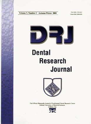
Dental Research Journal
Volume:5 Issue: 2, Mar 2008
- 58 صفحه،
- تاریخ انتشار: 1387/07/21
- تعداد عناوین: 8
-
Pages 47-52This study was designed in order to evaluate the microbiological effectiveness of a locally delivered xanthan-based CHLO-SITE gel as an adjunctive therapy to scaling and root planning in the treatment of moderate-advanced chronic periodontitis.
In a randomized controlled split-mouth clinical trial, 20 patients with chronic periodontitis and pocket depth of 4-6 mm were selected. One side of the mouth served as control where scaling and root planning was performed as routine while the other side of the same mouth (experimental site) was treated by an extra injection of xanthan-based CHLO-SITE gel. Samples were taken from both sites on baseline, 1 month and 3 months post-treatment periods. Samples were exposed to nutrient material followed by colony counts. Data were then analyzed using Wilcoxon signed ranks and Friedman tests.
At baseline, there was no significant difference in colony counts between the control and the experimental sites (P = 0.36). However, after 1 month follow up, the mean colony count was 13850 ± 2253 and 151610 ± 11248 at experimental and control sites, respectively, with a significant difference between the two sites (P < 0.05). A significant difference was also found in colony count between the two groups in 3 months follow up episode (P < 0.05).
It seems that subgingival injection of xanthan-based CHLO-SITE gel could cause a significantly higher decrease in colony count than that of scaling and root planning therapy alone in chronic periodontitis.Keywords: Drugs, gels, periodontitis, xanthan gum -
Pages 53-60There is a variation of management of third molars, from conservative to surgical management. The objective of the study was to determine the prevalence and the factors associated with third molars treated in the dental department of the Penang hospital, Malaysia.
This was a descriptive case series analysis of all cases reported to Penang hospital, Malaysia from January 2000 to December 2005. A specially designed questionnaire was used. Descriptive statistics and chi square test were used to explore the data. Data were analyzed using SPSS version 13.0. The study was conducted ethically
The six year prevalence rate was 1.75%. The majority of patients were Malays and Chinese. Most were under the age of 25. There were a total of 261 cases of lower third molars. Mesial presentation was common among the races (P < 0.05). The number of cases increased from the year 2000 to 2005 (P < 0.05). Most clinical diagnoses of lower third molar presentations were confirmed radiologically (P < 0.05). There were many defaulters (those who did not return for definitive treatment or for follow up) and the number of cases treated surgically under anesthesia increased as the years progressed among all age groups (P < 0.05). There were a total of 11 cases of upper third molars. Similarly, most clinical diagnoses of the third molar presentations were confirmed radiologically (P < 0.05).
The high rate of defaulters indicates the need for pre-treatment counseling. An increasing congruence indicates an improvement in clinical competence of the dentistsKeywords: Epidemiology, impacted tooth, third molars, tooth extraction -
Pages 61-64p16 protein acts as a tumor suppressor and its functional loss seems to be one of the most frequent genetic alterations in human tumors. Because there is a lack of information, the expression of p16 is examined in different odontogenic cysts to clarify the possible role of this factor to determine the biological and clinical behavior of these lesions.
Eighteen radicular cysts (RC), 9 follicular cysts (FC), and 15 keratocystic odontogenic tumors (KCOT) were immunohistochemically evaluated. Chi-square test was used to compare data.
The cysts showed a different pattern of p16 expression according to the histologic type. All RCs or FCs were strongly or moderately positive for p16, whereas all KCOT cases exhibited loss of p16 expression or a low immunoreactivity.
KCOT is characterized by an aggressive potential of growth and recurrences after removal. Here, a dysregulation of the p16 pathway in KCOT was demonstrated. This lack of expression of positivity for p16 in KCOT could help explain the differences in the clinical and pathological behavior of KCOT and could be related to the increased aggressive behavior, invasiveness and high frequency of recurrences found in KCOT.Keywords: Cell cycle proteins, immunohistochemistry, odontogenic tumors, p16 genes -
Pages 65-69Many post systems are available to clinicians, yet no consensus exists regarding the superiority of any one in restoring endodontically treated teeth. The aim of this in vitro study was to compare the fracture resistance and failure mode of endodontically treated teeth restored with a cast metal post and crown with quartz fiber post and composite crown build-up.
Forty extracted maxillary canine teeth with similar size were chosen and randomly divided into 2 groups. After cutting the crowns and endodontic therapy, the teeth were restored with a quartz fiber post and composite crown build-up, or a cast metal post and crown in group 1 and 2, respectively. Fiber posts were cemented with dual cured resin cement and cast posts were luted using zinc phosphate cement. After thermocycling, a compressive load was applied at 135˚C to the long axis of the tooth at a crosshead speed of 1 mm/min and fracture loads and fracture modes were recorded. Mann-Whitney and t tests were used to determine the significance of the failure load values between the two groups.
The mean values for fracture strength in groups 1 and 2 were 344 and 446 N, respectively. The teeth in group 2 exhibited significantly higher resistance to fracture (P < 0.01); however, all failures occurred in the tooth structure.
In spite of the significantly lower failure loads achieved for the teeth restored with fiber posts, all of the fractures in this group were repairable.Keywords: Endodontically, treated teeth, post and core technique, restorations -
Pages 71-79External prostheses exhibit an unwanted color change over time. Color deterioration of prosthetic elastomers affects the life expectancy of facial prostheses in a service environment. The effect of different pigmentation and irradiation duration on color stability of four silicone elastomers after artificial weathering was investigated in this study.
The materials used included four different pigmented industrially synthesized RTV (room temperature vulcanizing) silicones. The materials chosen in this study were representative silicone prosthetics that are widely used in the last decade in maxillofacial prostheses. Artificial weathering was performed in a weatherometer of total radiant energy 1.35 W/m2 (UVA - UVB). The samples were exposed in eight different periods (8, 24, 48, 72, 96, 120, 144, 168 hours). L, a, b readings were obtained before and after weathering from a spectrophotometer to define color changes. Color changes were calculated from the following equation: ΔE = (ΔL2 Δa2 Δb2)½. The data were subjected to two-way analysis of variance at a significance level of α = 0.05. Also, simple mathematical models were developed for color changes.
The results showed that color changes depend on irradiation time and initial color of samples. Episil Europe 1 and Episil Africa 3 were identified as the most stable materials since their color changes were not eye detectable. Contrary to materials Episil Europe 2, 3 that showed significant color changes.
Artificial weathering caused significant, eye detectable, but yet still clinically acceptable color changes in the examined prosthetic silicone elastomers due to deterioration that occurs through irradiation.Keywords: Color, degradation, elastomers, prostheses, silicones -
Pages 81-87Bone regeneration in the defects around oral implants using substitutes may improve long-term prognosis of the implant. Hydroxyapatite (HA) is a good candidate for bone substitutes due to its similarity to bone minerals. Nanostructured hydroxyapatite is also expected to have better bioactivity than coarser crystals. The aim of this work was to synthesize and evaluate the bioactivity of HA.
Nanocrystalline HA was synthesized via mechanical activation method. Fourier transform infrared spectroscopy (FTIR) was utilized to identify the functional groups of the prepared HA. Transmission electron microscopy (TEM) technique was utilized to evaluate the shape and size of prepared HA powder. The synthesized powder was soaked in stimulated body fluid (SBF) medium for various periods of time in order to evaluate its bioactivity. The changes of the pH of SBF medium were measured. Atomic absorption analysis (AAS) was used to determine the dissolution of calcium ion in the SBF environment and scanning electron microscopy (SEM) was utilized to evaluate the surface morphology of nanocrystalline HA powder after immersion in SBF.
The prepared HA powder had nano-scale morphological structure with the mean crystallite size of 29 nm in diameter and bone-like composition. The ionic dissolution rate of prepared nanocrystalline HA was higher than that in conventional HA and was similar to that of biological apatite of bone. High bioactivity of prepared nanocrystalline HA powder due to the formation of apatite on its surface was observed.
Prepared nanocrystalline HA could be more useful for treatment of oral bone defects in comparison with conventional HA, and could be more effective as a bone replacement material to promote bone formation.Keywords: Bone substitutes, hydroxyapatite, nano, material, nanostructured material -
Pages 89-93The secretory immunoglobulin A (IgA) is the first line of defense against pathogens that invade mucosal surfaces. It has been reported that the immune system exhibits profound age-related changes. The aim of this study was to investigate the age-dependent changes of salivary IgA and IgE levels among healthy subjects.
Saliva samples were collected from 203 healthy individuals (aged 1-70 years). The salivary IgA and IgE concentrations were measured by use of ELISA technique and analyzed using the Mann-Whitney U, Kruskal-Wallis and Chi-Square tests.
The mean salivary IgA levels were 42.67 μg/ml at age 1-10 years, 82.44 μg/ml at age 11-20 years, 93.5 μg/ml at age 21-30 years, 97.58 μg/ml at age 31-40 years, 106.45 μg/ml at age 41-50 years, 113.47 μg/ml at age 51-60 years and 92.95 μg/ml at age 61-70 years. There was significant difference among mean salivary IgA levels of different age groups (P < 0.001). The frequency of subjects with detectable concentrations of salivary IgE increased with increasing age up to 40 years and thereafter decreased. There was also significant difference among the mean salivary IgE levels of different age groups (P < 0.001). In adults, the mean salivary levels of IgA and IgE were significantly higher than those observed in children (P < 0.0001 and P < 0.002, respectively).
These results showed that the salivary IgA and IgE levels exhibit age-related changes. Oral immunization may be considered to improve oral immunity when the salivary concentrations of IgA begin to decrease during lifetime.Keywords: Adult, immunoglobulin A, immunoglobulin E, saliva -
Pages 95-98Radicular cysts are considered rare in the primary dentition, comprising only 0.5-3.3% of the total number of radicular cysts in both primary and permanent dentitions. The aim of this case report is to present the clinical, radiographic and histological characteristics of radicular cyst associated with first primary molar following formocresol pulpotomy. Extraction and enucleation of the cyst was carried out under local anesthesia after elevation of the mucoperiosteal flap, which led to uneventful healing and the space of the missing primary molar was maintained using a band and loop space maintainer. The relationship between the intracanal medicaments used for pulp therapy and the rapid growth of these cysts that had been enumerated in the literature was noticed in this case. This does not imply that prohibition of medicaments for pulp treatment of primary teeth is necessary, but pulpotomy treated primary molars should receive periodic postoperative radiographic examination and absence of clinical symptoms does not mean that a pulpotomy treated tooth is healthy.Keywords: Periodontal cyst, primary teeth, pulpotomy, radicular cyst

