فهرست مطالب

Jundishapur Journal of Microbiology
Volume:2 Issue: 4, Oct-Dec 2009
- تاریخ انتشار: 1388/08/11
- تعداد عناوین: 7
-
-
Study on growth of Toxoplasma gondii tissue cyst in laboratory mousePage 3Introduction andObjectiveToxoplasma gondii is a widespread protozoan parasite that infects human and animals. Tissue cyst of T. gondii is an important source of human and other hosts infection including cats as definitive host. This study was carried out to determine growth of T. gondii tissue cyst in laboratory syurian mouse.Materials And MethodsFifteen mice in three groups were infected with T. gondii by intraperitoneally inoculation. The mice of each group killed after two, three and four months. Ten smears of brains were prepared from each group and examined microscopically for Toxoplasma tissue cyst. The diameter of 17 cysts from each group was determined and recorded and the mean size in each group calculated.ResultsThe mean diameters of tissue cysts were 49.4×44.8μm after two months and 64.5×55.7μ after three months of inoculation. After four months the mean diameter of tissue cyst reached to 60.6×53.48μm. The volume of tissue cysts grew within two and three months after inoculation but after four months the growth leveled off.ConclusionThe present study showed that the mean diameter of tissue cyst grew during the first three months and after four months became stable and some may get compacted and lesser.Keywords: Toxoplasma gondii, Tissue cyst, Mouse
-
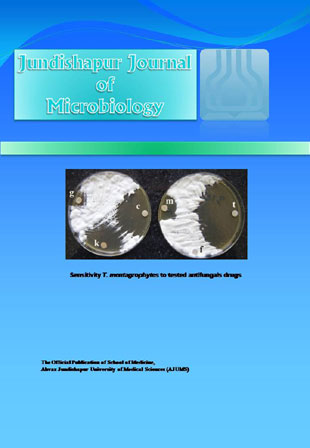
In vitro activity of six antifungal drugs against clinically important dermatophytesPage 7Introduction andObjectiveDermatophytosis is a common fungal disease which involves the keratinized tissue. Several antifungal agents can be used to manage these infections. Unfortunately, drug resistant can result in treatment failure. The disk diffusion in vitro assay is a simple method that can be used to evaluate antifungal susceptibility in dermatophytes. The main aim of this study was to evaluate the antifungal activity of six antifungal drugs against several fresh clinical dermatophyte Iranian isolates..Materials And MethodsForty clinical dermatophytes were isolated from patients suspected of having active dermatophytosis. Paper disks containing terbinafine, griseofulvin, clotrimazole, miconazole, fluconazole and ketoconazole were used in the disk diffusion method to evaluate the in vitro activity of the antifungal agents by measuring the mean diameter of inhibition around the disks..ResultsThe isolates belong to three genera and eight species as: Trichophyton mentagrophytes 13(32.5%), T. rubrum 8(20%), Epidermophyton floccosum 7(17.5%), T. violaceum 4(10%), Microsporum gypseum 3(7.5%), T. tonsurans 2(5%), T. verrucosum 2(5%), T. schoenleinii 1(2.5%), and an unknown dermatophyte 1(2.5%). No isolates were resistant to clotrimazole and miconazole..ConclusionThis study revealed that clotrimazole, miconazole, terbinafine, and griseofulvin were the most ideal antifungal drugs for the treatment of dermatophytosis. Disk diffusion method is a simple and valuable method for the evaluation of antifungal susceptibility of dermatophytes..Keywords: Dermatophytosis, Disk diffusion, Antifungal agents, Dermatophyte
-
Pages 124-131Introduction andObjectiveInfection with Salmonella is the most frequently reported cause of bacterial food-borne illness worldwide. Raw meat samples are a common source and, in recent years, much attention has been focused in determining the prevalence of Salmonella during the different stages in the poultry and beef production chain. This study was conducted to examine the prevalence of Salmonella contamination, and the antibiotic resistance characteristics of isolated strains, from raw samples of packed and unpacked beef and chicken collected randomly from retail stores in Tehran.Materials And MethodsA total of one hundred and thirty three samples were collected from 27 meat providing retail stores in Tehran. Salmonella strains were isolated and identified according to the techniques recommended by the International Organization for Standardization (ISO 6579, 1998). Antimicrobial resistance test was performed by disk diffusion method using 13 antibiotics.ResultsOut of one hundred and thirty three samples tested, fifty one (38.3%) were identified as Salmonella strains. The percentages of Salmonella in chicken and beef samples were 62.7% and 37.3% respectively. The sereotyping results showed that isolated strains belonged to 10 different serotypes, and the most dominated serotype was Salmonella thompson (54.9%). Among the variety of antibiotics tested, the highest resistance was found with nalidixic acid followed by tetracycline, trimethoprim, and streptomycin. The percentages resistance of isolates from chicken samples to nalidixic acid, tetracycline, trimethoprim, and streptomycin were 90.6%, 71.9%, 56.6%, and 25%, and the isolates from meat samples were 36.8%, 21%, 26.3%, and 5.3% respectively. About 23.5% of theSalmonella strains were multiresistant to two or more antibiotic families. Finally, six resistance profiles have been identified. In overall, the degree of resistance of serotypes to nalidixic acid was greater than other tested antibiotics.ConclusionOur results indicate that antimicrobial resistant Salmonella strains were widely spread among raw chicken and beef meats samples.Keywords: Salmonella, Serotype, Meat, Chicken, Antibiotic resistance
-
Pages 132-139Introduction andObjectiveTrichomonas vaginalis, a flagellate’s pathogen protozoon, that is the cause of most common types of vaginitis may serve as a cofactor in HIV, associated with adverse pregnancy outcomes and predispose pregnant women to premature rupture of membranes and early labour. The prevalence range of disease is from 5% to more than 50% in different populations. In this study, we documented the prevalence of it in the enrolled population and determined the frequency distribution of trichomoniasis in health centers of Ardakan, Meibod, Yazd cities, and evaluation of diagnosis methods in 2006-2008.Materials And MethodsA total of 551 pregnant women were studied in health centers of Ardakan, Meibod and Yazd cities. Two sterile swabs were used to collect vaginal samples from each subject. The first one was used for making smear for Giemsa staining method and the second swab was specifically meant for culture. For obtaining some demographic information about the age, gender and marital status of the patients, structured questionnaires were administered to all the subjects examined, while in depth interviews were conducted on some subjects where questionnaires were not helpful. Data obtained were analyzed statistically by using chi-squared test (χ2) and students'' T-test.ResultsOf 270 subjects studied in Ardakan, 16 cases (5.9%) had infection. In addition, of 181 subjects studied in Meibod, nine cases (5%), showed infection, and of 100 subjects studied in Yazd, two cases (2%) had infection. Clinically, 307 cases out of 551 subjects (55.7%) lacked any type of clinical symptoms. The rest of the patients showed clinical demonstration of whom 244 cases (44.3%) had vaginal discharge. There was no statistically significant correlation between trichomoniasis and factors such as gender, level of literacy, and number of pregnancies (P value=0.05). Most of the subjects belonged to the age group 21-25 year, this being consistent with more sexual activity. In addition all of the studied cases were at pregnancy age that, the incidence of infection is naturally insignificant for those at the middle years of pregnancy age range.ConclusionMere microscopic diagnosis should be avoided since inexperiencedpathologists readily mistake white or colorless vaginal discharge for semen. Additionally,obstetricians and midwives should instruct their patients in this regard and notify thesexuality transmitted disease pathogens to medical lab personnel.Keywords: Trichomoniasis, Pregnant women, Trichomonas vaginalis, Yazd
-
Pages 144-147Introduction andObjectiveThe World Health Organization estimates that about 8 to 10 million new Tuberculosis (TB) cases occur annually worldwide and its incidence is currently increasing. There are two million deaths from TB each year. The plants are an important source of new antimicrobial agents. In this study, antibacterial activity of Allium ascalonicum against Mycobacterium tuberculosis was evaluated..Materials And MethodsTo extraction antibacterial agent from this plant, 100g of underground root of A. ascalonicum was mixed with 100ml ethyl acetate and shacked gently. Partially purified antibacterial compound was isolated by organic solvents. Antibacterial activity of this fraction against M. tuberculosis was performed using the E test..ResultsShallot extract showed antimycobacterial activity with a minimum inhibitory concentration (MIC) value of 500μg/ml..ConclusionIt is implied that A. ascalonicum extract could be used as an effective antibacterial agent against M. tuberculosis, which is a resistant infection in pulmonary tuberculosis..Keywords: Antibacterial effect, Allium ascalonicum, Mycobacterium tuberculosis
-
Pages 148-151Introduction andObjectiveA large number of microorganisms live on normal skin as commensals. When the skin is inflamed or otherwise abnormal, bacteria usually regarded as non-pathogenic on body surface may assume the role of opportunist pathogens. In this study we evaluated bacterial colonization on skin lesions of admitted patients in dermatology department in a teaching hospital..Materials And MethodsSamples were collected from lesions and sent to medical microbiology laboratory for microbial study. Samples were cultured on suitable media and incubated at 37°C. All isolated bacteria were identified using microscopic and biochemical tests..ResultsAmong 42 patients, who took part in the study, 24 were females and 18 were males. The most common microorganism found was Staphylococcus coagulase positive followed by was Staphylococcus coagulase positive (24) followed by Enterobacter species (5) and Pseudomonas aeroginosa (3). Four patients had infectious diseases. Other diagnosed diseases in the study groups were; pemphigus vulgaris, Stevenson Johonson syndrome, psoriasis, erythema multiforme, and erythroderma..ConclusionThe role of normal skin resident flora in diseased conditions has been goal of many studies. This study is important became it was for the first time which has been carried out in Ahvaz area with a huge sampling..Keywords: Skin lesions, Bacterial colonization, Staphylococcus aureus
-
Pages 152-157Introduction andObjectiveFever of unknown origin (FUO) is still an important problem in clinical practice and is a challenging problem worldwide. The objective was to define the clinical spectrum, categories of the diseases and diagnostic tools..Materials And MethodsThis retrospective study was undertaken from 2006 to 2008. All patients fulfilling the modified criteria for FUO, hospitalized in infectious disease ward of Razi Hospital in Ahvaz, were enrolled for analysis. Extracted data of patient's medical files including variables such as final diagnosis, diagnostic tools, and ESR values were analyzed in SPSS 11.5..ResultsThe etiology of FUO was infectious diseases in 48.9% of the patients, collagen-vascular diseases in 17.8%, neoplasm in 8.3% and miscellaneous diseases in 8.3%. In 16.7% of the cases, the etiology could not be found. The two leading diseases were extra pulmonary tuberculosis (29.3%) and osteomyelitis (26.9%). Culturing, biopsy and CT-scan with the frequency of 31%, 16.7%, and 19.5% respectively were the frequent diagnostic tools. ESR with more than 50mm/h was associated with higher rate of serious disease..ConclusionIn conclusion, tuberculosis was still the most important cause of FUO in our study. Culturing, biopsy and CT-scan were appropriate diagnostic tools. ESR with high value is a clue to the existence of a serious disease..Keywords: Fever of unknown origin, Infectious diseases, Tuberculosis


