فهرست مطالب

Iranian Journal of Kidney Diseases
Volume:5 Issue: 5, Sep 2011
- تاریخ انتشار: 1390/05/28
- تعداد عناوین: 14
-
-
Pages 285-299Vascular calcification is a well-known complication of chronic kidney disease and one of the main predictors for increased cardiovascular morbidity and mortality in these patients. It may happen in 2 main types of intimal calcification, as a part of diffuse atherosclerosis, and medial calcification, which is generally focal in distribution, unrelated to atherosclerotic risk factors, and seen in younger hemodialysis patients. Pathogenesis may be genetic, mineral metabolism related, or nonmineral metabolism related. Increased calcium, phosphorus, and calcium- phosphorus product; decreased parathyroid hormone level; and overzealous use of active vitamin D supplements are the main mineral metabolism-related mechanisms of vascular calcification. Other mechanisms are formation of matrix vesicles and cellular apoptosis, with generation of hydroxyapatite crystals within vesicles and apoptotic bodies. The interplay of various activator proteins of vascular calcification such as bone morphogenetic proteins and receptor activator of nuclear factor-kappa B ligand, or inhibitor proteins like matrix Gla protein, bone morphogenetic protein-7, osteopontin, osteoprotegerin, fetuin-A, Smad6, and pyrophosphate are important in establishment of vascular calcification. Vascular calcification is related to all-cause and cardiovascular mortality both in general population and dialysis patients. Minimizing traditional risk factors of vascular calcification, prevention of hypercalcemia, and avoidance of high doses of calcium-based phosphate binders and vitamin D analogues are important measures for prevention or attenuation of progression of vascular calcification. Sevelamer and cinacalcet may prevent progression of vascular calcification. With the evolving knowledge of the pathogenesis of vascular calcification, we can look forward to emergence of novel therapies for this complication in the future.
-
Pages 300-308The history of kidney and urologic disorders dates back to the dawn of civilization. Throughout history of medicine, urine, the first bodily fluid to be examined, has continuously been studied as a means of understanding inner bodily function. The purpose of this review was to appraise the contributions of the ancient Iranian physician pioneers in the field of kidney and urological disorders, and to compare their beliefs and clinical methods with the modern medicine. We searched all available reliable electronic and published sources for the views of ancient Iranian physicians, Avicenna, Rhazes, Al-Akhawayni, and Jorjani, and compared them with recent medical literature. Our findings showed that ancient Iranian physicians described the symptoms, signs, and treatment of kidney and urological disorders; addressed bladder anatomy and physiology; and performed bladder catheterization and stone removal procedures in accordance with contemporary medicine. Ancient Iranian physicians pursued a comprehensive scientific methodology based on experiment, which is in compliance with the bases of modern medicine.
-
Pages 309-313Introduction. The aim of this study was to evaluate the clinical features and metabolic and anatomic risk factors of urolithiasis in children.Materials and Methods. Between 2004 and 2009, a total of 84 children (35 girls and 49 boys) had been treated because of urolithiasis. Clinical presentation, urinary tract infection, calculus localization, family history, presence of anatomic abnormalities, and urinary metabolic risk factors were evaluated, retrospectively.Results. The children were between 6 months and 16 years of age (mean age, 5.25 ± 3.61 years). The calculus diameter was 3.2 mm to 31 mm (mean, 7.31 ± 4.64 mm). In 90.6% of the cases, the calculus was located only in the kidneys and in 2.4% it was only in the bladder. The most common presentations were urinary tract infection, restlessness, and abdominal pain. A positive family history of urinary calculi was detected in 27.3%; urinary tract infection, in 23.8%; and anatomic abnormality, in 10.7% of the patients. Metabolic evaluation, which was carried out in 78 patients, revealed that 52.6% of them had a metabolic risk factor including normocalcemic hypercalciuria (21.7%), hyperuricosuria (11.5%), cystinuria (3.8%), and hyperoxaluria (5.1%).Conclusions. We think that urolithiasis remains a serious problem in children in our country. Family history of urolithiasis, urologic abnormalities, especially under the age of 5 years, metabolic disorders, and urinary tract infections tend to be associated with childhood urolithiasis.
-
Pages 314-319Introduction. Electron microscopy (EM) has been widely utilized in the evaluation of kidney biopsies. However, few recent reports have critically assessed its diagnostic value. The aim of this study is to assess the role and value of EM in the evaluation of native kidney biopsies at our institution.Materials and Methods. A retrospective evaluation of 273 native kidney biopsies performed at our institution over 7 years was done by 2 renal pathologists in order to assess the contribution of EM to the final diagnosis in the knowledge of the light microscopy and immunofluorescence findings.Results. Electron microscopy had an important diagnostic contribution in 39% of cases, in 17% of which EM was essential for diagnosis. Electron microscopy was essential in the diagnosis of minimal change disease, hereditary nephritis, fibrillary glomerulonephritis, and certain classes of lupus nephritis.Conclusions. In a great percentage of kidney biopsies, it was possible to make the diagnosis with certainty based on light microscopy and immunofluorescence findings alone. However, still there are numbers of cases in which EM is essentially needed to reach definitive diagnosis. Therefore, at least a piece of tissue should be kept for EM in appropriate fixative in each case, which could then be performed at the discretion of the pathologist.
-
Pages 320-323Introduction. The role of vitamin A in re-epithelialization of the damaged mucosal surfaces has been documented. The aim of this study was to evaluate the role of vitamin A in preventing renal scaring after acute pyelonephritis in children.Materials and Methods. This clinical trial study was conducted in children with acute pyelonephritis in Mofid Children Hospital (Tehran, Iran). Patients were randomly divided into two groups to receive ceftriaxone and vitamin A or ceftriaxone only. Dimercaptosuccinic acid (DMSA) renal scintigraphy was performed before the start of the treatment and 6 months later. Results were compared for renal scaring between the two groups.Results. Seventy-six patients (11 boys and 65 girls) were enrolled. The mean age was 25 ± 24 months and 54 patients (71.1%) were under 2 years old. The average vitamin A level was 71 ± 24 microg/dL in the treatment group and it was 62 ± 18 µg/dL in the control group. Baseline DMSA scans were comparable between the two groups in terms of scarring (P =. 53), but the second DMSA scans showed a significant change in progression of the renal injury and scaring in the control group compared to those treated with vitamin A as well as antibiotic (P <. 001).Conclusions. We found administration of the vitamin A was useful in decreasing the amount of the injury and scarring following the pyelonephritis. Based on our study, vitamin A can be used in conjunction with other treatments in the management of acute pyelonephritis in children.
-
Pages 324-327Introduction. Pre-eclampsia is part of a spectrum of conditions known as the hypertensive disorders of pregnancy. It is claimed that pregnant women with pre-eclampsia or eclampsia are at increased risk of kidney disease and hypertension later in life. We investigated whether Iranian women with a history of pre-eclampsia had higher rates of hypertension and microalbuminuria compared with women with uneventful pregnancy.Materials and Methods. Medical records of pregnancies delivered at two hospitals in Ahvaz, between March 2001 and February 2003 were reviewed. Thirty-five pre-eclamptic women were identified and contacted for assessment of hypertension and albuminuria. They were compared with 35 women matched for year of delivery and age who had a pregnancy uncomplicated by hypertension.Results. The mean follow-up from the index pregnancy was 5.7 years (range, 5.2 to 7.3 years). While only 1 woman (2.9%) in the control group was currently hypertensive, 28.6% of those with a history of pre-eclampsia (n = 10) were hypertensive (P =. 003; relative risk, 10.0; 95% confidence interval, 1.35 to 74.00), 7 of whom were receiving antihypertensive medication at the time of evaluation. Among the formerly pre-eclamptic women, 7 had albuminuria (20.0%), whereas none of the controls were albuminuric (P <. 001). Microalbuminuria was present in all hypertensive women in the pre-eclampsia group, but not in the only women in the control group with hypertension.Conclusions. We showed that in patients with a history of pre-eclampsia, there are increased risks of hypertension and microalbuminuria in the long term after pregnancy.
-
Pages 328-331Introduction. Catheter-related infection is associated with increased all-cause mortality and morbidity in hemodialysis patients. This study aimed to evaluate an antimicrobial lock solution (cloxacillin and heparin) in temporary noncuffed double-lumen catheters for long-term intermittent hemodialysis as a method of preventing catheter-related infection.Materials and Methods. Patients on hemodialysis with noncuffed temporary double lumen catheter were randomly divided into 2 groups. Fifty patients received a solution containing cloxacillin, 100 mg/mL, plus heparin, 1000 IU/mL as a 2.5-mL solution instilled in each of catheter lumens after dialysis session. Another 50 patients received only heparin. They were allowed to dwell until the next session of dialysis.Results. One catheter-related bacteremia was observed in the antibiotic group whereas catheter-related bacteremia was observed in 8 of those who received heparin only. The rate of catheter-related bacteremia episodes were 0.5 per 1000 catheter-days in the antibiotic group versus 7.8 per 1000 catheter-days in the control group (P =. 02).Conclusions. In the present study, application of cloxacillin as antibiotic lock solution for dialysis catheters resulted in a considerable reduction in catheter-related bacteremia rate.
-
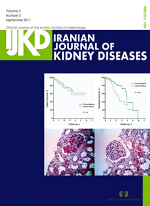
Pages 332-337Introduction. Culture-negative peritonitis is a major challenge in the treatment of peritonitis in continuous ambulatory peritoneal dialysis (CAPD). This study aimed to evaluate the culture-negative peritonitis in patients from the Iranian CAPD Registry.Materials and Methods. Data of 1472 patients from 26 CAPD centers were analysed. Peritonitis was defined as any clinical suspicion together with peritoneal leukocyte count of 100/mL and more.Results. The patients had been on PD for a mean of 500 ± 402 days. There were a total of 660 episodes of peritonitis observed among 299 patients (peritonitis rate of 1 episode in 34.1 patient-months). Excluding patients with both negative and positive culture results, there were 391 episodes of peritonitis in 220 patients (174 culture-positive episodes in 97 patients and 217 culture-negative episodes in 123). The 1- to 4-year patient survival rates were 85%, 75%, 69%, and 59% for the patients with culture-positive peritonitis, and 92%, 78%, 73% and 63% for the patients with culture-negative peritonitis, respectively (P =. 34). The technique survival rates were 90%, 57%, 42%, and 27% and 95%, 85%, 74%, and 40%, respectively (P =. 001). On follow-up, there were higher rates of active PD patients, lower rates of PD dropouts, and higher rates of kidney transplantation in patients with culture-negative peritonitis compared to those with culture-positive peritonitis. Conclusions. In our patients, the prevalence of culture-negative peritonitis was high (55.9%). Patient survival with culture-negative peritonitis was comparable to those with culture-positive peritonitis and technique survival was higher among those with culture-negative peritonitis.
-
Pages 338-341Introduction. Hepatitis B virus (HBV) infection is much more common in hemodialysis patients than the general population. Up to half of hemodialysis patients do not have adequate protective HBV antibodies after HBV vaccination. We studied the effects of adding levamisole, as an immunomodulator and adjuvant agent, on seroconversion response to HBV vaccination in hemodialysis patients.Materials and Methods. Thirty-six hemodialysis patients were divided into 2 groups. The first group received 40 microg of HBV vaccine intramuscularly at 0, 1, and 6 months plus 100 mg of oral levamisole per day for 12 days. The second group received the same amount and method of vaccine and placebo. Serum antibody levels were measured in each group after 0, 2, and 4 months after the last dose of vaccination.Results. Anti-HBV antibody level in the patients who received levamisole was lower than that in the control group. Antibody levels in the levamisole group at 0, 2, and 4 months after the last dose of vaccination were 44.4%, 77.8%, and 77.7%, respectively. In the control group, response rates at 0, 2, and 4 months were 55.6%, 72.2%, and 77.8% respectively (P =. 04, P =. 12, and P =. 08, respectively).Conclusions. Anti-HBV antibody level was significantly lower immediately after HBV vaccination when it was accompanied by levamisole administration. However, no significant differences were observed between the two groups at 2 and 4 months. Further evaluation is recommended to assess the effect of adding levamisole on Hepatitis B surface antibody titer in hemodialysis patients.
-
Pages 342-346Introduction. Both atorvastatin and mycophenolate mofetil (MMF) have been used for panel reactive antibodies (PRA) reduction in transplant candidates. The purpose of this study was to compare the effect of low-dose MMF and atorvastatin on PRA in sensitized hemodialysis patients waiting for kidney transplantation.Materials and Methods. A total of 40 adult patients with end-stage renal disease who were highly sensitized to human leukocyte antigens (PRA > 40%) were enrolled and randomly assigned into atorvastatin or low-dose MMF groups. All of the patients received the treatments for 2 months. The PRA status was determined at the end of the 1st and 2nd month.Results. Forty percent of the patients in the atorvastatin group compared with 5% in the low-dose MMF group showed complete response, defined as a minimum 50% reduction in PRA (P =. 02). Reduction of PRA in the atorvastatin group was significantly higher than that in the low-dose MMF group (P =. 01). No major infectious or other complications occurred in our patients.Conclusions. Atorvastatin has a significant effect on lowering of PRA in sensitized hemodialysis patients waiting for kidney transplantation. In addition, a short course of low-dose MMF is safe in ESRD patients; however, it has no effect on reduction of PRA.
-
Pages 347-350A 60-year-old man was admitted to our clinic with dyspnea, hemoptesis, anuria, nephritic syndrome, and a positive myeloperoxidase antineutrophil cytoplasmic antibody titer. He was diagnosed with antineutrophil cytoplasmic antibody-associated vasculitis due to Wegener granulomatosis, microscopic polyangiitis, or drug induction. Unexpectedly, histopathologic examination of the kidney biopsy specimen revealed the diagnosis of noncrescentic and nonnecrotizing glomerulonephritis. We report this case because of the unusual histologic type of renal involvement.
-
Pages 351-353Dermatological complications, especially skin infections, are very common following organ transplantation, and result in a lot of distress in the recipient. Herpes zoster, herpes simplex, and human papillomavirus infections are common infections in kidney transplant recipients, and therapeutic management is usually disappointing in immunosuppression state. We report here 2 cases of kidney transplant recipients who developed diffuse human papillomavirus-induced cutaneous warts with no response to conventional treatments. According to similar reports in organ transplant recipients, we modified the immunosuppressive regimen by converting to sirolimus, which led to a rapid relief from cutaneous warts in both patients. This evidence along with other case reports suggest that conversion to sirolimus may be considered as an effective strategy in cases of giant or multiple viral warts in kidney and perhaps other transplant recipients.
-
Page 355


