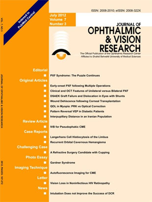
فهرست مطالب

Journal of Ophthalmic and Vision Research
Volume:7 Issue: 3, Jul-Sep 2012
- 94 صفحه،
- تاریخ انتشار: 1391/08/01
- تعداد عناوین: 16
-
-
Page 190PurposeTo present early-onset ocular manifestations of pseudoexfoliation syndrome in young patients who had undergone multiple intraocular procedures.MethodsThis is an observational case series, introducing four cases with histories of multiple intraocular procedures for glaucoma.ResultsAll reported cases demonstrated typical manifestations of pseudoexfoliation unilaterally in the eye that had undergone multiple surgeries. The diagnosis of pseudoexfoliation was made prior to the age of 50 in all subjects and the earliest manifestation was at the age of 18 in a case with primary congenital glaucoma.ConclusionThe role of multiple surgical procedures, in addition to genetic predisposition, should be further investigated as a possible inciting factor predisposing to pseudoexfoliation in younger individuals.
-
Page 197PurposeTo compare clinical findings and peripapillary retinal nerve fiber layer (RNFL) thickness using optical coherence tomography (OCT) in affected and fellow eyes of patients with unilateral pseudoexfoliation (PXF) syndrome with that of bilateral cases.MethodsThis cross-sectional study enrolled 91 subjects with PXF including 32 unilateral and 59 bilateral cases. Subjects with elevated intraocular pressure or findings suggestive of glaucoma were excluded. RNFL thickness and optic nerve head profile were studied in all eyes using the RNFL and optic nerve head analysis OCT protocol. Clinical and OCT features were compared in affected and unaffected eyes of unilateral PXF subjects to bilateral cases.ResultsBilateral cases with PXF were older
-
Page 203PurposeTo investigate the rates of Descemet''s stripping automated endothelial keratoplasty (DSAEK) graft dislocation and failure in glaucomatous eyes, including eyes with history of trabeculectomy and/or aqueous shunts.MethodsA retrospective, case-control study on a total of 424 consecutive eyes undergoing DSAEK at an academic setting compared 96 glaucomatous eyes to a control group of 328 eyes. Pre- and post DSAEK procedure data was aggregated for up to 2 years (mean follow-up, 6.5±6.9 months) including rates of graft dislocation and failure.ResultsOut of 96 glaucomatous eyes, 20 had undergone trabeculectomy, 27 had received one or more aqueous shunts, 12 had undergone both procedures and 37 were on medical therapy. Complete DSAEK graft dislocation and failure occurred in 2.7% and 3% of non-glaucomatous patients, respectively. Eyes with history of aqueous shunt surgery experienced graft dislocation and failure rates of 26.0% (OR=4.6, 95% CI 1.5-13.7, p=0.0067) and 26.0% (OR=10.3, 95% CI 3.8-27.1, p0.40) had no significant increase in graft dislocation or failure rates.ConclusionEyes with medically controlled glaucoma or prior trabeculectomy demonstrated comparable rates of graft dislocation and failure as compared to controls. Aqueous shunt surgery was associated with increased rates of graft dislocation and failure after DSAEK.
-
Page 214PurposeTo investigate the incidence، mechanisms، characteristics، and visual outcomes of traumatic wound dehiscence following keratoplasty.MethodsMedical records of 32 consecutive patients with traumatic globe rupture following keratoplasty who had been treated at our center from 2001 to 2009 were retrospectively reviewed.ResultsThe study population consisted of 32 eyes of 32 patients including 25 men and 7 women with history of corneal transplantation who had sustained eye trauma leading to globe rupture. Mean patient age was 38. 1 (range، 8 to 87) years and median interval between keratoplasty and the traumatic event was 9 months (range، 30 days to 20 years). Associated anterior segment findings included iris prolapse in 71. 9%، lens extrusion in 34. 4%، and hyphema in 40. 6% of eyes. Posterior segment complications included vitreous prolapse (56%)، vitreous hemorrhage (28%) and retinal detachment (18%). Eyes which had undergone deep anterior lamellar keratoplasty (DALK; 5 cases، 15. 6%) tended to have less severe presentation and better final visual acuity. There was no correlation between the time interval from keratoplasty to the traumatic event، and final visual outcomes.ConclusionThe host-graft interface demonstrates decreased stability long after surgery and the visual prognosis of traumatic wound dehiscence is poor in many cases. An intact Descemet''s membrane in DALK may mitigate the severity of ocular injuries، but even in these cases، the visual outcome of globe rupture is not good and prevention of ocular trauma should be emphasized to all patients undergoing any kind of keratoplasty.
-
Page 219PurposeTo compare quality of life (QOL) in myopic patients who underwent photorefractive keratectomy (PRK) with that of myopic spectacle or contact lens users.MethodsThis observational comparative study was performed on 102 low to moderate myopic patients who had undergone PRK at least 6 months ago and 106 myopic spectacle or contact lens wearers. Vision related QOL and its correlation with demographic variables, visual acuity and refractive status were compared between the two groups. QOL was measured using a validated translated version of the Visual Function Questionnaire (VFQ-25) which contains 25 questions in 12 subscales with a total score of zero to 100.ResultsMean total QOL score was 97.0±4.4 and 86.1±10.7 in PRK and nonsurgical groups respectively [mean difference (d)=11, P0.05). Overall, 10 out of 12 QOL subscales were significantly higher in the PRK group (P0.9) and ocular pain (d=3.1, P=0.3) were not significantly different between the study groups.ConclusionCorrection of myopia using PRK is associated with higher QOL scores in most subscales as compared to spectacle or contact lens wear.
-
Page 225PurposeTo evaluate cortical and retinal activity by pattern visual evoked potentials (PVEP) in patients with type II diabetes mellitus.MethodsPVEP was recorded in 40 diabetic patients including 20 subjects with non-proliferative diabetic retinopathy (NPDR) and 20 others without any retinopathy on fundus photography, and compared to 40 age- and sex-matched normal non-diabetic controls.ResultsP100 wave latency was significantly longer in diabetic patients as compared to normal controls.
-
Page 231PurposeTo report normal interpupillary distance (IPD) values in different age groups of an Iranian population.MethodsThis study was performed on 1,500 randomly selected subjects from 3,260 consecutive out-patients with refractive errors referred to Farabi Eye Hospital, Isfahan,Iran over a period of two years (2008 to 2010). Measurement of refractive errors and IPD for far distance were performed using an autorefractometer (RMA-3000 autorefractometer,Topcon, Tokyo, Japan).ResultsMean IPD in adult subjects was 61.1±3.5 mm in women and 63.6±3.9 mm inmen.
-
Page 235Cystoid macular edema (CME) is a major cause of decreased vision after complicated or uncomplicated cataract surgery. This paper reviews the use of intravitreal bevacizumab (IVB) injection for treatment of pseudophakic CME. In a literature search of all articles available on Medline and Scopus databases, 11 studies including one prospective and 4 retrospective studies, 4 case reports, one letter to editor and one review article were identified. All articles except one, reported the use of IVB for chronic CME unresponsive to at least one conventional treatment modality. The level of evidence for all studies was categorized as low or very low. Although intravitreal bevacizumab might be effective for many cases of pseudophakic CME, its use should be reserved for eyes unresponsive to conventional treatment modalities.
-
Page 240PurposeTo report a rare presentation of unifocal Langerhans cell histiocytosis (LCH) simulating a limbal papilloma. Case report: A 24-year-old man presented with a limbal mass in his left eye which had initially been suspected to be a papilloma based on clinical findings. The mass was excised and a histopathological diagnosis of «acute bullous inflammation with granulation tissue» was made. The lesion relapsed 10 months later which necessitated repeat resection along with corneoscleral patch grafting. Histopathological studies of the excised lesion led to a final diagnosis of LCH.ConclusionTo the best of our knowledge, this is the second report of a rare presentation of LCH in the limbus which recurred after excision of the primary mass. The recurrent lesion was diagnosed based on histopathology and managed accordingly.
-
Page 244PurposeTo report late recurrence of orbital cavernous hemangioma in a patient ten years after complete resection of the primary tumor. Case Report: A 32-year-old woman with a history of progressive visual loss and proptosis underwent lateral orbitotomy for resection of a large cavernous hemangioma. Ten years later, proptosis recurred and the patient developed progressive ocular deviation. Imaging studies were in favor of a recurrent cavernous hemangioma and the tumor was excised via the previous incision site. Reassessment of previous orbital images suggested the presence of two separate tumors, only one of which had been excised at the time of initial surgery.ConclusionRecurrent orbital cavernous hemangioma may follow incomplete excision of multiple orbital lesions with gradual growth of unidentified residual tumors. Accordingly, when an encapsulated cavernous hemangioma is removed, exploration is recommended to rule out multiple lesions.
-
Page 257
-
Page 261Lipofuscin results from digestion of photoreceptor outer segments by the retinal pigment epithelium (RPE) and is the principal compound that causes RPE fluorescence during autofluorescence imaging. Absorption of the 488-nanometer blue light by macular pigments, especially by the carotenoids lutein and zeaxanthin, causes normal macular hypo-autofluorescence. Fundus autofluorescence imaging is being increasingly employedin ophthalmic practice to diagnose and monitor patients with a variety of retinal disorders. In macular edema for example, areas of hyper-autofluorescence are usually present which are postulated to be due to dispersion of macular pigments by pockets of intraretinal fluid. For this reason, the masking effect of macular pigments is reduced and the natural autofluorescence of lipofuscin can be observed without interference. In cystic types of macular edema, e.g. cystoid macular edema due to retinal vein occlusion, diabetic macular edema and post cataract surgery, hyper-autofluorescent regions corresponding to cystic spaces of fluid accumulation can be identified. In addition, the amount of hyper-autofluorescence seems to correspond to the severity of edema. Hence, autofluorescence imaging, as a noninvasive technique, can provide valuable information on cystoid macular edema in terms of diagnosis, follow-up and efficacy of treatment.

