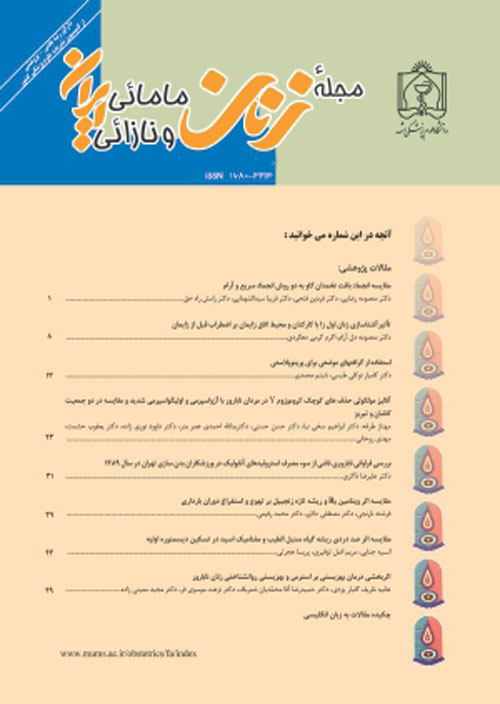
فهرست مطالب
مجله زنان مامائی و نازائی ایران
سال پانزدهم شماره 25 (هفته سوم آبان 1391)
- تاریخ انتشار: 1391/08/19
- تعداد عناوین: 4
-
-
صفحه 1مقدمهیکی از مهم ترین درمان های زوج های نابارور، انتقال جنین فریز شده می باشد. اگر چه انتقال جنین تازه مورد تایید و توجه است، اما مطالعات اندکی در مورد عوامل بالینی موثر در میزان لقاح و میزان بارداری در انتقال جنین فریز شده وجود دارد. مطالعه حاضر با هدف بررسی برخی عوامل بالینی موثر در پیش آگهی انتقال جنین فریز شده انجام شد.روش کاراین مطالعه گذشته نگر با بررسی 372 پرونده بیمارانی که بین فروردین 1388 لغایت فروردین 1390 به مرکز تحقیقاتی درمانی ناباروری یزد مراجعه و از جنین فریز شده استفاده کرده بودند، انجام شد. کلیه اطلاعات مربوط به پرونده بیماران در پرسشنامه ثبت شد. برای تجزیه و تحلیل داده ها از نرم افزار آماری SPSS (نسخه 15) و آزمون های کولموگروف- اسمیرنوف و من ویتنی استفاده شد. میزان p کمتر از 05/0 معنی دار در نظر گرفته شد.یافته هامیزان بارداری بالینی در زنان زیر 35 سال 7/57 درصد و بالای 35 سال 2/29 درصد و همچنین میزان بارداری بالینی در زنان با هورمون محرک فولیکول روز سوم زیر 10، 3/56 درصد و هورمون محرک فولیکول بالای 10، 5/17 درصد بود که این نتایج از نظر آماری معنی دار بود (0001/0=p). اما سایر عوامل مانند علت فریز جنین، پروتکل اولیه لقاح آزمایشگاهی، روش لقاح آزمایشگاهی، ضخامت اندومتر و طول سیکل درمان تا روز انتقال جنین از نظر آماری در انتقال جنین فریز شده موثر نبودند (05/0نتیجه گیریسن زن و هورمون محرک فولیکول روز سوم یکی از مهمترین عوامل موثر در میزان بارداری بالینی در انتقال جنین فریز شده می باشد.
کلیدواژگان: انتقال جنین، جنین فریز شده، لقاح آزمایشگاهی، میزان بارداری بالینی -
صفحه 8مقدمهپره اکلامپسی یکی از عوارض دوران بارداری است که باعث به خطر افتادن زندگی مادر و جنین می شود. مطالعات مختلف نشان دهنده نقش مهم پلاکت ها در پاتوژنز پره اکلامپسی می باشند. مطالعه حاضر با هدف مقایسه شاخص های پلاکتی در زنان باردار سالم و زنان پره اکلامپسی و ارزش پیشگویی آن ها در مورد شدت پره اکلامپسی انجام شد.روش کاراین مطالعه مورد - شاهدی در فاصله زمانی تیر ماه 1387 تا دی ماه 1389، بر روی زنان باردار مراجعه کننده به بیمارستان امام رضا (ع) کرمانشاه در هنگام وضع حمل انجام شد. افراد در سه گروه مبتلا به پره اکلامپسی خفیف (57 نفر)، مبتلا به پره اکلامپسی شدید (50 نفر) و زنان باردار سالم (102 نفر) قرار گرفتند. در حوالی زایمان، 5 میلی لیتر از خون وریدی افراد هر سه گروه در شرایط استاندارد آزمایشگاهی گرفته شد تا شاخص های پلاکتی آن اندازه گیری شود. برای سنجش شاخص های پلاکتی که شامل تعداد پلاکت، میانگین حجم پلاکتی، گستره توزیع پلاکتی و نسبت سلول های بزرگ پلاکتی بودند از دستگاه شمارش سلول های خونی استفاده شد. تجزیه و تحلیل داده ها با استفاده از نرم افزار آماری SPSS (نسخه 16) و روش آماری ANOVA (آنالیز واریانس) انجام شد. جهت بررسی تفاوت بین گروه ها از آزمون آماری توکی استفاده شد. میزان p کمتر از 05/0 معنی دار در نظر گرفته شد.یافته هادر این مطالعه بین زنان پره اکلامپسی و باردار سالم از نظر تعداد پلاکت و شاخص های پلاکتی اختلاف آماری معنی داری وجود نداشت (05/0نتیجه گیریشاخص های پلاکتی در دوران بارداری تحت تاثیر پره اکلامپسی قرار نمی گیرند. همچنین بین تعداد پلاکت ها و نیز شاخص های پلاکتی در زنان مبتلا به پره اکلامپسی خفیف و شدید تفاوتی وجود ندارد.
کلیدواژگان: پره اکلامپسی، پلاکت، شاخص های پلاکتی -
صفحه 14مقدمهامروزه مسئله زایمان و کنترل مناسب آن، امر بسیار مهمی است زیرا حدود 50% سزارین های انجام شده به دلیل پیشرفت غیرطبیعی در مرحله اول زایمان می باشد. در مامایی مدرن به طور وسیعی از انفوزیون اکسی توسین برای تحریک و تسریع زایمان استفاده می شود. از آنجایی که در عدم تناسب سری- لگنی شکل انقباضات رحم تغییر می کند، مطالعه حاضر با هدف بررسی شکل انقباضات رحمی با تجویز اکسی توسین در فاز فعال مرحله اول زایمان انجام شد.روش کاردر این مطالعه کوهورت، ابتدا وضعیت انقباضات رحمی 100 زن نخست زا با بارداری تک قلو و نمایش قله سر به مدت 30 دقیقه در اتساع 4-3 سانتی متر مانیتور شد. طی لیبر در 53 زن به دلیل عدم پیشرفت لیبر، اکسی توسین تجویز شد. نهایتا در اتساع 10-8 سانتی متری دهانه رحم به مدت 30 دقیقه الگوی انقباضات ثبت شد و نسبت طول زمان برگشت یک انقباض از قله به خط پایه به زمان رسیدن از خط پایه به قله در دو مرحله شروع و انتهای مرحله اول زایمان در دو گروه (با و بدون تجویز اکسی توسین) محاسبه و مقایسه شد. تجزیه و تحلیل داده ها با استفاده از نرم افزار آماری SPSS (نسخه 5/11) و آزمون آماری تی زوجی، تی مستقل و کای اسکوئر انجام شد. میزان کمتر یا مساوی 05/0 معنی دار در نظر گرفته شد.یافته هانسبت طول زمان برگشت یک انقباض از قله به خط پایه به زمان رسیدن از خط پایه به قله در ابتدا و انتهای لیبر در دو گروه با و بدون تجویز اکسی توسین به ترتیب 203/0 ± 164/1، 223/0 ±150/1 (687/0=p) و 199/0 ±161/1 و 214/0 ±091/1 بود (059/0=p) که تفاوت مشاهده شده در دو گروه از نظر آماری معنی داری نبود.نتیجه گیریاستفاده از اکسی توسین جهت تقویت انقباضات رحمی شکل انقباضات رحمی را تغییر نمی دهد.
کلیدواژگان: اکسی توسین، شکل انقباضات رحمی، مرحله اول زایمان، نسبت F:R -
صفحه 21مقدمهطول مدت زایمان از عوامل موثر بر نتایج بارداری و آسیب های وارده بر مادر و جنین می باشد. به گونه ای که با طولانی شدن بیش از حد زایمان، احتمال عفونت، کمبود اکسیژن، صدمات جسمی- عصبی و مرگ در جنین و نوزاد، همچنین خونریزی و عفونت بعد از زایمان و آشفتگی حاصل از اضطراب، بی خوابی و خستگی در مادر افزایش می یابد. مراقبت مطلوب از مادران در هنگام زایمان از اهداف نظام بهداشتی- درمانی می باشد. مطالعه حاضر با هدف تعیین تاثیر فشار در نقاط سانینجیائو- هوگو بر طول مدت زایمان در زنان نخست زا انجام شد.روش کاراین مطالعه کارآزمایی بالینی در سال 91-1390 بر روی 84 زن نخست زای واجد شرایط مراجعه کننده به بیمارستان های سبلان و علوی اردبیل انجام شد. افراد توسط بلوک بندی تصادفی 4 و 6 تایی به دو گروه مداخله و کنترل تقسیم شدند. مداخله به صورت اعمال فشار در نقاط سانینجیائو و هوگو در دیلاتاسیون های 4، 6، 8 و 10 سانتی متری سرویکس بود. طول مدت فاز فعال و مرحله دوم از طریق معاینه واژینال توسط پژوهشگر در پرسشنامه حاوی اطلاعات فردی ثبت شد. داده ها پس از گردآوری با استفاده از نرم افزار آماری SPSS (نسخه 16) و روش های آماری توصیفی - تحلیلی مورد تجزیه و تحلیل قرار گرفت. میزان p کمتر از 05/0 معنی دار در نظر گرفته شد.یافته هامیانگین سنی شرکت کنندگان 97/3 ± 9/21 سال بود. میانگین طول مدت فاز فعال زایمان در دو گروه مداخله و کنترل به ترتیب 1:19 ± 2:15 ساعت و 1:59 ± 4:10 ساعت (001/0=p) و میانگین طول مدت مرحله دوم زایمان در دو گروه مداخله و کنترل به ترتیب 00:25±00:45 دقیقه و 00:39 ±1:04 ساعت بود (008/0=p).نتیجه گیریفشار بر نقاط سانینجیائو- هوگو در دیلاتاسیون های مختلف باعث کاهش طول مدت فاز فعال و مرحله دوم زایمان می شود.
کلیدواژگان: زنان نخست زا، سانینجیائو، طب فشاری، طول مدت زایمان، هوگو
-
Page 1IntroductionCryopreserved-embryo transfer is one of the most important treatments for infertile couples. While fresh-embryo transfer is mostly approved and concerned، few studies have evaluated the potential effect of clinical factor on the implantation and pregnancy rates in frozen embryo transfer. The aim of this study was to investigate some clinical factors that potentially influence the outcome of cryopreserved-embryo transfer.MethodsIn this retrospective investigation، 372 patients'' records that referred to Research and Clinical Center of Infertility and had frozen-embryo transfer cycles were studied from 2009 to 2011. All data of patient`s files fill in the questionnaire form. Data were analyzed by SPSS software version 15 and Kolmogorov-Smirnov، Paired t and t student tests. P value less than 0. 05 was considered statistically significant.ResultsClinical pregnancy-rate in patients under 35 years old was significantly higher than patients aged more than 35 years old (57. 7% versus 29. 2%). Also، clinical pregnancy-rate in women with FSH more and less than 10 IU/L were 56. 3% and 17. 5%، respectively (PV=0. 0001) which were statistically significant. Other clinical factors such as: the causes of embryo freezing، primary protocols of IVF /ICSI، endometrial thickness and duration of cycle up to day of embryo transfer had no significant effects on frozen-embryo transfer (P>0. 05).ConclusionAge of women and FSH in third day were the most important factors influencing the clinical pregnancy rate following frozen embryo transfer.Keywords: Clinical pregnancy rate, Embryo transfer, Frozen embryo, IVF
-
Page 8IntroductionPreeclampsia is one of the complications of pregnancy period that threatens the life of both mother and fetus. Various studies have shown that platelets play a major role in the pathogenesis of preeclampsia. The aim of this study is to compare platelet indices in preeclamptic and normal pregnant women and to evaluate whether these parameters have a predictive significance in determining the severity of preeclampsia.MethodsThis case-control study was held on pregnant women who referred to Imam Reza Hospital of Kermanshah city for delivery from 2008 to 2010. Participants were classified into three groups of mild preeclamptic (57 person)، severe preeclamptic (50 persons)، and normal pregnant women (102). Under standard conditions، 5 ml of individual''s venous blood of each group was taken prenatally. Automated blood cell counter XT-1800 was used to measure platelet indices that include platelet number، mean platelet volume (MPV)، Platelet distribution width (PDW)، and Platelet large cell ratio (P-LCR). Data analysis was performed by using ANOVA، SPSS statistical software version 16. Tukey test was used to assess differences between groups. P value less than 0. 05 was considered statistically significant.ResultsThere was no statistically significant difference in platelet number and indices between preeclampsia and healthy pregnant women. (P>0. 05) As a result، no significant difference of platelet number (Pand indices was observed in patients with preeclampsia (P>0. 05).ConclusionThe results showed that platelet indices were not affected by preeclampsia during pregnancy. Also، no difference was observed in the term of platelet number and indices in mild and severe preeclamptic cases.Keywords: Platelet, Platelet indices, Preeclampsia
-
Page 14IntroductionAbout 50% of cesarean sections are performed because of abnormal development of first stage، therefore، delivery and its control is very important nowadays. For induction or augmentation of labor، oxytocin infusion is widely used in modern obstetric. Since the contraction shape varies by cephalo-pelvic disproportion، this study was conducted to evaluate the shape of uterine contractions with oxytocin in the active phase of labor.MethodsIn this Cohort study 100 nulliparous women with singleton pregnancy and cephalic presentation was monitored in dilation 3-4 cm for 30 minutes. In order to precede labor، oxytocin was prescribed to 53 women. Finally، the pattern of contractions was recorded in 10-8 cm cervical dilation for 30 minutes. The ratio of spending time of contraction from peak to baseline (F: Fall) and vice versa (R: Rise) in two phases of beginning and end of the first stage of labor in two groups (with and without prescription of Oxytocin) was calculated and compared. Data were analyzed by using SPSS statistical software version 11. 5، paired T-test، independent T-test، and chi-square. P value less than 0. 05 was considered statistically significant.ResultsThe ratio of F and R was 1. 164±0. 203 and 1. 150±0. 223 at the beginning and the end of labor in oxytocin received group. The F: R ratio was 1. 161±0. 203 and 1. 091±1. 161±0. 214 at the beginning and the end of labor in the other group. No statistically significant difference was observed in the ratio of F and R between with and without oxytocin groups.ConclusionUsing Oxytocin for augmentation of uterine contractions does not change the shape of the uterine contractions.Keywords: Fall to Rise ratio, First stage of labor, Oxytocin, Uterine contractions
-
Page 21IntroductionDelivery duration is one of the factors that affect pregnancy outcomes، and damages of mother and fetus. Excessive prolongation of labor increases possibility of infection، hypoxia، physical-neural damages، newborn''s death، postpartum hemorrhage and infection، anxiety، insomnia، and mother''s fatigue. One of the most important goals of healthcare system is optimal care of mother during childbirth. This research has been done in order to determine the effects of acupressure at Sanyinjiao (SP6) and Hugo (LI4) points in nulliparous women.MethodsThis clinical trial was held on 84 nulliparous eligible women who attended to Sabalan and Alavi hospitals in Ardabil in 2011-2012. The subjects were allocated into two groups (intervention- control) with random block-size of 4 and 6. Pressures at Sanyinjiao (SP6) and Hugo (LI4) points in 4، 6، 8، 10 cm of cervical dilatation were applied in intervention group. Through vaginal examination، active phase duration and second stage were recorded in the demographic questionnaire by the researcher. Gathered data were analyzed by the means of SPSS statistical software version 16 and descriptive-analytic statistical methods. P value less than 0. 05 was considered statistically significant.ResultsThe mean age of participants was 21. 9± 3. 97 years. The mean duration of the active phase in intervention and control groups were respectively 2:15±1:19 and 4:10±1:59 hours (p=0. 001). The mean duration of the second stage in two intervention and control groups were respectively 00:45±00:25 minutes and 1:04±00:39 hour (p=0. 008).ConclusionAcupressure at Sanyinjiao (SP6) and Hugo (LI4) points in different dilatation reduces the length of active phase and second stage of delivery time.Keywords: Acupressure, Length of delivery, Nulliparous women, Sanyinjiao (SP6), Hugo (LI4)


