فهرست مطالب

Cell Journal (Yakhteh)
Volume:16 Issue: 3, Autumn 2014
- تاریخ انتشار: 1393/07/10
- تعداد عناوین: 20
-
-
Page 235ObjectiveHuman induced pluripotent stem cells (iPSCs) have been shown to have promising capacity for stem cell therapy and tissue engineering applications. Therefore, it is essential to compare the ability of these cells with the commonly used mesenchymal stem cells (MSC) for bone tissue engineering in vitro.Materials And MethodsIn this experimental study, the biological behavior and osteogenic capacity of the iPSCs were compared with MSCs isolated from human adipose tissue (AT-MSCs) using 3-(4,5-di-methylthiazol-2-yl)-2,5-diphenyltetrazolium bromide (MTT) assay, Alizarin red staining, alkaline phosphatase (ALP) activity measurements, calcium content assay and common osteogenic-related genes. Data were reported as the mean ± SD. One-way analysis of variance (ANOVA) was used to compare the results. A p value of less than 0.05 was considered statistically significant.ResultsThere was a significant difference between the rate of proliferation of the two types of stem cells; iPSCs showed increased proliferation compared to AT-MSCs. During osteogenic differentiation, ALP activity and mineralization were demonstrated to be significantly higher in iPSCs. Although AT-MSCs expressed higher levels of Runx2, iPSCs expressed higher levels of osteonection and osteocalcin during differentiation.ConclusioniPSCs showed a higher capacity for osteogenic differentiation and hold promising potential for bone tissue engineering and cell therapy applications.Keywords: Osteogenic, Tissue Engineering, Mesenchymal Stem Cells, Flow Cytometry, Gene Expression
-
Page 245ObjectiveAssessments of cell reactions such as motility, orientation and activation to the topography of the substratum will assist with the fabrication of a proper implantable scaffold for future tissue engineering applications.The current challenge is to analyze the orientation effect of elecrospun nanofibers of poly (ε-caprolactone) (PCL) on viability and proliferation of mouse embryonic stem cells (mESCs).Materials And MethodsIn this experimental study, we used the electrospinning method to fabricate nanofibrous PCL scaffolds. Chemical and mechanical characterizations were specified by the contact angle and tensile test. O2 plasma treatment was used to improve surface hydrophilicity. We used the 3-(4,5-dimethylthiazol-2-yl)-2,5-diphenyltetrazolium bromide (MTT) assay to evaluate mESCs adhesion and proliferation before and after surface modification. The influence of the orientation of the nanofibers on mESCs growth was evaluated by scanning electron microscopy (SEM). Statistical analysis was performed using one-way analysis of variance (ANOVA) With differences considered statistically significant at p≤0.05.ResultsThe results showed that plasma treatment improved the hydrophilic property of PCL scaffolds. MTT assay showed a significant increase in proliferation of mESCs on plasma treated PCL (p-PCL) scaffolds compared to non-treated PCL (p≤0.05). However gelatin coated tissue culture plate (TCP) had a better effect in initial cell attachment after one day of cell seeding. There was more cell proliferation on day 3 in aligned plasma treated (AP) nanofibers compared to the TCP. SEM showed optical density of the cell colonies. Aligned nanofibrous scaffolds had larger colony sizes and spread more than random nanofibrous scaffolds.ConclusionThis study showed that plasma treating of scaffolds was a more suitable substrate for growth and cell attachment. In addition, aligned nanofibrous scaffolds highly supported the proliferation and spreading of mESCs when compared to random nanofibrous scaffolds and TCP.Keywords: Embryonic Stem Cells, Nanofibers, Poly (εcaprolactone), Surface Modification, Cell Proliferation
-
Page 255ObjectiveAutoimmune diseases precede a complex dysregulation of the immune system. T helper17 (Th17) and interleukin (IL)-17 have central roles in initiation of inflammation and subsequent autoimmune diseases. IL-27 significantly controls autoimmune diseases by Th17 and IL-17 suppression. In the present study we have created genetic engineered mesenchymal stem cells (MSCs) that mediate with lentiviral vectors to release IL-27 as an adequate vehicle for ex vivo gene therapy in the reduction of inflammation and autoimmune diseases.Materials And MethodsIn this experimental study, we isolated adipose-derived MSCs (AD-MSCs) from lipoaspirate and subsequently characterized them by differentiation. Two subunits of IL-27 (p28 and EBI3) were cloned in a pCDH-513B-1 lentiviral vector. Expressions of p28 and EBI3 (Epstein-Barr virus induced gene 3) were determined by real time polymerase chain reaction (PCR). MSCs were transduced by a pCDH-CMV-p28-IRESEBI3- EF-copGFP-Pur lentiviral vector and the bioassay of IL-27 was evaluated by IL-10 expression.ResultsCell differentiation confirmed true isolation of MSCs from lipoaspirate. Restriction enzyme digestion and sequencing verified successful cloning of both p28 and EBI3 in the pCDH-513B-1 lentiviral vector. Real time PCR showed high expressions level of IL-27 and IL-10 as well as accurate activity of IL-27.ConclusionThe results showed transduction of functional IL-27 to AD-MSCs by means of a lentiviral vector. The lentiviral vector did not impact MSC characteristics.Keywords: Autoimmune Disease, Gene Therapy, IL, 27, Mesenchymal Stem Cells
-
Page 263ObjectiveTendon never returns to its complete biological and mechanical properties after repair. Bone marrow and, recently, adipose tissue have been used as sources of mesenchymal stem cells which have been proven to enhance tendon healing. In the present study, we compared the effects of allotransplantation of bone marrow derived mesenchymal stromal cells (BMSCs) and adipose derived stromal vascular fraction (SVF) on tendon mechanical properties after experimentally induced flexor tendon transection.Materials And MethodsIn this experimental study, we used 48 adult male New Zealand white rabbits. Twelve of rabbits were used as donors of bone marrow and adipose tissue, the rest were divided into control and treatment groups. The injury model was a unilateral complete transection of the deep digital flexor tendon. Immediately after suture repair, 4×10⁶ cells of either fresh SVF from enzymatic digestion of adipose tissue or cultured BMSCs were intratendinously injected into tendon stumps in the treatment groups. Controls received phosphate-buffered saline (PBS). Immobilization with a cast was continued for two weeks after surgery. Animals were sacrificed three and eight weeks after surgery and tendons underwent mechanical evaluations. The differences among the groups were analyzed using the analysis of variance (ANOVA) test followed by Tukey’s multiple comparisons test.ResultsStromal cell transplantation resulted in a significant increase in ultimate and yield loads, energy absorption, and stress of repairs compared to the controls. However, there were no statistically significant changes detected in terms of stiffness. In comparison, we observed no significant differences at the third week between SVF and BMSCs treated tendons in terms of all load related properties. However, at the eighth week SVF transplantation resulted in significantly increased energy absorption, stress and stiffness compared to BMSCs.ConclusionThe enhanced biomechanical properties of repairs in this study advocates the application of adipose derived SVF as an excellent source of multipotent cells instead of traditional BMSCs and may seem more encouraging in cell-based therapy for tendon injuries.Keywords: Tendon, Tensile Strength, Adipose Tissue, Bone Marrow, Transplantation
-
Page 271ObjectiveCryopreservation of ovarian tissue or follicles has been proposed as an alternative method for fertility preservation. Although successful vitrification of follicles has been reported in several mammalian species, the survival rate is generally low. The aim of this study was to investigate the effects of fibroblast growth factor (FGF) and epidermal growth factor (EGF) on in vitro preantral follicle development after vitrification.Materials And MethodsIn this experimental study, preantral follicles with diameter of 150-180 μm were mechanically isolated from ovaries of 18-21 days old NMRI mice. Follicles were vitrified and warmed, then cultured in α-minimal essential medium (α-MEM) without growth factor supplementation as control group (group I), while supplemented with 20 ng/ml FGF (group II), 20 ng/ml EGF (group III), and 20 ng/ml FGF +20 ng/ml EGF (group IV). After 12 days, human chorionic gonadotrophin (hCG)/EGF was added to culture medium, and after 18-20 hours, the presence of cumulus oocyte complexes (COCs) and oocyte maturation were assessed. The chi-square (χ2) test was used to analyze survival and ovulation rates of the follicles.ResultsOur results showed that the rate of metaphase II (MII) oocytes in FGF group increased in comparison with control and other treatment groups (p<0.027), but there was no difference between control with EGF and EGF+FGF groups in oocyte maturation rate (p>0.05). There was a significant decrease in survival rate of follicles in EGF+FGE group in comparison with other groups (p<0.008). After in vitro ovulation induction, the follicles in EGF group showed a higher ovulation rate (p<0.008) than those cultured in other groups.ConclusionFGF has beneficial effect on oocyte maturation, and EGF increases COCs number in vitro. Combination of EGF and FGE decreases the number of survived follicles.Keywords: Vitrification, Mouse Preantral Follicle, In Vitro Maturation, Epidermal Growth Factor, Fibroblast Growth Factor
-
Page 279ObjectiveThe aim of the present study was to investigate the effects of four equilibration times (2, 4, 8 and 16 hours) and two extenders (tris or Bioxcell®) on cryopreservation of buffalo semen.Materials And MethodsIn this experimental study, split pooled ejaculates (n=4), possessing more than 70% visual sperm motility were divided in two aliquots and diluted in Bioxcell ® and tris-citric egg yolk (TCE) extenders. Semen was cooled to 4˚C within 2 hours, equilibrated at 4˚C for 2, 4, 8 and 16 hours, then transferred into 0.5 ml French straws, and frozen in a programmable cell freezer before being plunged into liquid nitrogen. Postthaw motility characteristics, plasma membrane integrity, acrosome morphology and DNA integrity of the buffalo sperm were studied after thawing.ResultsThere were significant interactions between equilibration times and extendersfor sperm motility and membrane integrity. Post thaw sperm motility (PMOT), progressivemotile spermatozoa (PROG), plasma membrane integrity (PMI) and normal apical ridge(NAR) measures were lower for sperm equilibrated for 2 hours in both TCE and Bioxcell®extender compared to others equilibration times. PMOT, PMI and NAR for sperm equilibrated for 4, 8 and 16 hours showed no significant differences in either extender, although PROG measures were superior in Bioxcell® compared to TCE at all equilibration times (p<0.05). Kinematic parameters such as average path velocity, curvilinear velocity and linearity in the Bioxcell® extender were superior to those in the TCE extender studied. In contrast to motility and viability, the DNA integrity of post thaw spermatozoa remained unaffected by different equilibration times.ConclusionEquilibration time is necessary for preservation of the motility and integrity of buffalo sperm membranes. Equilibration times of over than 2 hours resulted in the greatest preservation of total semen parameters during cryopreservation. There were no significant interactions between equilibration times over 4 hours and type of extender which lead to greater post thaw sperm survival.Keywords: Buffalo, Sperm, Cryopreservation, Extender, Chromatin
-
Page 289ObjectiveThe effects of dietary fish oil on semen quality and sperm fatty acid profiles during consumption of n-3 fatty acids as well as the persistency of fatty acids in ram’s sperm after removing dietary oil from the diet were investigated.Materials And MethodsIn this experimental study, we randomly assigned 9 Zandi rams to two groups (isoenergetic and isonitrogenous diets): control (CTR; n=5) and fish oil (FO; n=4) for 70 days with a constant level of vitamin E in both groups. Semen was collected at the first week and at the last week of the feeding period (phase 1). After the feeding period, all rams were fed a conventional diet and semen samples were collected one and two months after removal of FO (phase 2). The sperm parameters and fatty acid profiles were measured by computer assisted semen analyzer (CASA) and gas chromatography (GC), respectively. The completely randomized design was used and data were analyzed with SPSS version 16.ResultsDietary FO had significant positive effects on all sperm quality and quantity parameters compared with the CTR during the feeding period (p<0.05). The positive effects of FO on sperm concentration and total sperm output were observed at one and two months after removal of FO (p<0.05), whereas other sperm parameters were unaffected. Before feeding, C14 (myristic acid), C16 (palmitic acid), C18 (stearic acid), C18:1 (oleic acid) and C22:6 (docosahexaenoic acid: DHA) were the primary sperm FA. FO in the diet increased sperm DHA, C14:0 and C18:0 during the feeding period (p<0.05).ConclusionThe present study showed not only manipulation of ram sperm fatty acid profiles by dietary FO and sperm parameters during the feeding period, but also the persistency of unique effects of dietary omega-3 fatty acids up to two months following its removal from the diet. Also, we recommend that sperm fatty acid profiles should be comprehensively analyzed and monitored.Keywords: : Fish Oil, Ovine, Spermatozoa
-
Page 299ObjectiveSilybin is a polyphenol with anti-oxidant and anti-cancer properties. The poor bioavailability of some polyphenols can be improved by binding to phosphatidylcholine. In recent years, studies have been conducted to evaluate the anti-cancer effect of silybin. We studied the effect of silybin and silybin-phosphatidylcholine on ESR1 and ESR2 gene expression and viability in the T47D breast cancer cell line.Materials And MethodsIn this experimental study, a 3-(4,5-Dimethylthiazol-2-Yl)-2,5- Diphenyltetrazolium Bromide test (MTT test) was used to determine doses for cell treatment, and the gene expression was analyzed by real-time reverse transcriptase-polymerase chain reaction (real-time RT- PCR).ResultsSignificant dose- and time-dependent cell growth inhibitory effects of silybin and silybin-phosphatidylcholine along with ESR1 down-regulation were observed in T47D cells. In contrast to ESR1, the T47D cell line showed negligible ESR2 expression.ConclusionThis study suggests that silybin and silybin-phosphatidylcholine down-regulate ESR1 in ER+ breast cancers. Results also show that in the T47D cell line, silybinphosphatidylcholine has a much higher growth inhibitory effect and a more significant down-regulation of ESR1 compared with silybin.Keywords: Silybin, Silybin, Phosphatidylcholine, Breast Cancer, ESR1
-
Page 309ObjectivePeople are usually susceptible to carcinogenic aromatic amines, present in cigarrette smoke and polluted environment, which can cause DNA damage. Therefore, maintenance of genomic DNA integrity is a direct result of proper function of DNA repair enzymes. Polymorphic diversity could affect the function of repair enzymes andthus augment the risk of different cancers. Xeroderma pigmentosum group D (XPD) gene encodes one of the most prominent repair enzymes and the polymorphisms of this gene are thought to be of importance in lung cancer risk. This gene encodes the helicase, which is a component of transcription factor IIH and an important part of the nucleotide excision repair system. Studies reveal that individuals with Lys751Gln polymorphism of XPD gene have a low repairing capacity to delete the damages ofultraviolet light among other XPD polymorphisms.Materials And MethodsIn this case-control study, first Lys751Gln polymorphism was genotyped, then its association with lung cancer risk was analyzed. Genomic DNA wasextracted from the whole blood sample of 640 individuals from Iran (352 healthy individuals and 288 patients). Allele frequencies and heterozygosity of Lys751Gln polymorphism were determined using polymerase chain reaction-restriction fragment length polymorphism method.ResultsAccording to statistical analyses, lung cancer risk in individuals with Lys751Gln polymorphism (Odd Ratio=1.8, 95% Confidence Interval 0.848-3.819) is approximately twice as high as that of Lys/Lys genotype, however 751Gln/Gln genotype did not relate to lung cancer risk (Odd Ratio=0.7, 95% Confidence Interval 0/307-1/595).ConclusionThis study suggests that heterozygous polymorphism (Lys/Gln) increases the sensitivity of lung cancer risk, while homozygous polymorphism (Lys/Lys) probably decreases its risk and C allele frequency shows no remarkable increase in the patients.Keywords: XPD, Lung Cancer, Polymorphism, Iranian, RFLP, NER
-
Page 315ObjectiveStroke is most important cause of death and disability in adults. The hippocampal CA1 and sub-ventricular zone neurons are vulnerable to ischemia that can impair memory and learning functions. Although neurogenesis normally occurs in the dentate gyrus (DG) of the hippocampus and sub-ventricular zone (SVZ) following brain damage, this response is unable to compensate for severely damaged areas. This study aims to assess both neurogenesis and the neuroprotective effects of transforming growth factor-alpha (TGF-α) on the hippocampus and SVZ following ischemia-reperfusion.Materials And MethodsIn this experimental study, a total of 48 male Wistar rats were divided into the following groups: surgical (n=12), phosphate buffered saline (PBS) treated vehicle shams (n=12), ischemia (n=12) and treatment (n=12) groups. Ischemia was induced by common carotid occlusion for 30 minutes followed by reperfusion, and TGF-α was then injected into the right lateral ventricle. Spatial memory was assessed using Morris water maze (MWM). Nestin and Bcl-2 family protein expressions were studied by immunohistochemistry (IHC) and Western blot methods, respectively. Finally, data were analyzed using Statistical Package for the Social Sciences (SPSS, SPSS Inc., Chicago, USA) version 16 and one-way analysis of variance (ANOVA).ResultsTGF-α injection significantly increased nestin expression in both the hippocampalDG and SVZ areas. TGF-α treatment caused a significant decrease in Bax expression and an increase in Bcl-2 anti-apoptotic protein expression in the hippocampus. Our results showed a significant increase in the number of pyramidal neurons. Memory also improvedsignificantly following TGF-α treatment.ConclusionOur findings proved that TGF-α reduced ischemic injury and played a neuroprotective role in the pathogenesis of ischemic injury.Keywords: Ischemia Reperfusion, Hippocampus, Spatial Memory Disorder, TGF, α
-
Page 325ObjectiveThe cerebellum is a key structure involved in coordinated motor planning, cognition, learning and memory functions. This study presents a permanent model of a toxin produced cerebellar lesion characterized according to contemporary motor and cognitive abnormalities.Materials And MethodsIn this experimental study, slow administration of quinolinic acid (QA, 5 μl of 200 μmol, 1 μl/minute) in the right cerebellar hemisphere (lobule VI) caused noticeable motor and cognitive disturbances along with cellular degeneration in all treated animals. We assessed behavioral and histopathological studies over ten weeks after QA treatment. The data were analyzed with ANOVA and the student’s t test.ResultsThe QA treated group showed marked motor learning deficits on the rotating rod test (p≤0.0001), locomotor asymmetry on the cylinder test (p≤0.0001), dysmetria on the beam balance test (p≤0.0001), abnormalities in neuromuscular strength on the hang wire test (p≤0.0001), spatial memory deficits in the Morris water maze (MWM, p≤0.001) and fear conditioned memory on the passive avoidance test (p≤0.01) over a ten-week period compared with the control animals. Histopathological analysis showed loss of Purkinje cells (p≤0.001) and granular cell density (p≤0.0001) in the lesioned hemisphere of the cerebellum.ConclusionResults of the present study show that QA can remove numerous cells which respond to this toxin in hemispheric lobule VI and thus provide a potential model for functional and cell-based studies.Keywords: Quinolinic Acid, Cerebellum, Cognition, Purkinje Cell, Granular Cell
-
Page 335ObjectiveIn radiation treatment, the irradiation which is effective enough to control the tumors far exceeds normal-tissues tolerance. Thus to avoid such unfavourable outcomes, some methods sensitizing the tumor cells to radiation are used. Iododeoxyuridine (IUdR) is a halogenated thymidine analogue that known to be effective as a radiosensitizer in human cancer therapy. Improving the potential efficacy of radiation therapy after combining to hyperthermia depends on the magnitude of the differential sensitization of the hyperthermic effects or on the differential cytotoxicity of the radiation effects on the tumor cells. In this study, we evaluated the combined effects of IUdR, hyperthermia and gamma rays of 60Co on human glioblastoma spheroids culture.Materials And MethodsIn this experimental study,the cultured spheroids with 100μm diameter were treated by 1 μM IUdR, 43˚C hyperthermia for an hour and 2 Gy gamma rays, respectively. The DNA damages induced in cells were compared using alkaline comet assay method, and dosimetry was then performed by TLD-100. Comet scores were calculated as mean ± standard error of mean (SEM) using one-way ANOVA.ResultsComparison of DNA damages induced by IUdR and hyperthermia + gamma treatment showed 2.67- and 1.92-fold enhancement, respectively, as compared to the damages induced by radiation alone or radiation combined IUdR. Dosimetry results showed the accurate dose delivered to cells.ConclusionAnalysis of the comet tail moments of spheroids showed that the radiation treatments combined with hyperthermia and IUdR caused significant radiosensitization when compared to related results of irradiation alone or of irradiation with IUdR. These results suggest a potential clinical advantage of combining radiation with hyperthermia and indicate effectiveness of hyperthermia treatment in inducing cytotoxicity of tumor cells.Keywords: Radiation, Hyperthermia, IUdR, Glioblastoma, Comet Assay
-
Page 343ObjectiveTh17 cells are known to be involved in some types of inflammations and autoimmune disorders. RORC2 is the key transcription factor coordinating Th17 celldifferentiation. Thus, blocking RORC2 may be useful in suppressing Th17-dependentinflammatory processes. The aim was to silence RORC2 by specific siRNAs in naïve T cells differentiating to Th17. Time-dependent expression of RORC2 as well as IL-17 and IL-23R were considered before and after RORC2 silencing.Materials And MethodsIn this experimental study, naïve CD4+ T cells were isolated from human cord blood samples. Cytokines TGFβ plus IL-6 and IL-23 were used to polarize the naïve T cells to Th17 cells in X-VIVO 15 serum free medium. A mixture of three siRNAs specific for RORC2 was applied for blocking its expression. RORC2, IL-17 and IL-23R mRNA and protein levels were measured using qRT-PCR, ELISA and flow cytometry techniques. Pearson correlation and one-way ANOVA were used for statistical analyses.ResultsSignificant correlations were obtained in time-dependent analysis of IL-17 and IL-23R expression in relation with RORC2 (R=0.87 and 0.89 respectively, p<0.05). Silencing of RORC2 was accompanied with almost complete suppression of IL-17 (99.3%; p<0.05) and significant decrease in IL-23R gene expression (77.2%, p<0.05).ConclusionOur results showed that RORC2 is the main and the primary trigger for upregulation of IL-17 and IL-23R genes in human Th17 cell differentiation. Moreover, we show that day 3 could be considered as the key day in the Th17 differentiation process.Keywords: IL, 17, IL, 23R, RORC2, siRNA, Th17
-
Page 353One of the most important applications of transgenic animals for medical purposes is to transplant their organs into human’s body, an issue which has caused a lot of ethical and scientific discussions. we can divide the ethical arguments to two comprehensive groups; the first group which is known as deontological critiques (related to the action itself regardless of any results pointing the human or animal) and the second group, called the consequentialist critiques (which are directly pointing the consequences of the action). The latter arguments also can be divided to two subgroups. In the first one which named anthropocentrism, just humankind has inherent value in the moral society, and it studies the problem just from a human-based point of view while in second named, biocentrism all the living organism have this value and it deals specially with the problem from the animal-based viewpoint. In this descriptive-analytic study, ethical issues were retrieved from books, papers, international guidelines, thesis, declarations and instructions, and even some weekly journals using keywords related to transgenic animals, organ, and transplantation. According to the precautionary principle with the strong legal and ethical background, due to lack of accepted scientific certainties about the safety of the procedure, in this phase, transplanting animal’s organs into human beings have the potential harm and danger for both human and animals, and application of this procedure is unethical until the safety to human will be proven.Keywords: Transgenic Animals, Organ Transplantation, Animal Welfare, Xenotransplantation
-
Page 361CD34 is a type I membrane protein with a molecular mass of approximately 110 kDa. This antigen is associated with human hematopoietic progenitor cells and is a differentiation stage-specific leukocyte antigen. In this study we have generated and characterized monoclonal antibodies (mAbs) directed against a CD34 marker. Mice were immunized with two keyhole lympet hemocyanin (KLH)-conjugated CD34 peptides. Fused cells were grown in hypoxanthine, aminopterine and thymidine (HAT) selective medium and cloned by the limiting dilution (L.D) method. Several monoclones were isolated by three rounds of limited dilutions. From these, we chose stable clones that presented sustained antibody production for subsequent characterization. Antibodies were tested for their reactivity and specificity to recognize the CD34 peptides and further screened by enzyme-linked immunosorbent assay (ELISA) and Western blotting analyses. One of the mAbs (3D5) was strongly reactive against the CD34 peptide and with native CD34 from human umbilical cord blood cells (UCB) in ELISA and Western blotting analyses. The results have shown that this antibody is highly specific and functional in biomedical applications such as ELISA and Western blot assays. This monoclonalantibodies(mAb) can be a useful tool for isolation and purification of human hematopoietic stem cells (HSCs).Keywords: Monoclonal Antibody, CD34, Hematopoietic Stem Cells, Isolation
-
Page 367Vaspin as a novel adipokine has insulin-sensitizing effects, which may be associated with decreased blood glucose concentration. In this study, we aimed to investigate the effects of resistance exercise training on plasma vaspin concentrations and its relation to plasma levels of insulin and glucose in patients with type 2 diabetes (T2D). In a quasi-experimental study, 18 male patients with T2D (mean age, 48.50 ± 7.73 years, mean weight, 79.41 ± 12.60 kg) were divided into 2 groups as follows: control (n=9), and resistance training (RT; n=9) groups. Resistance training was performed 3 times weekly for 8 weeks. Anthropometric, metabolic parameters and plasma vaspin levels were measured at baseline and at the end of study. Within-group data were analyzed with the paired t test, and between-group effects were analyzed with the independent t test. Waist-hip ratio (WHR), glucose, insulin of plasma and insulin resistance [homeostasis model assessment of insulin resistance (HOMA-IR) score] were all significantly decreased, whereas levels of vaspin and plasma lipids [cholesterol, triglycerides (TG), high-density lipoprotein (HDL), low-density lipoprotein (LDL) and very low-density lipoproteins (VLDL)] showed no significant changes in RT group as compared with the related values of control groups. Serum vaspin levels did not correlate with anthropometric and metabolic parameters at the assigned times. Our findings suggest that 8-week of resistance trainingsignificantly improved insulin resistance index; however, this form of exercise failed to result in significant changes in serum vaspin concentration and lipid profiles. Further research is needed to investigate the role of vaspin in human physiology and to elucidate the effect(s) of exercise intervention on serum vaspin concentrations (Registration Number: IRCT2013060911772N1).Keywords: Resistance Training_Vaspin_Lipid_Type 2 Diabetes
-
Page 375Cytomorphological changes of mitomycin C on urothelial cells may be misinterpreted as a neoplastic process. A 60-year old male patient who was given an eight-week course of intravesical mitomycin C due to non-invasive low grade transitional cell carcinoma. During his follow-up care, the findings of a urine cytology exam were as follows: nuclear enlargement of cells, wrinkled nuclear membranes, little hyperchromasia, pleomorphism, abnormal nuclear morphology and disordered orientation of the urothelium. Furthermore, there were eosinophils nearby the atypical cells. This report aimed at reminding the cytomorphologic changes of mitomycin C may be misinterpreted as carcinoma, so the presence of eosinophils is required to predict the drug-induced changes.Keywords: Mitomycine C, Urothelial Cell, Eosinophil
-
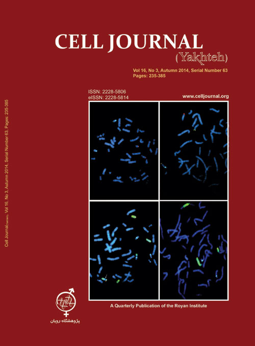
Page 377Complex chromosomal rearrangements (CCRs) are rare events involving more than two chromosomes and over two breakpoints. They are usually associated with infertility or sub fertility in male carriers. Here we report a novel case of a CCR in a 30-year-old oligoasthenosperm man with a history of varicocelectomy, normal testes size and normal endocrinology profile referred for chromosome analysis to the Genetics unit of Royan Reproductive Biomedicine Research Center. Chromosomal analysis was performed using peripheral blood lymphocyte cultures and analyzed by GTG banding. Additional tests such as C-banding and multicolor fluorescence in situ hybridization (FISH) procedure for each of the involved chromosomes were performed to determine the patterns of the segregations. Y chromosome microdeletions in the azoospermia factor (AZF) region were analyzed with multiplex polymerase chain reaction. To identify the history and origin of this CCR, all the family members were analyzed. No micro deletion in Y chromosome was detected. The same de novo reciprocal exchange was also found in his monozygous twin brother. The other siblings and parents were normal. CCRs are associated with male infertility as a result of spermatogenic disruption due to complex meiotic configurations and the production of chromosomally abnormal sperms. These chromosomal rearrangements might have an influence on decreasing the number of sperms.Keywords: Complex Chromosomal Rearrangement (CCR), Infertility, Karyotype, FISH
-
Page 383World Cancer Day (WCD) is celebrated on February 4th each year around the worldto remind the efforts done by united nations, World Health Organization (WHO), andother governmental and non-governmental health organizations with the aim of deliveringthe real message about cancer and its treatments to fight against this fataldisease through uniting all the people on global basis (1-4). In brief, cancer is a largegroup of different disorders involving unregulated cell growth. In malignancy, cellsdivide and grow uncontrollably to form malignant tumors and to invade neighboringparts of the body. The cancer tumor may spread to more distant parts of the bodythrough blood or lymphatic systems (5-8). It has been observed that most of the cancercases and related deaths happen in less developed countries that this situation isexpected to get worse by 2030. Therefore, it is very crucial to get control over suchcondition throughout the world. Furthermore, WCD aims to save millions of preventabledeaths every year by raising alertness and by educating people about malignancy, while forcing the governments throughout the world to take action against thisdisease (2-8). The day is also a key chance for cancer patients to ensure that worldleaders stick to the promises they made at the United Nations Summit for reducingthe cancer and its impacts. During this particular occasion, participants try to promotehealthy lifestyles, balanced diet, regular physical activity, weight management, aswell as using antioxidants in order to diminish the risk of malignancies (1-8). Furthermore, this day is celebrated to plan certain new strategies and to imply various newprograms in order to make people aware of this disorder. WCD is also celebrated tomake non-patient people aware about the preventive methods and the risk factorsof cancer (2-8). The theme of WCD of 2014 is «Debunk the Myths» because somepeople believe that if they contact or live with a patient who has cancer, they wouldget cancer as well. The day is, therefore, distinguished to eradicate such types of thesocial myths (5-13) and to make certain concepts about different aspects of the malignancy, such as its symptoms, causing factors and treatment (1, 3, 9). Furthermore, on this day, some of activities are organized to indicate that cancer patients shouldnot be treated distinctly, while they are entitled to the same rights as normal peoplein the society (14-19). Although they have less chances of existence, they should befulfilled their wishes by their relatives. It should not make them sense that the treatmentremedies are given them for existence as they are dying. In this regards, it isvery important to make them feel better like a normal person. They should be alsoprepared a normal environment in their home and society. It is essential that normalpeople avoid being overly sympathetic to them (20-27).Keywords: World Health Organization, Cancer, Patients


