فهرست مطالب

International Journal Of Fertility and Sterility
Volume:1 Issue: 4, Feb-Mar 2008
- 50 صفحه،
- تاریخ انتشار: 1387/06/15
- تعداد عناوین: 10
-
-
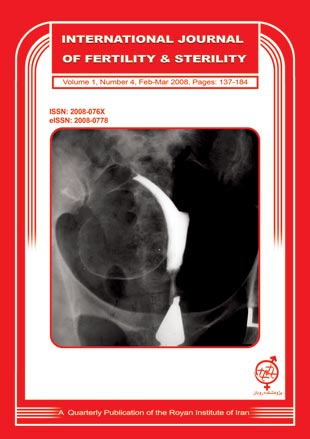
Page 137BackgroundUterine leiomyoma is the most common benign tumor of genital tract. The etiology of myomas is unknown. Leiomyoma shows a broad spectrum of radiographic appearances depending on the number, size, and location of the tumor. The diagnostic method for uterine leiomyomas is based primarily on the clinical situation. Despite of the varied diagnostic options such as; transvaginal sonography, sonohysterography, hysteroscopy, laparoscopy and MRI; hysterosalpingography is still one of the valuable imaging methods for identification of uterine leiomyoma. The various features of the proved leiomyoma are illustrated in this pictorial review. The incidence, risk factors and clinical features will also be discussed briefly.
-
Page 145BackgroundThe present study describes the susceptibility of prepubertal testis of mice to prooxidant induced oxidative impairments both under in vitro and in vivo exposure conditions.Material And MethodsFollowing in vitro exposure to iron (5,10 and 25 μM), oxidative response measured in terms of lipid peroxidation and hydroperoxide levels in testis of pre pubertal mice (4 wk) was more robust compared to that of pubertal mice (6 wk).ResultsFurther, in an in vivo study, pre pubertal mice administered (i.p) sub lethal doses (12.5, 25 and 50mg/100g bw/d, 5d) of Iron dextran, showed significant induction of oxidative stress response in testis cytosol and mitochondria manifested as lipid peroxidation, generation of reactive oxygen species, hydroperoxide levels and enhanced protein carbonyl levels (a measure of protein oxidation). Diminished levels of GSH and total thiols in both cytosol and mitochondria of testis suggested an altered redox state. Significant perturbations in the activities of antioxidant enzymes such as glutathione transferase, glutathione peroxidase and SOD were discernible suggesting the ongoing oxidative stress in vivo. These oxidative impairments were accompanied by functional implications in testis as reflected in the altered activities of dehydrogenases and reduced activities of both 3 β- and 17 β-hydroxysteriod dehydrogenase.ConclusionCollectively, these data provide an account of the susceptibility of prepubertal testis to iron-induced oxidative stress, associated functional consequences and this model is being further exploited for understanding the implications on the physiology of testis and consequent effect on fertility.
-
Page 155BackgroundThe aim of present study is to evaluate the effect of PolyVinyl Pyrrolidone (PVP) routinly used during ICSI procedure on sperm membrane integrity, and sperm chromatin status.Material And MethodsThis study was carried out on 21 semen samples from the infertile men referring to Isfahan Fertility and Infertility Center. The processed semen samples were divided into two portions. One portion was added to Ham’s F10+ 10% PVP, and the other portion was added to Ham’s F10 as a control group. Hypo-osmotic swelling test (HOST), SDS, and SDS+EDTA tests were carried out on the control and PVP groups at 15, 30, and 60min.ResultsThe results show that sperm membrane integrity measured by HOST, and sperm chromatin stability measured by SDS test, reduces by increasing the exposure time to PVP. However, the ability of sperm chromatin undergoing decondensation (that has been assessed by SDS+EDTA), does not show any changes by increasing the exposure time to PVP.ConclusionThe results of current study shows that reducing the exposure time of sperm to PVP may protect sperm membrane, and chromatin integrity.
-
Page 159BackgroundTolnidamine-induced changes have been reported earlier on spermatogenesis, fertility and sperm count in rat, rabbit and langur monkey. The aim of this study is to assess the response of these aspects to tolnidamine in the laboratory mouse.Material And MethodsAdult male mice (12-14 weeks old) of Parkes (P) strain were used in the present study. All the animals were divided into five groups. Groups I, II and V were taken as untreated, vehicle-treated initial and vehicle-treated terminal controls, respectively. Meanwhile, animals of Group III were administered with tolnidamine (100mg/kg BW, twice a week) orally for 3, 5 and 7 weeks and killed 24 hrs. after the last injection. Animals of Group IV were administered with the same dose of the tolnidamine for 7 weeks and then sacrificed 5 and 7 weeks after withdrawal of the drug. Tolnidamine-induced changes were evaluated on spermatogenesis, motility and count of epididymal spermatozoa, fertility and accessory sex glands and compared with the untreated and vehicle-treated controls.ResultsTolnidamine treatment induced significant decrease in the weights of the testis and epididymis; however, the weights of the accessory sex glands remained unaltered following the treatment. Duration-dependent degenerative changes were noticed in the testicular germinal epithelium showing vacuolization and loosening of the germ cells and Sertoli cells. Percentage motility and count of epididymal spermatozoa declined significantly following administration of tolnidamine. Likewise fertility of the treated males as well as number of the live blastocysts in females impregnated with such males also exhibited a significant decrease when compared with the controls. However, no change was noticed in the mating ability of the mice treated with tolnidamine. The level of seminal vesicular fructose also remained unaltered after the treatment. Withdrawal studies revealed duration-dependent recovery in spermatogenesis, percentage motility and count of spermatozoa and fertility.ConclusionThe findings of the present study, therefore, reveal that tonidamine administration in P mice induces reversible inhibition of spermatogenesis, motility and count of spermatozoa and fertility without affecting the androgen-dependent parameters.
-
Page 165BackgroundTo evaluate the possible association between phthalate esters (PEs) and the occurrence of endometriosis. Blood samples were collected from 99 infertile women with endometriosis (study group); 135 age-matched women without endometriosis (control group) but with infertility related to tubal defects, fibroids, polycystic ovaries, idiopathic infertility and pelvic inflammatory diseases diagnosed by laparoscopy with no evidence of endometriosis or other gynecological disorders during laparoscopic sterilization.Material And MethodsThis is a prospective case-control study, which recruited women undergoing infertility treatment at three collaborating centers (BMMHRC: Bhagwan Mahavir Medical Hospital and Research Centre, MHRT: Maternal Health and Research Trust, and Owaisi Hospital and Research Center) of Reproductive Medicine Hyderabad, which receives cases from all over the region of Andhra Pradesh, India. The concentrations of Phthalate Esters were measured by using the High Performance Liquid Chromatography (HPLC). Evaluation of Phthalate Esters concentrations in women with endometriosis compared with women who are free from the disease.ResultsWomen with endometriosis showed significantly higher concentrations of Phthalate esters (Dimethyl phthalate (DMP), Diethyl phthalate (DEP), Di-n-butyl phthalate (DnBP), Butyl benzyl phthalate (BBP) and Bis (2-ethylhexyl) phthalate (BEHP)) compared with control group. We found that (38%) of the cases with endometriosis and (21%) of the control group. The correlation between the concentrations of Phthalate esters and different severity of endometriosis was strong and statistically significant at p<0.05 for all five compounds (DMP): r=+0.57, p<0.0001; DnBP r=+0.39, p<0.0001; BBP: r=+0.89, p<0.0001; DnOP: r=+0.66, p<0.0001 and BEHP: r=+0.33, p<0.0014.ConclusionThis study for the first time from Indian subcontinent demonstrates that possibly Phthalate Esters might have a role in etiology of endometriosis.
-
The Presence of Anti Thyroid and Anti Ovarian Auto-Antibodies in Familial Pre-mature Ovarian FailurePage 171BackgroundTo evaluate the possible association between phthalate esters (PEs) and the occurrence of endometriosis. Blood samples were collected from 99 infertile women with endometriosis (study group); 135 age-matched women without endometriosis (control group) but with infertility related to tubal defects, fibroids, polycystic ovaries, idiopathic infertility and pelvic inflammatory diseases diagnosed by laparoscopy with no evidence of endometriosis or other gynecological disorders during laparoscopic sterilization.Material And MethodsThis is a prospective case-control study, which recruited women undergoing infertility treatment at three collaborating centers (BMMHRC: Bhagwan Mahavir Medical Hospital and Research Centre, MHRT: Maternal Health and Research Trust, and Owaisi Hospital and Research Center) of Reproductive Medicine Hyderabad, which receives cases from all over the region of Andhra Pradesh, India. The concentrations of Phthalate Esters were measured by using the High Performance Liquid Chromatography (HPLC). Evaluation of Phthalate Esters concentrations in women with endometriosis compared with women who are free from the disease.ResultsWomen with endometriosis showed significantly higher concentrations of Phthalate esters (Dimethyl phthalate (DMP), Diethyl phthalate (DEP), Di-n-butyl phthalate (DnBP), Butyl benzyl phthalate (BBP) and Bis (2-ethylhexyl) phthalate (BEHP)) compared with control group. We found that (38%) of the cases with endometriosis and (21%) of the control group. The correlation between the concentrations of Phthalate esters and different severity of endometriosis was strong and statistically significant at p<0.05 for all five compounds (DMP): r=+0.57, p<0.0001; DnBP r=+0.39, p<0.0001; BBP: r=+0.89, p<0.0001; DnOP: r=+0.66, p<0.0001 and BEHP: r=+0.33, p<0.0014.ConclusionThis study for the first time from Indian subcontinent demonstrates that possibly Phthalate Esters might have a role in etiology of endometriosis.
-
Page 175BackgroundAbnormal uterine bleeding (AUB) is one of the most common clinical problems in gynecology. Transvaginal sonography (TVS) and hysteroscopy are two diagnostic methods for patients with AUB. For most of the patients with AUB, diagnostic hysteroscopy can be done in clinic with minimal discomfort and much lower expense than operative room.Material And MethodsIn our clinical trial study, from March 21, 2005 to March 20, 2007, patients with AUB in Ahwaz Imam Khomayni hospital, after history and physical examinations underwent TVS. Of those, 147 patients with normal TVS entered the study and were considered for outpatient hysteroscopy. Patients with endometrial cavity lesion were scheduled for operation room, and those with empty endometrial cavity aspiration biopsy were done outpatiently. Specimens were sent to pathologist for examination.ResultsAll the patients were divided into three groups: group 1 or minority was under 30 years old (7 women), group 2 was 30-40 years, and group 3 or majority was over 40 years old (96 women). 115 patients (78.2%) had normal and 32 patients (21.8%) had abnormal hysteroscopic results. 116 patients (78.8%) had normal and 31 patients (21.2%) had abnormal pathologic results; moreover, cervical canal polyp was the most common lesion hysteroscopically and pathologically in all groups.ConclusionOf 147 patients (100%) with AUB and normal TVS, 32 patients (21.8%) were abnormal hysteroscopically. Cervical canal polyps may be missed by transvaginal sonography, but can be diagnosed by hysteroscopy. In patients with AUB and normal TVS, hysteroscopy can be used as the second step.
-
Page 179BackgroundThe Arnold-Chiari malformation is a congenital abnormality of CNS, characterized by downward displacement the parts of the cerebellum, fourth ventricle, pons and medulla oblongata into the spinal canal. This malformation is one of causative factor of death in neonates and infants. A thorough understanding of the direct and indirect sonographic findings is necessary for diagnosis of Chiari II malformation in the developing fetus. In this case report, we present a Chiari malformation II detected at 23 weeks of gestation by routinely sonographic screening. The Role of prenatal sonography in recognition of the malformation and prognostic value of these features are discussed.
-
Page 183
-
Advisory BoardsPage 184


