فهرست مطالب

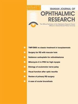
Journal of Ophthalmic and Vision Research
Volume:1 Issue: 1, Spring and Summer 2006
- 66 صفحه،
- تاریخ انتشار: 1385/05/04
- تعداد عناوین: 10
-
-
From the EditorPage 7
-
Page 9Purpose
To compare the efficacy of classic treatment for ocular toxoplasmosis (pyrimethamine, sulfadiazine and predinsolone) with a regimen consisting of trimethoprim/sulfamethoxazole (TMP/SMX) [co-trimoxazole] plus predinsolone.
MethodsIn a prospective randomized single-blind clinical trial, 59 patients with active ocular toxoplasmosis were randomly assigned to two treatment groups: 29 were treated with pyrimethamine/sulfadiazine and 30 patients received TMP/SMX. Treatment consisted of six weeks treatment with antibiotics plus steroids. Anti-toxoplasmosis antibodies (IgM and IgG) were measured using ELISA. Outcome measures included changes in retinochoroidal lesion size after six weeks of treatment, visual acuity before and after intervention, adverse drug reactions during follow up and rate of recurrence.
ResultsActive toxoplasmosis retinochoroiditis resolved in all patients over six weeks of treatment with no significant difference in mean reduction in retinochoroidal lesion size between the two treatment groups (61% reduction in the classic treatment group and 59% in the TMP/SMX group, P=0.75). Similarly no significant difference was found in visual acuity after treatment between the two groups [mean visual acuity after treatment was 0.12 LogMAR (20/25) in classic treatment group and 0.09 LogMAR (20/25) in TMP/SMX group, P = 0.56]. Adverse events were similar in both groups with one patient in each suffering from any significant drug side effects. The overall recurrence rate after 14 months of follow up was 6.7% with no significant difference between the treatment groups (P = 0.48).
ConclusionDrug efficacy in terms of reduction in retinal lesion size and improvement in visual acuity was similar between a regimen of TMP/SMX and the classic treatment of ocular toxoplasmosis with pyrimethamine and sulfadiazine. Therapy with TMP/SMX appears to be an acceptable alternative for the treatment of ocular toxoplasmosis.
-
Page 17Purpose
To determine the type and outcome of surgery for retinal detachment resulting from macular hole in highly myopic eyes.
MethodsThis retrospective analysis was performed on the medical records of highly myopic patients who underwent surgery for retinal detachment (RD) resulting from macular hole at Labbafinejad Hospital, Tehran-Iran from 1992 to 2001. Variables included age, gender, number and type of operations, visual acuity before and after the procedures and surgical success rate.
ResultsOverall, 28 eyes of 27 patients (26 female and one male) with mean age of 59.8±11 years were included. Mean follow-up was 17.3 (range 3-72) months. Mean axial length was 29±2.74mm (range: 24 to 35mm) and mean degree of myopia was -16.4±3.1 D (range -10 to -22 D). Posterior staphyloma was present in 20 eyes (71%). Seven eyes had undergone failed scleral buckling as the primary procedure prior to referral. Intravitreal SF6 injection was the primary procedure in 12 eyes with localized detachments; the retina became attached in 5 (41.6%) of these eyes, however redetachment occurred in 7 (58.4%) eyes. Overall, 23 eyes (including 7 failed scleral buckling cases, 7 redetachments following SF6 injection and 9 cases of primary surgery) underwent vitrectomy with use of high viscosity silicone oil. No major complications occurred during the operations. Overall, final anatomical success was 92.9% and visual improvement occurred in 85.7% of the eyes.
ConclusionIn highly myopic eyes with RD due to macular hole, less invasive procedures such as SF6 injection seem to be appropriate for eyes with localized detachment. In cases of total or subtotal RD and posterior staphyloma, pars plana vitrectomy and silicone oil tamponade seem to be the preferred procedure.
-
Page 23Purpose
To evaluate the efficacy of adjuvant subtenon carboplatin in the management of intraocular retinoblastoma.
MethodsThis study was conducted as a randomized, double-masked clinical trial. A diagnosis of intraocular retinoblastoma was made based on clinical examination, ultrasonography and orbital CT-scanning. The greatest basal dimension of the tumors was estimated in disc diameter (DD) by indirect ophthalmoscopy. Tumor thickness was determined by ultrasonography. Each eye was assigned to one of 10 blocks based on tumor stage (Reese-Ellsworth classification) and randomly received systemic chemotherapy alone (control group) or systemic chemotherapy plus 20mg subtenon carboplatin (case group). Indirect laser photocoagulation or cryotherapy was performed as additional treatment.
ResultsThe study included 35 tumors in 17 eyes of 14 patients (19 tumors in 8 eyes in the control group and 16 tumors in 9 eyes in the case group). There was 57.22% and 61.73% decrease in tumor thickness in the control and case groups, respectively. This difference was not statistically significant (P=0.12). The decrease in greatest basal tumor dimension in the control group (47.32%) was not significantly different from that in the case group (38.80%). One eye (12.5%) in the control group and 3 eyes (33.3%) in the case group were enucleated.
ConclusionAdjuvant subtenon carboplatin does not seem to increase the efficacy of systemic chemotherapy in the treatment of intraocular retinoblastoma.
-
Page 31PurposeTo study the effect of prophylactic application of mitomycin-C on regression and corneal haze formation after photorefractive keratectomy (PRK) for high myopia.MethodsFifty-four eyes of 28 high myopic patients were enrolled in this prospective study. All eyes underwent PRK with application of 0.02% mitomycin-C for two minutes and irrigation with 15-20 ml of normal saline. Follow-up visits were scheduled for the first 7 days and 1, 3 and 6 months after surgery. Hanna grading (in the scale of 0 to 4+) was used to assess corneal haze.ResultsMean spherical equivalent refraction (SE) was -7.08 ± 1.11 diopters (D), preoperatively. All eyes were examined on the first 7 days and one month after surgery; 48 eyes (88.9%) were evaluated 3 and 6 months post-surgery. Six months after surgery, all eyes had uncorrected visual acuity (UCVA) of 20/40 or better and 37 eyes (77.1 %) achieved UCVA of 20/20 or better, 45 eyes (93.7%) had SE within ±1.00D of emmetropia. One month postoperatively, 2 eyes (3.7%) had grade 0.5 haze, while at 3 and 6 months after surgery no visited eye had haze at all. There was no decrease in best corrected visual acuity after 6 months. In spatial frequencies of 6 and 12 cycle/degree, contrast sensitivity decreased immediately after PRK but increased to the preoperative values by the 6th postoperative month.ConclusionMitomycin-C can prevent the development of corneal haze when treating high myopia with PRK. In patients with insufficient corneal thickness for laser in situ keratomileusis (LASIK), mitomycin-C makes a useful adjunct to PRK to provide an alternative treatment for myopia. However, further research with longer follow-up is suggested.
-
Page 37PurposeTo determine the etiology of oculomotor nerve paralysis over a one year period at a university-based hospital.MethodsThis observational case series was conducted on consecutive patients with a clinical diagnosis of isolated oculomotor nerve paresis who were referred to the neuro-ophthalmology clinic at Farabi Eye Hospital, Tehran, Iran during 2001-2002. All patients were evaluated for hypertension and diabetes. In patients with confirmed diabetes mellitus or hypertension, oculomotor nerve palsy was diagnosed as ischemic. However if no recovery was observed up to four months, the patient underwent MRI and MRA. The etiology of oculomotor nerve palsy was classified into six categories including ischemia, trauma, aneurysm, neoplasm, miscellaneous and idiopathic.ResultsDuring the period of the study, 28 eyes of 28 patients (17 male and 11 female subjects) with mean age of 50.5 years were enrolled. Blepharoptosis was observed in 89.3%. Pupil reaction was normal in 50%, sluggish in 14.3% and absent in 35.7%. Pupil size was normal in 57.1% and mydriatic in 42.9%. The paralysis was ischemic in 42.8%, traumatic in 14.3%, aneurysmal in 7.1%, neoplastic in 7.1%, miscellaneous in 10.7% and idiopathic in 17.8% of the cases.ConclusionIn the present series, ischemia was the most common cause of oculomotor nerve palsy in which the most prevalent underlying disorder was diabetes mellitus.
-
Page 41Purpose
To evaluate changes in different aspects of visual function including visual acuity, visual field, contrast sensitivity, colour vision and stereopsis in patients with optic neuritis before and after medical intervention.
MethodsIn a noncomparative interventional case series on 31 eyes of 30 patients with optic neuritis, the following aspects of visual function were compared before and after treatment. Medical intervention was conducted following the Optic Neuritis Treatment Trial (ONTT) guidelines. Visual function was assessed by evaluating changes in visual acuity, visual field (Goldmann in all patients and automated in some patients), contrast sensitivity using Cambridge low contrast grating, colour vision using Ishihara plates and stereopsis using Titmus stereoacuity test.
ResultsVisual acuity was significantly lower (3/10) in the affected eyes than unaffected eyes (8/10) [P < 0.001]. Contrast sensitivity was also significantly better in the unaffected eyes. Mean stereoacuity was 310 sec/arc. The visual field impairment was also significantly higher than that of the unaffected eye and also normal population sample. Weak deutan defects were present in 60% of the patients. After medical treatment, visual acuity, visual field defects, contrast sensitivity, colour and stereopsis were significantly improved.
ConclusionDifferent aspects of visual function including visual acuity, visual field, contrast sensitivity, colour vision, and stereopsis are impaired in optic neuritis. Medical treatment with intravenous methylprednisolone followed by oral steroids is effective in improving these parameters. However, some deficits may persist after therapy. Since spontaneous recovery after optic neuritis is common, clinical trials are needed to determine the true effect of treatment versus follow-up.
-
Page 47The evolution of current surgical approaches for reattaching a primary retinal detachment and issues that determine the various techniques are analyzed through a comprehensive literature review of retinal detachment surgery starting from 1929. Ongoing changes in treatment modalities during the past 35 years are discussed: a change from surgery on the entire retinal detachment to a surgery limited to the retinal break and a change from extraocular to intraocular surgery for achieving retinal reattachment.
-
Page 61
A 21-year-old female was referred for severe bilateral visual loss 3 weeks after a diagnosis of brucellosis. On ocular examination she had bilateral optic nerve head swelling, preretinal hemorrhages and retinal vasculitis. The patient was diagnosed with bilateral optic neuritis secondary to brucellosis and developed optic atrophy and severe visual loss despite medical treatment. Brucellosis can lead to various types of ocular involvement including vasculitis, optic neuritis and retinal hemorrhage
-
Instruction for AuthorsPage 64

