فهرست مطالب

Iranian Journal of Kidney Diseases
Volume:13 Issue: 3, May2019
- تاریخ انتشار: 1398/05/31
- تعداد عناوین: 9
-
-
Pages 139-150Shiga toxin induced Escherichia Coli (STEC) is associated with chronic kidney disease or neurologic disability. The aim of this study is to determine the prevalence of STEC identified in human studies in Iran. Search engines of PubMed, EMBASE, OVID, SCOPUS, Web of Science, Google Scholar, IranMedex, MagIran, SID and ganj.irandoc were used. All human studies with stool or rectal swap samples evaluated for STEC and the outcome of HUS in Iran, which had been published between 1985 and 2017, were included. Chi-square and I2 statistic tests were applied to assess between-studies heterogeneity. Pooled prevalence and odd ratio were calculated using random effect models. A total of 30 articles containing 23379 samples were included for the final analysis. The design of study was cross sectional in 16, case control in 13 and one was cohort. The pooled prevalence of STEC was 7% (95% CI, 5 - 11; I2 = 98.3%). In subgroup analysis, the pooled prevalence was 8% (95% CI, 4 - 13; I2 = 97.55%) in children but 4% (95% CI, 2 - 7; I2 = 97.66%) in adults. The odds of patients with diarrhea having had STEC were 7.06 times the odds of healthy subjects (pooled OR = 7.06, 95% CI: 3.66-13.61). Patients with bloody diarrhea less likely to have positive STEC than patients with non-bloody diarrhea (pooled OR = 0.33, 95% CI: 0.10-1.02). STEC was prevalent in diarrheic patients and the rate increased in recent years. The highest contamination was seen in East-South of Iran. Public health intervention is mandatory to eliminate it effectively.Keywords: gastroenteritis, humans, Iran, shiga toxin, shiga-toxigenic escherichia coli
-
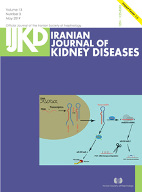
Pages 151-164The functional and structural disease with autosomal dominant inheritance (ADPKD) shows a poly cystic nature is described by the presence of epithelial cysts in the human renal parenchyma. There are no standard and reliable biomarkers for the detection of ADPKD in early stages which delays the therapeutic approaches. Ideal biomarkers of ADPKD, should have high sensitivity, specificity, and excellent association with disease pathogenesis and development. Both in vitro cellular and in vivo studies on animal models proved the significant roles of miRNAs in the course of ADPKD. In addition, different studies have explored miRNAs up or down regulation both in renal tissue and extracellular fluids in ADPKD which represent novel indicators applicable for diagnosis or targeted therapy. Since urine is a non-invasive, easily accessible sample for ADPKD, it could be the best sample for diagnosis. Additionally, due to early action of miRNAs for regulation the gene expression or because of their unique chemical properties, detectable urine miRNAs can be employed as appropriate biomarkers for timely diagnosis or intensive care of the progression of renal destruction or response to treatment. In this review, the specific microRNAs involved in the pathogenesis of PKD will be discussed with a particular focus on extracellular miRNAs with possibility for application as biomarkers.Keywords: microRNAs, circulating, extra cellularmicroRNAs, autosomaldominant polycystic kidneydisease, biomarker
-
Pages 165-172IntroductionNephrotic syndrome is a heterogeneous disease in children, with nearly 10% categorized as steroid-resistant. In this study we evaluated disease related mutations within NPHS1, NPHS2 and new potential variants in other genes.MethodsIn the first phase of study, 25 patients with SRNS were analyzed by Sanger sequencing for NPHS1 (exon 2, 26) and all exons of NPHS2 genes. In the next step, whole exome sequencing was performed on 10 patients with no mutation in NPHS1 and NPHS2.ResultsWES analysis revealed a novel mutation in FAT1 (c.10570C > A; Q3524K). We identified 4 pathogenic mutations, located in exon 4 and 5 of NPHS2 gene in 20% of patients (V180M, P118L, R168C and Leu156Phe). Also our study has contributed to the description of previously known pathogenic mutations across WT1 (R205C) and SMARCAL1 (R764Q) and a novel polymorphism in CRB2.ConclusionIt seems that NPHS2, especially exons 4 and 5, should be considered as the first step in genetic evaluation of Iranian patients. We suggest conducting WES after NPHS2 screening to identify the potential genes associated with SRNS, Further studies are required to examine more common genes in the first step and then designing native laboratory panels.Keywords: SRNS, NPHS1, NPHS2, WES, FAT1, nephroticsyndrome
-
Pages 173-181IntroductionIncreased glypican-5 expression in podocytes induces podocyte injury. Glypican-5 is shed into the urine, but its value in predicting progression of diabetic nephropathy (DN) has not been investigated.MethodsGlypican-5 was determined in spot urine from 20 type 2 diabetes mellitus (T2DM) patients, and 37 type 2 DN patients, and 20 healthy controls by enzyme-linked immunosorbent assay. The association of urinary glypican-5 with markers of renal function was evaluated.ResultsUrinary glypican-5 was significantly higher in DN patients than in both DM patients and controls. Glypican-5 level was not associated with baseline 24-hour urine protein/albumin excretion, serum creatinine, estimated glomerular filtration rate (eGFR), systolic blood pressure (SBP), or hemoglobin (Hb) A1c values in the DN patients. After 52 weeks follow-up, urinary glypican-5 level was associated with significant increases in urine protein and albumin excretion and a significant decline in eGFR in the DN patients. The decline in eGFR was independent of changes in urine protein and albumin excretion, SBP, or HbA1c. The results indicate that urinary glypican-5 was not only a biomarker but also contributed to the pathogenesis of DN in these patients.ConclusionUrinary glypican-5 was specifically elevated in type 2 diabetes patients with DN and it was associated with disease progression. Urinary glypican-5 may serve as a useful non-invasive marker for the progression of DN.Keywords: glypican-5_podocyte_estimated glomerularfiltration rate_diabeticnephropathy_type 2 diabetes
-
Pages 182-190IntroductionContrast-induced nephropathy (CIN) is a frequent complication of contrast exposure. A recent study suggested that Na/K citrate might have a preventive role. We investigated the efficacy of Na/K citrate to prevent CIN in patients with renal dysfunction undergoing coronary intervention.MethodsThe randomized, double-blind, placebo-controlled trial included 201 patients with estimated creatinine clearance < 90 mL/ min, randomized to receive oral Na/K citrate plus saline infusion (treatment group, 104 patients) or oral water plus saline infusion (placebo group, 97 patients). CIN was defined as an absolute increase of serum creatinine ≥ 0.5 mg/dL or a relative increase ≥ 25% or a relative decrease of estimated GFR ≥ 25% within 5 days.ResultsCIN occurred in 22 patients (12.29%); 10 (11%) in treatment group and 12 (13.6%) in placebo group (P > .05). Post-exposure Cr values were not significantly different between the two groups (1.18 ± 0.28 mg/dL in the placebo vs. 1.15 ± 0.29 mg/dL in the treatment group, P > .05). CIN-negative patients in the treatment group showed a significantly higher increase in urine pH than that of CIN-positive patients (1.642 ± 0.577 vs. 1.20 ± 0.422, P < .05).ConclusionNa/K citrate solution is not effective for prophylaxis of CIN in patients with renal dysfunction. However, a probable preventive effect might exist in a subgroup of patients with at least 1.6 units increase in urine pH values following Na/K citrate administration.Keywords: contrast-inducednephropathy, chronic kidneydisease, coronary intervention, sodium potassium citrate, randomized clinical trial
-
Pages 191-197IntroductionContrast-induced Acute Kidney Injury (CI-AKI) is a prevalent complication of chronic kidney disease (CKD) patients. The aim of this study was to evaluate the effects of periprocedural administration of trimetazidine as an anti-oxidant agent on the incidence of CI-AKI in CKD patients based on changes of Neutrophil Gelatinase-Associated Lipocalin (uNGAL) level, which has recently been introduced as an early predictor of CI-AKI.MethodsOne hundred CKD patients with a mean GFR of 50 ± 7 cc/min who were candidate for coronary angiography assigned randomly to receive (50 patients, intervention group) or not receive (50 patients, control group) trimetazidine (70mg/d) for 72 hours. CI-AKI was defined as 0.5 mg or 25% increase in serum creatinine. We also checked uNGAL before and 12h after angiography.ResultsSerum creatinine, showed a trend of less increment in the case group, although could not achieve a significant difference, there was a significant difference in urinary NGAL rise between two groups. CI-AKI was defined as 1.7 times increase in uNGAL level (12h after angiography to pre-procedurally uNGAL level ratio) according to the ROC curves. The incidence of CI-AKI according to urinary NGAL definition was 8% in the Trimetazidine group and 24% in the control group (P < .05).ConclusionWe concluded that Trimetazidine treatment before angiography may be effective in CI-AKI prevention. Moreover, it is shown that 1.7 times increase in urine NGAL after angiography is a valuable cut off point for clinicians to discriminate high risk patients for contrast nephropathy.Keywords: trimetazidine, neutrophil gelatinaseassociatedlipocalin, contrastinducedacute kidney injury
-
Pages 198-206IntroductionMicroRNAs (miRNA) are involved in the pathogenesis of systemic lupus erythematosus (SLE), an autoimmune disease, and can be considered as diagnostic and prognostic biomarkers. Lupus nephritis (LN) remains a major challenge of SLE since it damages the kidneys in the course of the disease.MethodsThe aim of this study was to investigate the diagnostic values of circulating miR-21, miR-148a, miR-150, and miR-423 involved in autoimmunity and kidney fibrosis in plasma samples of LN cases (N = 26) and healthy controls (N = 26) using quantitative- PCR (qPCR). The possible associations between the microRNAs and clinical parameters and their diagnostic values were also calculated.ResultsThe levels of circulating miR-21 (P < .001) and miR-423 (P < .05) significantly increased, while miR-150 decreased in LN (P > .05) patients as compared with healthy controls. Receiver operating characteristic (ROC) analysis indicated that miR-21 was superior in discriminating LN patients from controls with an Area Under Curve (AUC) of 0.912 [95% CI = 0.83 to 0.99, P < .001], whereas the multivariate ROC curve analysis revealed the high accuracy [AUC = 0.93, P < .001, 79% sensitivity and 83% specificity] of the miR-21, -150, and -423 to differentiate LN from controls.ConclusionThe involvement of the studied miRNAs in renal fibrosis and the obtained results make it rational to speculate that they may be used as potential biomarkers in LN.Keywords: microRNA, renalfibrosis, lupus, autoimmunity, diagnostic biomarker
-
Pages 207-210Known as systemic sclerosis (SSc), this autoimmune rheumatic disease has vast pathogenesis on many organs, including kidneys. It can lead to the point where the patient's survival relies entirely on dialysis. This report has basically focused on scleroderma renal crisis (SRC), which is the most serious renal manifestation of SSc, characterized by renal failure and sudden onset of hypertension. A 44-year-old man was hospitalized with hypertension, headache, vertigo, nausea, rhinorrhea, reflux, dysphagia, dyspnea (Fc II), visual impairment, mechanical arthralgia, and edema (+3) accompanied by a rare skin lesion. Raynaud's phenomenon was also remarkable in fingers and toes. According to signs and symptoms, SSc diagnosed and the proper treatment was applied. It is of great importance that in the case of malignant hypertension in patients with scleroderma, renal crisis always be kept in mind.Keywords: scleroderma, renalcrisis, hemodialysis
-
Page 211


