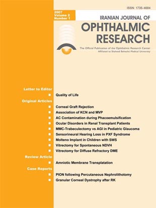
فهرست مطالب

Journal of Ophthalmic and Vision Research
Volume:2 Issue: 1, Spring and Summer 2007
- 84 صفحه،
- تاریخ انتشار: 1385/09/11
- تعداد عناوین: 13
-
-
Page 7PurposeTo determine the incidence and risk factors of late corneal graft rejection after penetrating keratoplasty (PKP).MethodsRecords of all patients who had undergone PKP from 2002 to 2004 without immunosuppressive therapy other than systemic steroids and with at least one year of follow up were reviewed. The role of possible risk factors such as demographic factors, other host factors, donor factors, indications for PKP as well as type of rejection were evaluated.ResultsDuring the study period, 295 PKPs were performed on 286 patients (176 male, 110 female). Mean age at the time of keratoplasty was 38±20 (range, 40 days to 90) years and mean follow up period was 20±10 (range 12-43) months. Graft rejection occurred in 94 eyes (31.8%) at an average of 7.3±6 months (range, 20 days to 39 months) after PKP. The most common type of rejection was endothelial (20.7%). Corneal vascularization, regrafting, anterior synechiae, irritating sutures, active inflammation, additional anterior segment procedures, history of trauma, uncontrolled glaucoma, prior graft rejection, recurrence of herpetic infection and eccentric grafting increased the rate of rejection. Patient age, donor size and bilateral transplantation had no significant influence on graft rejection.ConclusionSignificant risk factors for corneal graft rejection include corneal vascularization, anterior synechiae, irritating sutures, active inflammation, regrafting, additional surgery, trauma, uncontrolled intraocular pressure, history of graft rejection, recurrent herpetic infection, eccentric grafting and corneal scarring. Recipient age and donor cornea size do not seem to be risk factors for corneal graft rejection.
-
Page 15PurposeTo compare the prevalence of mitral valve prolapse (MVP) in patients with keratoconus (KCN) with that of normal subjects.MethodsThis study includes 62 individuals with KCN diagnosed by clinical findings and topographic criteria, and 167 age and sex matched controls with no clinical or topographic evidence of KCN. All participants were evaluated by two-dimensional M-mode and color doppler echocardiography. Perloff''s criteria were used for diagnosis of definite MVP.ResultsDefinite MVP was diagnosed in 22.6% of subjects with KCN and 6.6% of the control group (OR= 4.2; 95% CI, 1.93-11.3; P= 0.009). MVP was more prevalent in patients with KCN based on age and sex stratification. Odds ratio for MVP increased from 2.67 before the third decade of life to 33.44 in the third decade and slightly decreased to 16.52 in the fourth decade and above.ConclusionThis study disclosed an increased prevalence of MVP in individuals with keratoconus suggesting the necessity of cardiovascular evaluation in these patients.
-
Page 19PurposeTo evaluate anterior chamber aspirates at the conclusion of phacoemulsification and intraocular lens implantation (PE+IOL) for bacterial and fungal contamination.MethodsWe prospectively evaluated 80 eyes of 80 patients undergoing routine PE+IOL by performing bacterial and fungal culture on aspirates obtained from the anterior chamber at the end of the surgery.ResultsAnterior chamber fluid aspirates were positive for bacteria in 5 eyes (6.33%) with coagulase-negative staphylococcus being the most common organism (three eyes). No instance of positive fungus culture was observed. One of the culture-positive eyes developed postoperative uveitis which resolved during a week of treatment with topical corticosteroids and antibiotics. None of the eyes developed endophthalmitis.ConclusionIn the current series, the rate of anterior chamber contamination by bacteria at the end of phacoemulsification was in the lower range reported by previous studies
-
Page 23PurposeTo determine ocular findings in patients with renal transplants and to correlate them with clinical characteristics related to transplantation.MethodsThis cross-sectional study was performed on 150 patients who had received a renal transplant of at least three months'' duration with serum creatinine levels < 3 mg/dl. All patients underwent a complete ophthalmologic examination. Clinical variables related to the transplant included cause of renal failure, duration of hemodialysis prior to transplantation and immunosuppressive regimen.ResultsOverall, 91 male and 59 female subjects with mean age of 39±17.7 years were included. At least one ocular abnormality could be detected in 89.3% including visual acuity less than 20/25 (48.6%), conjunctival degeneration in the palpebral fissure (36.6%), posterior subcapsular cataracts (24%), pinguecula (17.3%), retinal pigment epitheliopathy (14%), arteriovenous crossing changes (8.6%), proliferative diabetic retinopathy (6%), central serous chorioretinopathy and retinal vein occlusions (each in 3.3%), and non-proliferative diabetic retinopathy, optic nerve atrophy and diabetic macular edema (each in 2.7%). Abnormal ocular findings were not correlated with the underlying renal disorder or use of cyclosporine and prednisolone, however they were positively correlated with transplant duration, pre-transplant dialysis duration and azathioprine or mycophenolate mofetil consumption.ConclusionOcular disorders are frequent among renal transplant patients especially with older transplants and those with a longer period of pre-transplant hemodialysis.
-
Page 28PurposeTo compare the outcomes and complications of mitomycin-C trabeculectomy (MMC-T) versus the Ahmed glaucoma implant (AGI) for treatment of pediatric aphakic glaucoma.MethodsIn a randomized clinical trial, 30 eyes of 28 children < 16 years of age who had undergone anterior lensectomy-vitrectomy for congenital cataract were assigned to MMC-T (15 eyes of 13 children) or AGI (15 eyes of 15 children). Surgical success was classified as complete (IOP 6-21 mmHg without any antiglaucoma medication) and partial (IOP 6-21 mmHg with < 2 topical antiglaucoma agents) in the absence of any sight-threatening complication or need for further glaucoma surgery, stable cup/disc ratios and visual loss < 2 Snellen lines. Overall success was defined as the sum of complete and partial success.ResultsMean patient age was 9.1±4.1 and 10.9±5.1 years in the MMC-T and AGI groups, respectively (P=0.29). After a mean follow up of 14.8±11 and 13.1±9.7 months; complete, partial and overall success rates were 33.3%, 40% and 73.3% in the MMC-T vs 20%, 66.7% and 86.7% in the AGI groups, respectively (P= 0.361). Complication and failure rates were 40% and 26.7% in the MMC-T group vs 26.7% and 13.3% in the AGI group, respectively (P= 0.439).ConclusionMMC-T and AGI seem to be comparable in terms of success and complications as the initial surgical procedure in pediatric aphakic glaucoma. Choice of either technique depends on surgeon''s experience and conjunctival quality and mobility.
-
Page 35PurposeTo determine hearing thresholds at sound frequencies important for speech comprehension in subjects with ocular pseudoexfoliation (PXF) and to compare them with that of controls without PXF.MethodsEighty-three subjects with ocular PXF and 83 age and sex matched controls without PXF were enrolled in this case-control study. Pure tone audiometry (bone conduction) was performed at 1, 2 and 3 kilohertz (KHz) in all subjects. Thresholds were compared to an age and sex stratified standard (ISO7029) and between study groups. Hearing loss was defined as sum of tested hearing thresholds (HTL-1,2,3) lower than the ISO7029 standard median.ResultsThe study included 60 male and 23 female subjects in each group. Hearing loss was present in 147 of 166 (88.6%) of examined ears in the case group vs 89 of 166 (53.6%) in the control group (P < 0.001; odds ratio [OR] = 6.69; 95% confidence interval [CI], 3.49-11.79). Overall 78 subjects (94.0%) in the case group vs 58 subjects (69.9%) in the control group had hearing loss in one or both ears (P < 0.001; OR=6.72; 95%CI, 2.42-18.62). Hearing thresholds at each of the examined frequencies and the HTL-1,2,3 were also significantly higher in individuals with PXF. Although glaucoma was significantly more common in subjects with PXF (51.8% vs 22.9%, P < 0.001), it was not associated with hearing loss in any of the study groups.ConclusionsHearing thresholds at frequencies which are important for speech comprehension are significantly worse in individuals with ocular PXF as compared to matched controls. This finding may support the multi-organ nature of PXF syndrome
-
Page 40PurposeTo evaluate the efficacy and safety of primary single-plate Molteno tube implantation for management of glaucoma in children with Sturge-Weber syndrome.MethodsPrimary single-plate Molteno tube implantation was performed in eyes of patients with Sturge-Weber syndrome aged < 17 years. Success was defined as intra-ocular pressure (IOP) higher than 22 mmHg with (relative success) or without (absolute success) antiglaucoma medications. Intra- and postoperative complications were also evaluated.ResultsOverall, nine eyes of seven patients with mean age of 9.6±3.7 (range 5-17) years were operated and followed for 32±4.7 (range 20-36) months. Mean IOP decreased from 34.2±8.3 mmHg preoperatively to 21.2±7.3 mmHg at final follow up (P=0.012). Mean number of antiglaucoma medications decreased from 3.4±0.5 preoperatively to 2.2±1.3 at final follow up (P=0.058). The cumulative probability of relative success was 97.2% (95% confidence interval [CI], 91.8-100%) at 12 months, 78.0% (95%CI, 60.4-95.7%) at 24 months and 43.3% (95%CI, 16.2-70.5%) at final follow up. Two eyes achieved absolute success during the first six months, however, at six months and later no eye achieved absolute success. No intraoperative complication occurred; postoperative complications included choroidal effusion necessitating drainage in three eyes (33.3%), and cataract formation and retinal detachment, each in one eye (11%). At final follow up, visual acuity remained unchanged in five eyes and deteriorated in four eyes.ConclusionThe outcomes of this small series revealed that primary single-plate Molteno tube implantation appears to be associated with limited success and a relatively high complication rate in pediatric glaucoma associated with Sturge-Weber syndrome.
-
Page 46PurposeTo determine the causes of non-traumatic non-diabetic vitreous hemorrhage (NDVH) and to report the visual and anatomical outcomes and complications of vitrectomy for this condition.MethodIn a retrospective case series, records of patients who had undergone vitrectomy for non-traumatic NDVH over a ten year period at Labbafinejad Medical Center, Tehran-Iran with at least six months of follow up were reviewed for causes of the condition and outcomes of surgery.ResultsFrom 1993 to 2003, 50 eyes of 49 patients (51% male) with mean age of 62.7±10.3 (range 35-87) years underwent vitrectomy for non-traumatic NDVH. Preoperatively, mean best-corrected visual acuity (BCVA) was 2.36±0.52 LogMAR and relative afferent pupillary defect was positive in 91.1% of the eyes. Mean BCVA increased significantly to 1.38±0.72 LogMAR at six months (P < 0.0001). Causes of non-traumatic NDVH included: branch retinal vein occlusion (56%), central retinal vein occlusion (16%), choroidal neovascularization (12%) and posterior vitreous detachment with retinal break, Eales'' disease, familial exudative vitreoretinopathy and Terson''s syndrome (each in 4%). The most common causes of poor visual outcomes were: macular pigmentary derangement (26%), optic atrophy (16%), severe lens opacity (12%) and epiretinal membrane (8%).ConclusionDespite the significant increase in VA following vitrectomy, irreversible macular or optic nerve pathology limits significant improvement in central visual acuity in several cases of non-traumatic NDVH. Vascular accidents were the most common cause of this condition.
-
Page 52PurposeTo evaluate the anatomic and visual results and complications of vitrectomy in eyes with diffuse refractory diabetic macular edema associated with a taut posterior hyaloid.MethodsThis prospective interventional case series was conducted on 25 eyes of 22 patients with diffuse refractory clinically significant diabetic macular edema, macular thickness greater than 250 mm on optic coherence tomography (OCT) and thickened posterior hyaloid. Best-corrected visual acuity (BCVA) and macular thickness measured by OCT were evaluated preoperatively and repeated 3 and 6 months postoperatively. Macular perfusion was evaluated by fluorescein angiography, pre- and six months postoperatively.ResultsMean BCVA was 1.14±0.51 LogMAR, preoperatively which improved to 0.89±0.53 LogMAR six months postoperatively (P=0.005). Mean preoperative macular thickness was 506±121.9 µm which decreased to 318±90.5 µm, six months postoperatively (P=0.001).ConclusionVitrectomy and removal of the posterior hyaloid membrane appears beneficial in eyes with diffuse diabetic macular edema unresponsive to laser therapy and a taut premacular posterior hyaloid.
-
Page 58The past decade has witnessed the revival of amniotic membrane transplantation (AMT) in ophthalmology. The importance of amniotic membrane lies in its ability to reduce inflammation and scarring, enhance epithelialization and wound healing, and in its antimicrobial properties. Amniotic membrane has recently been used as a substrate for culturing limbal stem cells for transplantation. It has also been used extensively in corneal conditions such as neurotrophic ulcers, persistent epithelial defects, shield ulcers, microbial keratitis, band keratopathy, bullous keratopathy, and following photorefractive keratectomy and chemical injuries. Other indications for AMT include ocular surface reconstruction surgery for conjunctival pathologies such as squamous neoplasia, pterygium, and symblepharon. In this review we describe the basic structure and properties of amniotic membrane, its preparation process and its applications in ophthalmology.
-
Page 76PurposeTo report a case of posterior ischemic optic neuropathy (PION) following percutaneous nephrolithotomy (PCNL). CASE REPORT: A 57-year-old man with history of diabetes mellitus, hyperlipidemia and mild anemia underwent PCNL for treatment of nephrolithiasis. He noticed painless visual loss in both eyes immediately after the procedure. Visual acuity was light perception, however ophthalmologic examinations were unremarkable and the optic discs were pink with no swelling. Visual fields were severely affected, but neuro-imaging was normal. Within three months, visual acuity and visual fields improved dramatically but the optic discs became slightly pale.ConclusionThis is the first report of PION following PCNL. PION is a rare cause of severe visual loss following surgery. Severe blood loss, hypotension, anemia and body position during surgery are the most important risk factors. Ophthalmologists, urologists and anesthesiologists should be aware of this condition and this rare possibility should be considered prior to surgery.
-
Page 81PurposeTo report manifestation of granular corneal dystrophy after radial keratotomy (RK). CASE REPORT: A 32-year-old man presented with white radial lines in both corneas. He had undergone uncomplicated RK in both eyes 8 years ago. Preoperative refraction had been OD: -3.5 -0.75@180 and OS: -3.0 -0.5@175. Uncorrected visual acuity was OD: 8/10 and OS: 7/10; best corrected visual acuity was 9/10 in both eyes with OD: -0.5 -0.5@60 and OS: -0.75 -0.5@80. Slit lamp examination revealed discrete well-demarcated whitish lesions with clear intervening stroma in the central anterior cornea consistent with granular dystrophy. Similar opacities were present within the RK incisions.ConclusionGranular dystrophy deposits may appear within RK incisions besides other previously reported locations

