فهرست مطالب

Journal of Dentistry, Shiraz University of Medical Sciences
Volume:23 Issue: 1, Mar 2022
- تاریخ انتشار: 1400/11/17
- تعداد عناوین: 12
-
-
Pages 1-6
Corona virus epidemic has caused a widespread disaster around the world. In studies, there are pieces of evidence of olfactory and taste dysfunction in patients with Covid-19. These symptoms occur independently or can be associated with other symptoms such as dry cough. The mechanism of the above-mentioned disorders and their clinical features in patients are not yet known. The rate of incidence of olfactory dysfunction in patients has been varied from 29.64% to 75.23% and the rate of incidence of taste dysfunction among the people can be different from 20.46% to 68.95%. Therefore, clinicians including ENT specialists and dentists should pay attention to the symptoms of anosmia and ageusia in these patients to prevent delayed diagnosis and inappropriate treatments. In this review article, data have been collected by searching the available articles in the domestic and foreign journals using databases such as PubMed, PubMed Central, Medline, EBSCO, Google Scholar, and Embase with key words of Anosmia, Ageusia, Dysgeusia, Covid-19, and Coronavirus from 2019 to 2020. Among the relevant references, 38 authoritative articles were chosen. The data showed that it seems olfactory and taste function disorders are the obvious symptoms of Covid-19, which can occur independently or with other symptoms, but the pathogenesis is not well specified yet. Therefore, further studies are required to achieve a reliable result in this area.
Keywords: Anosmia, Ageusia, Dysgeusia, COVID-19, coronavirus -
Pages 7-12Statement of the Problem
The castability of nonprecious gold color alloy using torch/ centrifugal and induction/vacuum-pressure casting techniques has not been studied yet.
PurposeThis study was conducted to compare the castability of nickel chromium, cobalt-chromium and nonprecious gold color alloy using torch/centrifugal and induction/ vacuum-pressure casting techniques.
Materials and MethodIn this in vitro study, a total number of 54 identical acrylic wax meshes were prepared and divided into 6 different groups of 9 each. Group 1: nickel-chromium alloy, which was casted with induction technique. Group 2: nickel-chromium alloy was casted with centrifugal technique. Group 3: cobalt-chromium alloy was casted with induction technique. Group 4: cobalt-chromium alloy was casted with centrifugal technique. Group 5: nonprecious gold color alloy was casted with induction technique. Group 6: nonprecious gold color alloy was casted with centrifugal technique. Then castability of specimens was measured using modified Whitlock’s method. The results were analyzed using two way ANOVA and post hoc tests.
ResultsANOVA test revealed no statistically significant difference between different alloys with a p Value of 0.313. Moreover, it represented no significant differences within the groups regarding alloy types and casting techniques with a p Value of 0.511 and 0.682, respectively.
ConclusionNo significant difference was found in the castability value of nickel-chromium, cobalt-chromium, and nonprecious gold color alloys. In addition, the castability value of three alloys tested in this study was not different by using torch/centrifugal or induction/vacuum-pressure casting machines.
Keywords: castability, Nickel-Chromium, Cobalt-Chromium, Non-Precious Gold Color (NPG) -
Pages 13-19Statement of the Problem
Endodontic materials that are placed in direct contact with living tissues should be biocompatible. The cytotoxicity of Nano Fast Cement (NFC) compared to ProRoot Mineral Trioxide Aggregate (ProRoot MTA) must be evaluated.
PurposeThisIn vitro study aimed to assess the cytotoxic effects of NFC in comparison to ProRoot MTA on L-929 mouse fibroblast cells.
Materials and MethodIn this animal study,L-929 mouse fibroblast cells were grown in Dulbecco's Modified Eagle's medium (DMEM) supplemented with 10% fetal bovine serum (FBS) in an atmosphere of 5% co₂/95% air at 37 C̊. A total of 10⁴ cells from the fourth collection were plated in each well of a 96-well micro-titer plate. Materials were mixed according to the manufacturer’s instruction and placed into the related plastic molds with 5 mm diameter and 3 mm height. After 24 hours and a complete setting, the extracts of the tested materials were produced at six different concentrations and placed in the related wells. Cells in DMEM served as the negative control group. DMEM alone was used as the positive control group. Methyl-thiazoltetrazolium (MTT) colorimetric assay was conducted after 24, 48, and 72 hours. The absorbance values were measured by ELISA plate reader at 540 nm wavelength. Three-way analysis of variance, post-hoc Tukey, LSD, and independent t-test were used for the statistical analyses using SPSS software, version 16.0.
ResultsThere was no statically significant difference between MTA and NFC in cell viability values at different concentrations and different time intervals (p= 0.649). Viability values were significantly decreased after 72 hours, but there was no significant difference between the first and second MTT assays (p= 0.987). Cytotoxicity significantly increased at concentrations higher than 6.25 µɡ/ml.
ConclusionCytotoxicity depends on time, concentration, and cement composition. There was no statistically significant difference between NFC and MTA concerning their cytotoxic effects on L-929 mouse fibroblast cells.
Keywords: Mineral Trioxide Aggregate, Cytotoxicity, Endodontics, Cement -
Pages 20-28Statement of the Problem
The first permanent molar (FPM) teeth are the most important elements of mastication and are crucial in the improvement of functionally proper occlusion. However, in childhood, these teeth are most susceptible to caries. The loss of an FPM in a child can cause changes in the dental arches. These changes can occur throughout a person’s life. In such cases, the dentists and dental specialists need to decide whether to preserve or extract the FPM.
PurposeThis study aimed to evaluate the extent of knowledge of dental specialists in Shiraz (Iran) on clinical guidelines for the preservation and extraction indications of FPMs.
Materials and MethodThe authors developed a dedicated questionnaire for the purpose of knowledge evaluation. A total of 6 orthodontists and 15 dental specialists, respectively confirmed the validity and reliability of the questionnaire. The 19-item questionnaire covered topics such as demographic data, preservation criteria for FPM teeth, and indications for FPM extraction. The survey was carried out across six dental disciplines in Shiraz (Iran) during July-August 2018. The data were analyzed using the SPSS software (version 22.0) with the dependent sample t test and one-way ANOVA. p Value< 0.05 was considered statistically significant.
ResultsOut of 89 dental specialists, 64 participants (53% male, 47% female) completed the questionnaire. The mean knowledge score for all participants was 10.09±3.93 (maximum of 19). The level of knowledge had a significant and inverse correlation with age (p< 0.001) and years of experience (p= 0.017). It also had a significant relationship with dental specialization (p< 0.001).
ConclusionThe overall level of knowledge of the specialists was insufficient, except for the pedodontists and orthodontists. A re-education training program for dental specialists is strongly recommended.
Keywords: Questionnaire, Survey, First Permanent Molar, Tooth Extraction, Knowledge -
Pages 29-32Statement of the Problem: Methemoglobinemia is a potentially life-threatening rare medical condition, which refers to an increase in the level of oxidized form of hemoglobin (methemoglobin). Excessive replacement of hemoglobin with methemoglobin leads to functional hypoxia and even fatal conditions.PurposeThe aim of this study was to evaluate the effect of two common local anesthetic agents namely lidocaine and articaine administered for hemostasis during surgery on methemoglobin level.Materials and MethodThis prospective cohort study was conducted from January 2017 to December 2019. Demographic data including age, gender, and weight of patients were collected. Sixty patients were randomly divided into three groups (n=20) regarding the local anesthetic agent administered for hemostasis during surgery as lidocaine group (group 1), articaine group (group 2), and control group (no local anesthetic; group 3). The patients were candidates for orthognathic surgery, reconstruction of the maxillary and mandibular atrophic ridges with autogenous grafts, and reconstruction of maxillofacial fractures. The methemoglobin level was measured before surgery and six hours after the initiation of surgery.ResultsThe mean age and weight of patients were not significantly different among the three groups (p= 0.891 and p= 0.416, respectively). No significant differences were observed among the three groups regarding the gender distribution (p= 0.343) or type of surgery (p= 0.990). Statistical analysis did not show any significant difference in the mean baseline methemoglobin level among the three groups (p= 0.109). Although the mean methemoglobin values increased in the three groups, paired sample t-test did not show any significant change in the values at six hours after the initiation of surgery compared with baseline in any of the three groups (p= 0.083 for group 1, p= 0.096 for group 2, and p= 0.104 for group 3).ConclusionThe results demonstrated that administration of lidocaine and articaine plus epinephrine for hemostasis during general anesthesia are equally safe.Keywords: Methemoglobinemia, Local anesthesia, General anesthesia, Methemoglobin
-
The Relationship between Oral and Dental Health Self-care and Hemoglobin A1c in Adults with DiabetesPages 33-39Statement of the Problem
Due to the mutual relationship between periodontal diseases and diabetes, it seems that adopting oral self-care in a way to prevent and control the progress of periodontal diseases, improves the oral health of diabetic patients as well as their general health.
PurposeThe aim of this study was to investigate the relationship between the oral self-care behaviors and the hemoglobin A1c (Hb A1c) levels in adults with diabetes.
Materials and MethodIn this cross-sectional study with convenience sampling, 120 adults between 18 to 50 years old, who had at least two healthy functional teeth, were selected from private endocrinology offices in Tehran in August 2019. The exclusion criteria were illiterate individuals and pregnant women. A standard questionnaire was used which included the information about demographic, diabetes, and self-care behaviors. The outcome variable was the latest Hb A1c rate.
ResultsThe mean age of participants was 35.8±10.5 years. The average Hb A1c was 7.4± 1.55%. 35.0% of participants brushed their teeth twice a day or more and 60.8% flossed rarely. The proportion of Hb A1c <7% was higher in three groups including the participants who had information about the effect of periodontal disease on diabetes (p= 0.032), participants who brushed twice a day or more (p= 0 .014), and those who used dental floss once a day or more (p< 0.001). The likelihood of having Hb A1c <7% in participants who had information about the effect of periodontal disease on diabetes was about three times more than those who had no information (OR= 3.05, p= 0.036). Furthermore, it was about six times higher in participants who used dental floss once a day or more than those who used rarely (OR= 5.66, p= 0.001).
ConclusionResults of the present study show that people who had better oral health self-care behaviors had better Hb A1c and diabetes control.
Keywords: Oral health, Self-Care Behaviors, Diabetes, Hemoglobin A1c -
Pages 40-50Statement of the Problem
Despite topical fluoride being used for over 50 years in caries prevention, its complete potential in terms of formation of fluorapatite enamel and prolonged surface retention has not been harnessed.
PurposeThis study aimed to assess the effectiveness of nanoparticle based acidulated phosphate fluoride (APF) gel on surface enamel fluoride uptake through split mouth design over a period of six months, on patients undergoing orthodontic treatment and indicated for bilateral extraction of premolars on at least one of the arch.
Materials and MethodA split mouth non randomized clinical trial was performed.Each of the 30 participants received one application of 4 minutes duration of both the intervention: Right half of the mouth received nanoparticle based APF gel, and left half of the mouth received conventional APF gel (16 Oz Pascal Corp.; strawberry flavor). Bilateral acid etch biopsy of enamel surface was take at 3 intervals- baseline,24 hours and 30 days. Using 1ml of 0.5M perchloric acid, acid etch enamel biopsy was taken for all the study subjects at 3 intervals of time (baseline, 24 hours and 30 days), bilaterally on the buccal and palatal/lingual surface of maxillary or mandibular premolars indicated for extraction, using 1 (l of 0.5M perchloric acid. After the premolars were extracted, scanning electron microscope (SEM) analysis was done to determine the surface characteristics of enamel in both groups.
ResultsOverall, both fluoride uptake and depth of biopsy remained significant even after controlling for the covariates (time, group) individually and simultaneously (p< 0.05). Further analysis showed that fluoride uptake was significantly increased and biopsy depth significantly decreased in the nanoparticle based APF gel group at 24 hour and 30 day evaluation respectively.
ConclusionBy reducing the size of sodium fluoride to increase the surface enamel uptake, our results support the use of this top down approach as a promising strategy for effective delivery of topical fluorides.This highlights that the top down approach of nanoscience to reduce the size active compound sodium fluoride has increased the uptake and retention of surface enamel fluoride.
Keywords: APF gel, Biopsy, Enamel fluoride, Nanoparticles -
Pages 51-57Statement of the Problem
Human papillomavirus (HPV) has a tropism for the squamous epithelium and cause a wide range of diseases, from benign lesions to invasive tumors that can affect the oral cavity.
PurposeThis study aimed to estimate HPV infection in compatible stomatological lesions.
Materials and MethodA cross-sectional study was carried out from March 2017 to August 2019, which included patients who attended the Oral Medicine Department of the School of Dentistry of the University of Buenos Aires who presented oral manifestations compatible with HPV infection that accepted to be studied by histopathology and determination of viral genotype by polymerase chain reaction (PCR). The study was carried out from the biopsy fixed in formalin and included in paraffin, for histopathological study and the genotypification of HPV by genotype-specific PCR and/or sequencing of the L1 fragment. To confirm the negative cases hybrid capture method was also used. The 95% OR-IC was estimated.
Results108 patients, 76 women and 32 men were studied, who underwent a clinical stomatological examination and genotyping of HPV (PCR-specific genotype), being positive for 60 patients and negative for 48. Among the positive cases (n= 60) 46.7% (n= 28) corresponded to elevated lesions infected with high-risk HPV genotypes, 43.3% (n= 26) to elevated lesions with low-risk HPV genotypes, regarding flat lesions it was found that 5% (n=3) corresponded with high-risk HPV genotypes and another 5% (n=3) with low-risk genotypes, with OR 1,076 95% CI (0.1993-5.818). The HPV genotypes found were 2, 6, 11, 13, 16, 18, 26, 31, 32, 33, 35, 51, 58, 64 and 72.
ConclusionOur results estimated an association between white, bright, and elevated oral lesions and the presence of high-risk HPV.
Keywords: Human papillomavirus, Oral, HPV, Oral Medicine -
Pages 58-64Statement of the Problem: One of the problems with light-cured composite resins is the limitation and inadequate depth of curing and polymerization, resulting in low surface microhardness and restoration failure.PurposeThe present study aimed to compare the surface microhardness of two different bulk-fill composite resins and one conventional composite resin using the Vickers microhardness test.Materials and MethodIn the present in vitro study, 108 samples from two different bulk-fill composite resins (Tetric N Ceram and Xtrafil) and one conventional composite resin (Filtek Z250) were prepared in metallic molds (2×4×10 mm) (n=36 for each composite resin). Six samples from each composite resin (n=6) underwent a hardness measurement test at specific depths (0.1, 1, 2, 3, 4 and 5mm). The samples were then stored at 37ºC for 24 hours, followed by a microhardness test at the depths mentioned above.ResultsIn all the composite resin samples, microhardness decreased with an increase in depth. The highest microhardness was recorded in Filtek Z250, followed by Xtrafil, with no significant difference. The lowest microhardness was recorded in Tetric N Ceram bulk-fill. Both bulk-fill composite resins at all the depths exhibited depth-to-surface standard microhardness (>80%).ConclusionAccording to the results, both evaluated bulk-fill composite resins exhibited favorable surface microhardness up to a depth of 5 mm.Keywords: Bulk-fill composite resin, curing depth, Microhardness, Polymerization
-
Pages 65-71Statement of the Problem
Oral health status has been suggested as one of the possible risk factors for oral human papillomavirus (OHPV) infection. There is inconsistent evidence concerning its relationship with the presence of OHPV.
PurposeThis study aimed to compare oral health status between two groups of patients with and without OHPV infection.
Materials and MethodThis cross-sectional study was performed on 272 oral rinse samples, collected from our previous study population. After signing the written informed consents, the oral examination was performed to determine some clinical parameters of oral health status including periodontal disease status, oral hygiene status, decayed, missing, and filled teeth (DMFT) of participants. Next, viral deoxyribonucleic acid (DNA) extraction, polymerase chain reaction (PCR), and HPV genotyping tests were performed on the samples.
ResultsOHPV DNA was detected in a total 31 (11.40%) samples that were considered as OHPV+ group. None ofthe determined clinical parameters of oral health status was significantly different between OHPV+ and OHPV DNA negative (OHPV-) groups (p> 0.05).
ConclusionThe findings of the current study did not indicate a significant association between oral health status and OHPV. Further studies with larger sample size are recommended to reach a definite conclusion.
Keywords: Oral health, Human papillomavirus, oral cavity, infection -
Pages 72-75
Nasolabial cyst or klestadt’s cyst is a rare nonodontogenic lesion occurring in the maxillofacial region. It is commonly seen in the nasoalveolar area, lateral to ala of nose. It is usually asymptomatic and is often ignored by the patient unless it enlarges in size resulting in cosmetic deformities. It is often challenging not only to maxillofacial surgeons but also to other head and neck surgeons. It is often misdiagnosed as other common dental related or odontogenic lesions and mistreated. Here we are describing such a surgically managed case as a case report and also discussing the etiopathogenesis and management.
Keywords: Nasolabial cyst, klestadt’s cyst, Nonodontogenic cyst -
Pages 76-79
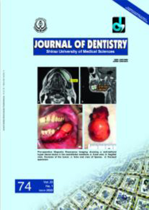
Despite being one of the most common mesenchymal benign tumors in the body, lipomas in the oral cavity constitute only 1-4% of all benign neoplasm. Buccal mucosa is the most common anatomic site within the oral cavity which is followed by tongue, lips, floor of the mouth, palate, vestibule, retromolar area and gingiva. The average tumor size ranges from 10 to 30mm. We present a rare case of unusually large sub-mucosal lipoma in lower buccal vestibule measuring about 52×29×22mm at its greatest dimension.
Keywords: Oral Lipoma, Buccal Vestibule, Soft tissue tumor

