فهرست مطالب
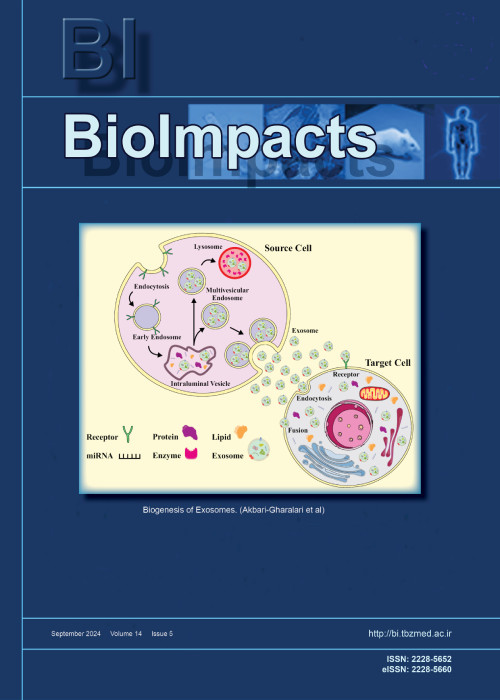
Biolmpacts
Volume:12 Issue: 6, Nov 2022
- تاریخ انتشار: 1401/08/18
- تعداد عناوین: 9
-
-
Pages 479-486Introduction
In targeted enzyme prodrug constructs, it is critical to control the bioactivity of the drug in its prodrug form. The preparation of such constructs often involves conjugation reactions directed to functional groups on amino acid side chains of the protein, which result in random conjugation and incomplete control of bioactivity of a prodrug, which may result in significant nontarget effect. Thus, more specific method of modification is desired. If the drug is a glycoprotein, enzymatic oxidation may offer an alternative approach for therapeutic glycoproteins.
MethodsTissue plasminogen activator (tPA), a model glycoprotein enzyme, was treated with galactose oxidase (GO) and horseradish peroxidase, followed by thiolation reaction and conjugation with low molecular weight heparin (LMWH). The LMWH-tPA conjugate was isolated by ion-exchange chromatography followed by centrifugal filtration. The conjugate was characterized for its fibrinolytic activity and for its plasminogen activation through an indirect amidolytic assay with a plasmin-specific substrate S-2251 when LMWH-tPA conjugate is complexed with protamine-albumin conjugate, followed by triggered activation in the presence of heparin.
ResultsLMWH-tPA conjugate prepared via enzymatic oxidation retained ~95% of its fibrinolytic activity with respect to native tPA. Upon complexation with protamine-albumin conjugate, the activity of LMWH-tPA was effectively inhibited (~90%) whereas the LMWH-tPA prepared by random thiolation exhibited ~55% inhibition. Addition of heparin fully generated the activities of both conjugates.
ConclusionThe tPA was successfully modified via enzymatic oxidation by GO, resulting in enhanced control of its activity in the prodrug construct. This approach can be applied to other therapeutic glycoproteins.
Keywords: Pretargeting, Thrombolytic drug, Triggered release, Glycoprotein oxidation, tPA -
Pages 487-499Introduction
Parkinson's disease (PD) is a chronic, devastating neurodegenerative disorder marked by the death of dopaminergic neurons in the midbrain's substantia nigra pars compacta (Snpc). In alpha-synuclein (α-Syn) self-aggregation, the existence of intracytoplasmic inclusion bodies called Lewy bodies (LBs) and Lewy neurites (LNs) causes PD, which is a cause of neuronal death.
MethodsThe present study is aimed at finding potential bioactive compounds from Cynodon dectylon that can degrade α-Syn aggregation in the brain, through in silico molecular docking investigations. Graph theoretical network analysis was used to identify the bioactive compounds that target α-Syn and decipher their network as a graph. From the data repository, twenty-nine bioactive chemicals from C. dactylon were chosen and their structures were retrieved from Pubchem. On the basis of their docking scores and binding energies, significant compounds were chosen for future investigation. The in silico prediction of chosen compounds, and their pharmacokinetic and physicochemical parameters were utilized to confirm their drug-likeness profile.
ResultsDuring molecular docking investigation the bioactive compounds vitexin (-7.3 kcal.mol-1) and homoorientin (-7.1 kcal.mol-1) showed significant binding energy against the α-Syn target protein. A computer investigation of molecular dynamics simulation study verifies the stability of the α-Syn-ligand complex. The intermolecular interactions assessed by the dynamic conditions indicate that the bioactive compound vitexin has the potency to prevent α-Syn aggregation.
ConclusionInterestingly, the observed results indicate that vitexin is a potential lead compound against α-Syn aggregation, and in vitro and in vivo studies are warranted to confirm the promising therapeutic capability.
Keywords: α-Synuclein, Cynodon dactylon, Neuroprotective agents, Molecular docking, Nolecular dynamics, In silico ADMET -
Pages 501-513Introduction
Glatiramer acetate (GA) is a newly emerged therapeutic peptide to reduce the frequency of relapses in multiple sclerosis (MS). Despite its good performance in controlling MS, it is not widely used due to daily or biweekly subcutaneous injections due to rapid degradation and body clearance. Therefore, implant design with sustained release leads to prolonged biological effects by gradually increasing drug exposure and protecting GA from rapid local degradation.
MethodsDifferent emulsion methods, PLGA type, surfactant concentration, drug/polymer ratio, drying processes, stirring method, and other variables in preliminary studies modified the final formulation. The release kinetics were studied through mechanistic kinetic models such as zero-order, Weibull, Higuchi, etc. In this study, all challenges for easy scale-up, methodological detail, and a simple, feasible setup in mass production were discussed.
ResultsThe optimized formulation was obtained by 1:6 drug/PLGA, 0.5% w/w polyvinyl alcohol, and 0.75% w/w NaCl in the external aqueous phase, 1:10 continuous phase to dispersed phase ratio, and without any surfactant in the primary emulsion. The final freeze-dried particles presented a narrow distributed size of 1-10 µm with 7.29% ± 0.51 drug loading and zero-order release behavior with appropriate regression correlation (R2 98.7), complete release, and only 7.1% initial burst release.
ConclusionTherefore, to achieve improvement in patient compliance through better and longer efficacy, designing the parenteral sustained release microspheres (MPSs) of this immune modulator is a promising approach that should be considered.
Keywords: Peptide, protein, Drug delivery, Polymeric microparticles, Multiple sclerosis, Controlled release, Poly(D, L-lactic-co-glycolic acid) -
Pages 515-531Introduction
Paclitaxel (PTX) is a cornerstone in the treatment of breast cancer, the most common type of cancer in women. However, this drug has serious limitations, including lack of tissue-specificity, poor water solubility, and the development of drug resistance. The transport of PTX in a polymeric nanoformulation could overcome these limitations.
MethodsIn this study, PLGA-PTX nanoparticles (NPs) were assayed in breast cancer cell lines, breast cancer stem cells (CSCs) and multicellular tumor spheroids (MTSs) analyzing cell cycle, cell uptake (Nile Red-NR-) and α-tubulin expression. In addition, PLGA-PTX NPs were tested in vivo using C57BL/6 mice, including a biodistribution assay.
ResultsPTX-PLGA NPs induced a significant decrease in the PTX IC50 of cancer cell lines (1.31 and 3.03-fold reduction in MDA-MB-231 and E0771 cells, respectively) and CSCs. In addition, MTSs treated with PTX-PLGA exhibited a more disorganized surface and significantly higher cell death rates compared to free PTX (27.9% and 16.3% less in MTSs from MCF-7 and E0771, respectively). PTX-PLGA nanoformulation preserved PTX’s mechanism of action and increased its cell internalization. Interestingly, PTX-PLGA NPs not only reduced the tumor volume of treated mice but also increased the antineoplastic drug accumulation in their lungs, liver, and spleen. In addition, mice treated with PTX-loaded NPs showed blood parameters similar to the control mice, in contrast with free PTX.
ConclusionThese results suggest that our PTX-PLGA NPs could be a suitable strategy for breast cancer therapy, improving antitumor drug efficiency and reducing systemic toxicity without altering its mechanism of action.
Keywords: Paclitaxel, PLGA, Breast cancer, Cancer stem cells, Mice xenografts -
Pages 533-548Introduction
Circulating tumor cells (CTCs) are the transformed tumor cells that can penetrate into the bloodstream and are available at concentrations as low as 1-100 cells per milliliter. To trap CTCs in the blood, one valid and mature technique that has been developed is the magnetophoresis-based separation in a microfluidic channel. Recently, nanostructured platforms have also been developed to trap specific targeted and marker cells in the blood. We aimed to integrate both in one platform to improve trapping.
MethodsHere, we developed a numerical scheme and an integrated device that considered the interaction between drag and magnetic forces on paramagnetic labeled cells in the fluid as well as interaction of these two forces with the adhesive force and the surface friction of the nanowires substrate. We aimed on developing a more advanced technique that integrated the magnetophoretic property of some Fe3O4 paramagnetic nanoparticles (PMNPs) with a silicon nanowires (SiNWs) substrate in a microfluidic device to trap MDA-MB231 cell lines as CTCs in the blood.
ResultsSimulation indicated assuming that the nanoparticles adhere perfectly to the white blood cells (WBCs) and the CTCs, the magnetic moment of the CTCs was almost one order of magnitude larger than that of the WBCs, so its attraction by the magnetic field was much higher. In general with significant statistics, the integrated device can trap almost all of the CTCs on the SiNWs substrate. In the experimental section, we took advantage of the integrated trapping techniques, including micropost barriers, magnetophoresis, and nanowires-based substrate to more effectively isolate the CTCs.
ConclusionThe simulation indicated that the proposed device could almost trap all of the CTCs onto the SiNWs substrate, whereas trapping in flat substrates with magnetophoretic force was very low. As a result of the magnetic field gradient, magnetophoretic force was applied to the cells through the nanoparticles, which would efficiently drive down the nanoparticle-tagged cells. For the experimental validation, anti-EpCAM antibodies for specific binding to tumor cells were used. Using this specific targeting method and by statistically counting, it was shown that the proposed technique has excellent performance and results in the trapping efficiency of above 90%.
Keywords: Well-aligned silicon nanowires, Fe3O4 paramagnetic nanoparticles, Magnetophoresis, Anti-EpCAM antibody, Circulating tumor cells -
Pages 549-559Introduction
Breast cancer cells produce exosomes that promote tumorigenesis. The anticancer properties of gallic acid have been reported. However, the mechanism underlying its anticancer effect on the exosomal secretory pathway is still unclear. We investigated the effect of gallic acid on exosome biogenesis in breast cancer cell lines.
MethodsThe cytotoxic effect of gallic acid on MCF-10a, MCF-7, and MDA-MD-231 cells was measured by MTT assay after 48 hours treatment. Expression of miRNAs including miRNA-21, -155, and 182 as well as exosomal genes such as Rab27a, b, Rab11, Alix, and CD63; along with HSP-70 (autophagy gene), was determined using Q-PCR. The subcellular distribution of it was monitored by flow cytometry analysis. Isolated exosomes were characterized by transmission and scanning electron microscopes and flow cytometry. Acetylcholinesterase activity is used to measure the number of exosomes in supernatants. In addition, autophagy markers including LC3 and P62 were measured by ELISA.
ResultsData showed that gallic acid was cytotoxic to cells (P < 0.05). Gallic acid modulated expression of miRNAs and down-regulated transcript levels of exosomal genes and up-regulated the HSP-70 gene in three cell lines (P < 0.05). The surface CD63/total CD63 ratio as well as acetylcholinesterase activity decreased in treated cells (P < 0.05). The protein level of LC3 was increased in three cell lines, while the expression of P62 increased in MCF-7 and MDA-MB-231 cancer cell lines.
ConclusionTogether, gallic acid decreased the activity of the exosomal secretory pathway in breast cancer cell lines, providing evidence for its anti-cancer effects.
Keywords: Breast cancer, Exosomes, Gallic acid, Autophagy, MCF-7 cells -
Pages 561-566Introduction
This study was proposed to assess the potential role of efflux transporters in reversing fluconazole resistance in Candida glabrata isolates treated with fluconazole loaded nanostructured lipid carriers (FLZ-NLCs).
MethodsThe ultrasound technique was used to synthesize the FLZ-NLCs. Four fluconazole-resistant, as well as one susceptible standard C. glabrata isolates, were applied and exposed to FLZ/ FLZ-NLCs for 20 h at 37°C. Real-time PCRs were done to estimate the likely changes in ATP-binding cassette transporter genes.
ResultsSimilar to the FLZ-exposed-susceptible standard strain which showed no alteration, the genes were not up-regulated significantly under the FLZ-NLCs treated condition. While they were over-expressed when the yeasts were treated with fluconazole.
ConclusionIt is highly suggested that due to the nature of the NLCs which shields the whole conformation of the drug, FLZ is not recognized by the efflux transporter subunits and consequently the translocation would not happen.
Keywords: Fluconazole, Nanostructured lipid carriers, Candida glabrata, Resistance -
Pages 567-588Introduction
Bacterial infections have always been a major threat to public health and humans' life, and fast detection of bacteria in various samples is significant to provide early and effective treatments. Cell-culture protocols, as well-established methods, involve labor-intensive and complicated preparation steps. For overcoming this drawback, electrochemical methods may provide promising alternative tools for fast and reliable detection of bacterial infections.
MethodsTherefore, this review study was done to present an overview of different electrochemical strategy based on recognition elements for detection of bacteria in the studies published during 2015-2020. For this purpose, many references in the field were reviewed, and the review covered several issues, including (a) enzymes, (b) receptors, (c) antimicrobial peptides, (d) lectins, (e) redox-active metabolites, (f) aptamer, (g) bacteriophage, (h) antibody, and (i) molecularly imprinted polymers.
ResultsDifferent analytical methods have developed are used to bacteria detection. However, most of these methods are highly time, and cost consuming, requiring trained personnel to perform the analysis. Among of these methods, electrochemical based methods are well accepted powerful tools for the detection of various analytes due to the inherent properties. Electrochemical sensors with different recognition elements can be used to design diagnostic system for bacterial infections. Recent studies have shown that electrochemical assay can provide promising reliable method for detection of bacteria.
ConclusionIn general, the field of bacterial detection by electrochemical sensors is continuously growing. It is believed that this field will focus on portable devices for detection of bacteria based on electrochemical methods. Development of these devices requires close collaboration of various disciplines, such as biology, electrochemistry, and biomaterial engineering.
Keywords: Bacterial infection, Electrochemical sensor, Rapid detection of infection, Bioreceptor, Bacteriophage -
Page 589
This corrects the article “Brain targeted delivery of rapamycin using transferrin decorated nanostructured lipid carriers published in 2022: Volume 12, Issue 1, Pages 21-32 (doi: 10.34172/bi.2021.23389). The original version of this article contained an error in Fig. 3. The confocal images for cellular uptake of coumarin-6 loaded bare NLCs (B-NLC), and transferrin decorated NLCs (Tf-NLC) after 2 hours incubation was mixed up and two different resolutions of the same specimen mistakenly named and reported unintentionally. This has now been corrected in the PDF and HTML versions of the article. Here the correct images are reported as below:


