فهرست مطالب

Iranian Endodontic Journal
Volume:18 Issue: 3, Summer 2023
- تاریخ انتشار: 1402/04/13
- تعداد عناوین: 11
-
-
Pages 126-133Introduction
Highly cited published articles play a critical role in shaping clinical practice, research directions, and advancements in a specific field of science. The current comprehensive scoping review aimed to provide an overview of highly cited articles published in the “Iranian Endodontic Journal” (IEJ), based on the IEJ’s H-index (=29); highlighting their key findings and prominent implications in the field of endodontics.
Materials and MethodsA systematic search was conducted in Scopus database to identify the top 29 highly cited published articles. The articles were selected based on their citation count (h-index); reflecting their impact and influence within the scientific community. Data extraction was performed to gather relevant information; including authors, titles, publication years, and the main topic(s) of each article.
ResultsThe selected highly cited published articles covered a broad range of endodontic topics; demonstrating the diversity and depth of research in the field. Key findings include significant contributions in vital pulp therapy, antimicrobial agents, root canal disinfection, regenerative techniques, cone-beam computed tomography applications, and intracanal medicaments. The distribution of research areas reflects the importance of evidence-based practice in clinical decision-making and patient care.
ConclusionsThese highly cited published articles have shown to have substantial impact on the field of endodontics. They have influenced clinical practice, guided research directions, and have improved patient care. The summary of key findings from each topic and the number of articles related to each area can provide readers with valuable insights into the distribution of research areas, and the significance of contributions made by the aforementioned highly cited published articles.
Keywords: Iranian Endodontic Journal, Highly cited published articles, Endodontics, Vital pulp therapy, Cone-beam computed tomography, Intracanal medicaments, Regenerative techniques -
Pages 134-144Introduction
To assess the methodological quality of systematic reviews (SRs) that evaluated the association between apical periodontitis (AP) and chronic diseases.
Materials and MethodsA systematic search was performed in the databases PubMed, Virtual Health Library, Scopus, Cochrane Library, Embase, Web of Science and Open Grey. SRs that evaluated the association between any chronic disease and AP, and that had performed a valid risk of bias assessment were included. The AMSTAR-2 tool was used for quality assessment and each included systematic review received a final categorization as having “high”, “moderate”, “low”, or “critically low” quality.
ResultsNine studies that met the eligibility criteria were included. The diseases investigated were cardiovascular diseases, diabetes mellitus, HIV, osteoporosis, chronic liver disease, blood disorders and autoimmune diseases. The systematic reviews included in this umbrella review showed a ‘low’ to ‘high’ quality of evidence.
ConclusionThere are substantial heterogeneity and several methodological concerns in the included studies. It was observed a positive association between diabetes mellitus and apical periodontitis with limited evidence, no association between HIV and apical periodontitis and a positive association between apical periodontitis and cardiovascular disease, blood disorders, chronic liver disease, osteoporosis and autoimmune diseases with moderate evidence.
Keywords: Apical Periodontitis, Endodontics, Chronic Disease, Systematic Review, Umbrella Review -
Pages 145-151Introduction
This randomized clinical trial aimed to determine whether the XP-endo finisher combined with or without foraminal enlargement has any significant effect on the incidence and intensity of postoperative pain in necrotic pulps.
Materials and MethodsClinical pain levels were measured after 6, 12, 24, 48, and 72 hours and at 7 postoperative days. All treatments were performed by an endodontist in a single visit. One hundred and twenty patients were included. All patients had a single tooth treated. The patients were divided into four groups: No FE (None Foraminal Enlargement) (n=30), FE (Foraminal Enlargement) (n=30), No FE+XPF (None Foraminal Enlargement+XP-endo Finisher) (n=30) and XPF+FE (XP-endo Finisher and Foraminal Enlargement) (n=30). The canals were irrigated with sodium hypochlorite, shaped using WaveOne Gold Medium file, and then filled by using a matching single cone and AH-Plus sealer. The cavity was filled using glass ionomer cement. Pain intensity was assessed using the visual analog scale. The data were analyzed with the ANOVA and Games-Howell test. The significance level was 5%.
ResultsThe XPF+FE group experienced a higher level of pain, being classified on the visual analog scale as moderate for 48 postoperative hours and mild for 7 postoperative days (P<0.05). In the other groups, the pain was mild, only with different time intervals (P>0.05).
ConclusionsForaminal enlargement associated with XP-endo Finisher may cause moderate postoperative pain.
Keywords: Foramen Enlargement, Postoperative Pain, Pulp Necrosis, Sodium Hypochlorite, XPendoFinisher Instrument -
Pages 152-158Introduction
The present study aimed to evaluate the effects of adding chicken eggshell powder (CESP) to calcium-enriched mixture (CEM) cement on its compressive strength (CS), solubility, and setting time.
Materials and MethodsIn this study, CESP was added at weight percentages of 3% and 5% to the powder component of the CEM cement. To measure the CS, a total of 36 samples (height, 6 mm; diameter, 4 mm) were tested in a universal testing machine. The setting time was assessed for 18 disk-shaped samples (diameter, 10 mm; height, 1 mm). Additionally, solubility test was performed on 18 samples (diameter, 8 mm; height, 1 mm) after 24 hours, 72 hours, seven days, and 14 days under dehydration conditions by calculating the weight changes; the results were then subjected to a normality test. Next, for the comparison of different test groups, parametric ANOVA test and post-hoc Tukey’s multiple comparison test were performed at a significance level of 0.05.
ResultsThe addition of 5% CESP to the CEM cement significantly reduced its setting time and water solubility (P=0.02 and P=0.01, respectively). Moreover, it significantly increased the CS over a 21-day period (P<0.001). Additionally, the addition of 3% CESP also resulted in a significant increase in CS (P<0.001). While 3% CESP reduced setting time and water solubility, the difference was not statistically significant.
ConclusionThe findings suggest that the addition of 5% CESP to CEM cement has the potential to improve its sealing ability, durability, and ability to withstand chewing forces in endodontic treatments. These results highlight the relevance of CESP as an additive for cement modifications and indicate its potential clinical implications.
Keywords: CEM Cement, Eggshell Powder, Compressive Strength, Solubility, Setting Time -
Pages 159-164Introduction
The purpose of this in vitro study was to investigate the effect of incorporating silver nanoparticles (AgNPs) of herbal origin into mineral trioxide aggregate (MTA) on the push-out bond strength (PBS) and compressive strength (CS) in simulated furcal area perforations.
Materials and MethodsIn this in vitro study, simulated furcal area perforations (1.3 mm in diameter and 2 mm in depth) were created in 40 extracted human lower molar teeth, which were divided into two groups (n=20): MTA alone and MTA combined with AgNPs (2% wt). Using a universal testing machine, PBS was evaluated by performing push-out tests, while CS was assessed using cylindrical specimens. The normal distribution of data was checked using the Kolmogorov-Smirnov test, and statistical analysis was performed using two-way ANOVA.
ResultsThe CS results showed no significant difference between the MTA group at 4 and 21 days (P=0.297), but a significant difference was observed in the nanosilver/MTA group (P=0.013). However, there was no significant difference in the push-out bond strength among the study groups (P>0.05).
ConclusionThe incorporation of herbal origin silver nanoparticles did not significantly affect the PBS or CS of MTA.
Keywords: Bond Strength, Compressive Strength, MTA, Nano Particle, Push-out -
Pages 165-167
Minimally invasive vital pulp therapy (VPT) techniques have become increasingly popular for treating mature permanent teeth with irreversible pulpitis. However, in cases where less invasive VPT approaches, such as miniature pulpotomy, fail to provide symptom relief and desired outcomes, alternative treatment strategies need to be explored. This case report presents the successful application of tampon pulpotomy, a modified full pulpotomy technique, in a vital molar tooth with irreversible pulpitis, after a previous miniature pulpotomy failure. The tampon pulpotomy procedure involved the placement of an endodontic biomaterial (i.e. calcium-enriched mixture cement) over the pulpal wound to stop bleeding and create a favorable environment for pulpal healing/regeneration. The patient was followed up for a period of 10 years, during which the tooth remained asymptomatic, functional, and exhibited normal periodontal ligament. This case report highlights the potential effectiveness of tampon/full pulpotomy as a retreatment option in cases where more conservative VPT techniques have shown limited success, offering a conservative approach to preserve tooth structure and pulpal vitality.
Keywords: Calcium-enriched Mixture Cement, Endodontics, Irreversible Pulpitis, Tampon Pulpotomy, Tricalcium Silicate, Vital Pulp Therapy -
Pages 168-173
The current study aims to report a case of invasive cervical resorption in a maxillary left central incisor with a history of dental trauma. After thorough clinical and tomographic evaluations, cervical cavitation, an irregularity in the gingival contour and crown discoloration were observed. Furthermore, presence of an extensive and well-defined area of invasive cervical resorption with pulp communication was discovered. The suggested diagnosis was asymptomatic irreversible pulpitis. The resorption area was treated with the complete removal of granulation tissue, sealed with light-curing glass ionomer cement. Then, the chemo-mechanical preparation and obturation of the root canal were performed. After two years of clinical follow-up and cone-beam computed tomography examination, there were no clinical signs and symptoms, the filling of the resorption area remained intact, and no hypodense image in the cervical region of tooth #21 could be detected. The management reported in this case presented a possible viable treatment for invasive cervical resorption, provided that correct diagnosis is made.
Keywords: Cone-beam Computed Tomography, Invasive Cervical Resorption, Resorption Treatment -
Pages 174-180
Maxillary incisors are typically straightforward cases for root canal therapy. While it is commonly assumed that maxillary central incisors have a single root canal, they may occasionally exhibit variations in their root canal system anatomy. In this report, we present a case of a maxillary central incisor with multiple root canals and provide a review of relevant literature on this anatomical variation. A 13-year-old female with deep carious lesion in tooth 11 was admitted in Department of Endodontics. Following a precise clinical and radiographic examination, a maxillary central incisor with necrotic pulp and chronic apical periodontitis along with unusual root anatomy was found and considered for non-surgical root canal treatment. Successful treatment results depend on various factors and awareness of root canal system anatomy is one of them. Due to an increasing number of reported cases of maxillary central incisors with different anatomy, it is imperative to consider anatomical variations even in the most routine cases.
Keywords: Anatomic Variation, Maxillary Central Incisor, Root Canal -
Root Canal Treatment of a Geminated Maxillary Second Molar with C-shaped Canal System: A Case ReportPages 181-185
Gemination is a rare phenomenon in the maxillary posterior teeth. Endodontic treatment of these teeth requires special care due to the bizarre anatomy particularly when it is accompanied by a C-shaped canal system. This report illustrates a patient with a rare geminated C-shaped maxillary second molar comprised of two sections in its crown, including a geminated section attached to a normal coronal of a second maxillary molar diagnosed with the pulpal status “necrosis” and “irreversible pulpitis” in geminated section and the molar respectively. Thus, endodontic treatment was performed on both parts of the tooth. Two months follow-up revealed well-functioning teeth with normal status of periapical tissue with no mobility or abnormality. Successful treatment of unusual anatomical teeth requires adherence to biomechanical principles of canal preparation and coronal restoration.
Keywords: Abnormalities, Gemination, Root Canal Treatment -
Pages 186-191
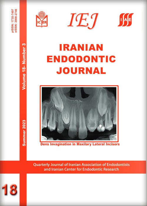
The superior lateral incisors are primarily affected by the developmental deformity known as dens invaginatus (DI). Oehler’s type III DI has the highest complexity rendering a root canal treatment (RCT) an arduous challenge for this type, so early diagnosis and treatment before pulp involvement are important. This report presents two maxillary lateral incisors with type IIIb DI, the left one being associated with a periapical lesion and the right one with normal pulp. A nine-year-old boy was referred to our clinic complaining of mobility of the maxillary left lateral incisor (LLI) associated with gumboil throughout the previous two months. Periapical radiolucency was visible on radiographs, as well as an invagination that crosses the apical foramen from the pulp chamber in both maxillary lateral incisors. The pulp of the main canal of LLI was vital and pseudo canals were necrotized and associated with chronic apical abscess. Based on the condition of the main pulp of maxillary lateral incisors, two separate treatments were carried out. RCT was done only for the pseudo canals in the LLI, while the main root canal was preserved. The right maxillary lateral incisor (RLI) had vital pulp with normal periapical tissue So the invagination was sealed as the tooth was erupting. During the oneyear follow-up period, the development of the root in LLI with a thick root wall and closed apex was observed in the periapical radiograph but pseudo canals became infected and the tooth became symptomatic, therefore retreatment for pseudo canals was carried out. The RLI root was developed and the tooth was clinically asymptomatic, so it didn’t need further treatment. Maintaining pulp vitality is crucial for type III Dens invaginated young permanent teeth since it could support root formation and improve long-term prognosis, and in cases with pulp involvement, non-surgical RCT is clinically predictable.
Keywords: Cone-beam Computed Tomography, Dens Invaginatus, Endodontic Treatment, Immature Permanent Teeth, Maxillary Lateral Incisors

