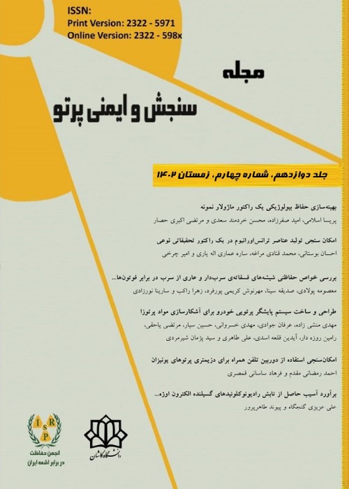
فهرست مطالب

نشریه سنجش و ایمنی پرتو
سال دوازدهم شماره 1 (پیاپی 45، بهار 1402)
- تاریخ انتشار: 1402/03/01
- تعداد عناوین: 6
-
-
صفحات 1-13
یکی از بهترین روش ها برای تشخیص سرطان سینه به خصوص در مراحل اولیه آن، ماموگرافی با پرتو ایکس است. با این وجود مطالعات کمی یافت می شود که خطر ابتلا به سرطان را در بافت هایی به جز سینه مورد بررسی قرار داده باشند. هدف پژوهش فعلی تعیین دز جذب شده در اندام های حساس بدن در حین ماموگرافی است. شبیه سازی با استفاده از کد GATE انجام شده است. در مرحله نخست یک تیوب پرتو ایکس برای ایجاد طیف های انرژی مناسب ماموگرافی ایجاد شده است. پس از آن به منظور بررسی کیفیت تصویر شامل تفکیک پذیری فضایی و نسبت کنتراست به نویز (CNR)، یک فانتوم کنترل کیفی طراحی شده است . یک فانتوم بزرگسال ORNL با اندام های مختلف طراحی و برای محاسبه دز در آن ها نیز هندسه سیستم یک دستگاه ماموگرافی دیجیتال در دو نمای اصلی ماموگرافی (CC و MLO) شبیه سازی شده است. در نهایت مشخص شد که دز تابش به بافت های بدن مانند رحم که در خارج از زمینه پرتو ایکس اولیه قرار دارند؛ کم (به طور متوسط 022/0) است. با این حال، بیشترین دز پس از سینه مورد معاینه و تومور، توسط سینه طرف مقابل در حدود 2735/5، در ریه ها 48/9، در قلب 7/6 و در معده 5/8 دریافت می شود. با توجه به نتایج به دست آمده، در مطالعه ما، انرژی 25 keV به عنوان انرژی بهینه معرفی می شود.
کلیدواژگان: ماموگرافی، سرطان سینه، کد GATE، دزیمتری، کیفیت تصویر -
صفحات 15-20در این پژوهش اثرات افزایش اسپین بر روی سد شکافت هسته ای در انرژی به جا گذاشته شده در واحد جرم سلول، ناشی از شکافت هسته ای برای هسته های مختلف با نتایج حاصل از شبیه سازی با کد MCNPX ، مورد مطالعه قرار گرفته است. با استفاده از تالی F7، میزان انرژی تخلیه شده ناشی از شکافت در سلول مورد نظر و همچنین میزان نوترون تولید شده توسط ایزوتوپ های Pu238،240،242،244، Cm242،244،246،248 و Cf252،254 اندازه گیری شد. در انرژی های نزدیک به اسپین های حالت پایه +4 و اسپین های بالاتر +12 و انرژی های نزدیک به اسپین های بازه سد شکافت در محدوده +12 تا +18 و از این محدوده تا اسپین های بالاتر، انرژی بجا گذاشته شده در سلول های حاوی ایزوتوپ های مختلف، افزایش یافت. میزان تولید نوترون در سلول های حاوی ایزوتوپ های مختلف نیز افزایش یافت. نتایج شبیه سازی نشان دادند که افزایش انرژی نوترون های فرودی، متناسب با افزایش اسپین هسته هدف می باشد. سپس اثر تغییر شکل هسته بر این نتایج و تقابل آن با افزایش اسپین، مورد بررسی قرار گرفت و مشخص گردید که نتایج به خوبی با نتایج حاصل از شبیه سازی ها، همخوانی دارد. یکی از نتایج مهم این تحقیق این است که نوترون با هر انرژی که به هسته برخورد کند، اگر هیچ واکنش هسته ای نیز برای آن رخ ندهد، حتما سبب افزایش اسپین آن هسته خواهد شد. سرانجام بر اساس مقایسه بین نتایج، معلوم گردید که نتایج شبیه سازی به وسیله کد MCNPX، نتایج حاصل از کد CNS را به خوبی تایید می کنند.کلیدواژگان: CNS، MCNPX، اسپین، سد شکافت، نوترون، تغییرات شکل هسته
-
صفحات 21-27عوارض شایع مربوط به بیماران با تومورهای سرطانی، متاسازهای استخوانی هستند. تاکنون رادیوداروهای استخوان خواه متعددی برای درمان متاستازهای استخوانی توسعه یافته است. ویژگی های مطلوب بیس فسفونات ها سبب شده است که در زمینه رادیوداروها مورد توجه قرار گیرند و مطالعات روی نسل های جدید آن ها انجام شود. هدف از این تحقیق تولید و کنترل کیفی رادیوداروی ساخته شده با ایبندرونیت به عنوان نسل سوم بیس فسفونات ها است. بدین منظور ایبندرونیت با رادیوایزوتوپ بتازای رنیم-188 نشاندار شد. خلوص رادیوایزوتوپی با استفاده از طیف سنجی گاما و خلوص رادیوشیمیایی به روش کروماتوگرافی لایه ی نازک مورد بررسی قرار گرفت. در نهایت توزیع بیولوژیکی این رادیودارو در موش ارزیابی شد. نتایج نشان داد که خلوص رادیوشیمیایی این ترکیب حدود 97% است. بیشترین جذب مربوط به اندام هدف (استخوان) با مقدار 11/7 % درصد در 12 ساعت بعد از تزریق رادیودارو بود. کلیه ها و معده دو اندامی بودند که بیشترین مقدار تجمع رادیودارو پس از استخوان را داشتند (9/2 % و 6/2 % در 4 ساعت). با توجه به امکان تولید این رادیودارو و همچنین توزیع مناسب آن در اندام های هدف و غیرهدف، می توان نتیجه گرفت که ایبندرنیت به عنوان نسل سوم بیس فسفونات ها جذب مطلوب و مناسبی دارد و این رادیودارو می تواند در درمان متاستازهای استخوانی مفید باشد.کلیدواژگان: کنترل کیفی، رادیودارو، توزیع زیستی، رنیم، ایبندرونیت
-
صفحات 27-35
برای انجام فرایندهای شیمی تابشی جهت استحصال رادیوایزوتوپ مدنظر از هدف داغ، به تجهیزاتی موسوم به اتاقک سربی و سلول داغ نیاز است. طراحی و حفاظ گذاری صحیح این تجهیزات، باهدف کاهش پرتوگیری کارکنان، از اهمیت ویژه ای برخوردار می باشد. در این مقاله بر اساس گامازایی هدف داغ مورداستفاده در فرایند تولید مولیبدن-99، محاسبات مربوط به طراحی حفاظ مناسب برای اتاقکی با ابعاد معین، از جنس بتون- باریت جهت ساخت سلول داغ و از جنس سرب جهت ساخت اتاقک سربی، انجام شده است. نتایج شبیه سازی با کد مونت کارلوی MCNP6.2 نشان می دهد که ضخامت حفاظ جهت محدود نمودن نرخ دز به µSv/h 10، به ترتیب برای ساخت سلول داغ و اتاقک سربی برابر cm 90 و cm 24 می باشد.
کلیدواژگان: حفاظ سازی، MCNP6.2، سلول داغ، اتاقک سربی، مولیبدن-99 -
صفحات 37-46میکرودوزیمتری، رهیافتی است که می تواند در ارتقاء کیفیت پرتودرمانی، بسیار موثر باشد. این دانش بر اساس مفهوم احتمالی انباشت انرژی در بافت ها و محیط های کوچک زیستی در ابعاد میکرومتر و کوچکتر، کمیت های مرتبط با اثر پرتوها را محاسبه و بررسی می کند. در این پژوهش، با استفاده از کد Geant4-DNA و سطح مقطع های تجربی و با به کارگیری روش تصادفی µ، کمیت های میکرودوزیمتری متوسط انرژی خطی و انرژی ویژه (فراوانی) الکترون های کم انرژی در استوانه هایی در ابعاد ساختارهای میکروسکوپی زنده مثل DNA، نوکلیوزوم و فیبر کروماتین در کره ای از آب با قطری در حدود میانگین اندازه هسته سلول های انسان محاسبه شده است. نتایج این پژوهش با کمیت های میکرودوزیمتری محاسبه شده با سطح مقطع های پیش فرض Geant4-DNA نیز مقایسه شده است. مشاهده شد که مقادیر متوسط انرژی خطی و انرژی ویژه (فراوانی) با سطح مقطع های تجربی بزرگتر از این مقادیر با سطح مقطع های پیش فرض Geant4-DNA هستند. بیشینه اختلاف این کمیت ها، در استوانه های با اندازه کوچک و متوسط در انرژی keV 1/0 و در استوانه بزرگ در انرژی keV 3/0 مشاهده شد. همچنین برای حجمی قابل قیاس با DNA، تناظر خوبی بین نتایج متوسط انرژی خطی فراوانی محاسبه شده با سطح مقطع های تجربی و نتایج آسیب الکترون ها در DNA سلول مشاهده شد.کلیدواژگان: کد Geant4-DNA، الکترون، سطح مقطع، میکرودوزیمتری، انرژی خطی، انرژی ویژه
-
صفحات 47-55
استفاده از براکی تراپی با استفاده از پلاک های چشمی به دلیل هزینه پایین و سهولت دسترسی نسبت به سایر روش های پرتودرمانی برای درمان انواع بدخیمی های چشم به ویژه ملانومای یووآ به طور گسترده ای مورد استفاده قرار می گیرد. اپلیکاتورهای گسیلنده بتا 106Ru/106Rh، کاربردهای فراوانی در براکی تراپی تومورهای چشمی پیدا کرده اند. تخمین توزیع دز ناشی از پلاک های چشمی با توجه به محل قرارگیری تومور از اهمیت بالایی برخوردار است. در این پژوهش، دو مدل پلاک چشمی مقعر 106Ru/106Rh، CCA و CCB، ساخت شرکت BEBIG Eckert & Ziegler BEBIG GmbH، با استفاده از کد مونت کارلوی GATE شبیه سازی شدند. آگاهی از توزیع دقیق دز در تومور و بافت های در معرض خطر چشم برای درمان ملانوما امری حیاتی محسوب می شود. در این راستا، یک فانتوم چشم شامل قسمت های مختلف صلبیه، مشیمیه، شبکیه، قرنیه، زجاجیه، عصب بینایی، عدسی، اتاقک قدامی به همراه یک تومور به ضخامت mm 3 و قطر پایه mm10 با استفاده از کد GATE مدل سازی شد. به منظور اهداف اعتبارسنجی، در ابتدا طیف انرژی چشمه 106Ru/106Rh مورد استفاده به صورت یک چشمه نقطه ای در مرکز یک فانتوم آب با بیشینه انرژی ذرات بتای MeV 54/3 مورد بررسی قرار گرفت که نتایج حاصل از آن در مقایسه با نتایج گزارش 72 ICRU با 2درصد اختلاف، تطابق خوبی را نشان می دهد. درصد دز عمقی در راستای محور مرکزی پلاک در فانتوم چشم و آب با استفاده از کد GATE محاسبه و با داده های موجود، مورد مقایسه قرار گرفت که حداکثر خطای نسبی ناشی از پلاک های CCA و CCB به ترتیب 8/6% و 4/4% می باشد. علاوه براین، ارزیابی تفاوت مابین میزان دز دریافتی اجزای مختلف فانتوم چشم نشان داد؛ پلاک چشمی CCA با توجه به دارا بودن ابعاد کوچک تر، نه تنها سبب تمرکز بیشتر دز در بافت تومور شده، بلکه دز رسیده به ساختارهایی نظیر عدسی (به عنوان یک حجم حساس) را نیز به شدت کاهش می دهد.
کلیدواژگان: براکی تراپی، پلاک های چشمی، ملانومای چشم، چشمه 106Ru، 106Rh، کد مونت کارلوی GATE
-
Pages 1-13
One of the best methods for detecting breast cancer, especially in its early stages, is X-ray mammography. However, few studies have examined the risk of developing cancer in tissues other than the breast. The aim of the current study is to determine the absorbed dose in sensitive body organs during mammography. Simulation was performed using the GATE code. In the first step, an X-ray tube was created to produce suitable energy spectra for mammography. Then, a quality control phantom was designed to assess image quality, including spatial resolution and contrast-to-noise ratio (CNR). An adult ORNL phantom with different organs was designed, and the geometry of a digital mammography device was simulated in two main mammography views (CC and MLO) to calculate the dose in the organs. Finally, it was found that the radiation dose to body tissues such as the uterus, which is outside the primary X-ray field, is low (on average 0.022 μGy). However, the highest dose is received by the contralateral breast, about 2735.5 μGy, in the lungs about 48.9 μGy, in the heart about 7.6 μGy and in the stomach about 5.8 μGy after the examined breast and tumor. Based on the results, in our study, the energy of 25 keV is introduced as the optimal energy.
Keywords: Mammography, breast cancer, GATE Code, Dosimetry, Image quality -
Pages 15-20In the present research, the increase of nuclear spin effects on the fission barrier was studied for different nuclei. Then, the deposited energy in the target was calculated using MCNPX code. Based on F7 tally, the released energies due to fission and the neutron production rate were measured for 238,240,242,244Pu, 242,244,246,248Cm and 252,254Cf isotopes. It was shown that by increasing the spin of nuclei from 4+ to 26+, the rate of neutron production for different isotopes also increases. The simulation results showed that the increase in the energy of incident neutrons is proportional to the increase in the spin of the target nucleus. At last, the mutual effect of increasing spin on the nuclear deformation was investigated which indicates a good agreement with the simulation results. One of the most important results of this work is that neutron collision with any energy increases the spin of nucleus. Finally, based on the comparison, it was found that the results of CNS (Cranked Nilsson-Strutinsky) code are confirmed with that of MCNPX.Keywords: CNS, MCNPX, spin, fission barrier, Neutron, nucleus deformation
-
Pages 21-27Skeletal metastases are prevalent complications related to patients with solid tumor cancer. Till now several boneseeking radiopharmaceuticals have been developed for bone metastases. Interesting features of bisphosphonates attracted attentions to them in the field of radiopharmaceutical therapy and studies on new generation of them have been doing too. The aim of this study was to produce and quality control of a radiopharmaceutical made with ibandronate as the third generation of bisphosphonates. For this purpose, Ibandronate was labeled with beta-emitting radionuclide of rhenium-188. Radionuclide purity was investigated using gamma spectrometry and radiochemical purity by thin layer chromatography. Finally, the biodistribution of this radiopharmaceutical was evaluated in mice. The results showed that the radiochemical purity of this compound is about 97%. the highest uptake (ID/g) was related to bone and the amount of this uptake was 7.11% at 12 hours after injection. The kidneys and stomach were the two organs that had the most activity after bone. Considering the possibility of producing the 188Re-IBA radiopharmaceutical, as well as the proper distribution of this radiopharmaceutical in target and non-target organs, it can be concluded that the ibandronate as the third generation of bisphosphonates this radiopharmaceutical can be useful in the bone metastases treatment.Keywords: Quality control, Radiopharmaceutical, Biodistribution, Re-IBA
-
Pages 27-35
Lead and hot cell equipment are needed to perform radiochemical processes to extract the desired radioisotope from the hot target. The proper design and shielding of this equipment are important to reduce the radiation exposure of employees. In this article, based on the gamma rays emitted from the hot target used in the molybdenum-99 production process, the calculations related to the design of a suitable shield for a chamber with specific dimensions made of Barite Concrete to make a hot cell or made of Lead to make a lead cell, has been done. The simulation results with MCNP6.2 Monte Carlo code show that the shield thickness to limit the dose rate to 10 µSv/h, for making hot cell or lead cells equals 90 cm and 24 cm, respectively.
Keywords: Shielding, MCNP6 Code, Hot Cell, Lead cell, Molybdenum-99 -
Pages 37-46Microdosimetry is an approach that can be very effective in improving the quality of radiation therapy. Based on the possible concept of deposited energy in microbiological tissues and environments, this knowledge can help to calculate and examine the quantities related to the effect of radiation. In this research, using the Geant4-DNA code in conjunction with experimental cross-sections and the µ-randomness method, the microdosimetry quantities known as the frequency-mean lineal energy and frequency-mean specific energy were calculated for low-energy electrons. The calculations concerned cylinders that are comparable in size with living organisms such as DNA, nucleosome, and chromatin fiber, distributed randomly in a sphere of water with a diameter of about the average size of the nucleus of human cells. The results were compared with the ones obtained based on the default cross-sections of Geant4-DNA. We found that the average values of (frequency-mean) lineal and specific energy calculated with experimental cross-sections were larger than those calculated with default cross sections. The maximum difference of these quantities was observed, in the so-called small and medium cylinders, at 0.1 keV and, in the large cylinder, at 0.3 keV. Also, for a volume comparable in size to DNA, a good correspondence between the results of frequency-mean lineal energy with experimental cross-sections and the electron DNA-damage in the cell was observed.Keywords: Geant4-DNA code, electron, cross-sections, Microdosimetry, lineal energy, Specific energy
-
Pages 47-55
The use of brachytherapy using ophthalmic plaques due to their lower cost, and ease of access compared to other radiotherapy methods is widely used to treat various types of eye malignancies, especially uvea melanoma (iris, ciliary body, and choroid). Beta emitter applicators of 106Ru/106Rh, have a lot of use in brachytherapy of intraocular tumors. Treating eye melanomas using beta-emitting 106Ru/106Rh plaques in Europe and Iran is popular. Estimating the dose distribution of eye plaques according to the location of the tumor is of great importance. In this study, two 106Ru/106Rh betta emitter concave eye plaque models, CCA and CCB, manufactured by the BEBIG Eckert & Ziegler BEBIG GmbH Company, were simulated using GATE Monte Carlo simulation code. Knowledge of the exact dose distributions in tumors and each organ at risk is critical in eye plaque brachytherapy for uveal melanoma treatment. In this regard, an eye phantom includes different parts sclera, choroid, retina, cornea, vitreous, optic nerve, lens, cornea, anterior chamber, and a tumor with thickness of 3 mm and a base diameter of 10 mm, were modeled using the GATE Monte Carlo simulation code. For validation purposes, at first, the energy spectrum of the 106Ru/106Rh source used in the study was verified as an isotropic point source centered in a water phantom using beta particles with a maximum energy of 3.54 MeV. Then the plaque central axis depth dose in the eye phantom was calculated using GATE and compared with available data. Furthermore, the difference between the deposited dose in the different components of the eye phantom shows that due to its smaller dimensions, the CCA eye plaque not only causes more concentration of the dose deposition in the tumor tissue but also greatly reduces the dose reaching structures such as the lens, as a sensitive volume.
Keywords: brachytherapy, ophthalmic plaques, eye melanoma, 106Ru, 106Rh source, GATE Monte Carlo code


