فهرست مطالب

Cell Journal (Yakhteh)
Volume:25 Issue: 10, Oct 2023
- تاریخ انتشار: 1402/08/02
- تعداد عناوین: 8
-
-
Pages 665-673Objective
Recessive dystrophic epidermolysis bullosa (RDEB) is a genetic skin fragility and ultimately lethal blistering disease caused by mutations in the COL7A1 gene which is responsible for coding type VII collagen. Investigating the pathological mechanisms and novel candidate therapies for RDEB could be fostered by new cellular models. The aim of this study was to employ CRISPR/Cas9 technology in the development of immortalized COL7A1-deficient keratinocyte cell lines intended for application as a cellular model for RDEB in ex vivo studies.
Materials and MethodsIn this experimental study, we used transient transfection to express COL7A1-targeting guide RNA (gRNA) and Cas9 in HEK001 immortalized keratinocyte cell line followed by enrichment with fluorescent-activated cell sorting (FACS) via GFP expressing cells (GFP+ HEK001). Homogenous single-cell clones were then isolated, genotyped, and evaluated for type VII collagen expression. We performed a scratch assay to confirm the functional effect of COL7A1 knockout.
ResultsWe achieved 46.1% (P<0.001) efficiency of in/del induction in the enriched transfected cell population. Except for 4% of single nucleotide insertions, the remaining in/dels were deletions of different sizes. Out of nine single expanded clones, two homozygous and two heterozygous COL7A1-deficient cell lines were obtained with defined mutation sequences. No off-target effect was detected in the knockout cell lines. Immunostaining and western blot analysis showed lack of type VII collagen (COL7A1) protein expression in these cell lines. We also showed that COL7A1-deficient cells had higher motility compared to their wild-type counterparts.
ConclusionWe reported the first isogenic immortalized COL7A1-deficient keratinocyte lines that provide a useful cell culture model to investigate aspects of RDEB biology and potential therapeutic options.
Keywords: COL7A1, CRISPR, Cas9, Keratinocyte, Recessive Dystrophic Epidermolysis Bullosa -
Pages 674-687Objective
Chimeric antigen receptor (CAR) T cell therapy has recently emerged as a promising approach for the treatment of different types of cancer. Improving CAR T cell manufacturing in terms of costs and product quality is an important concern for expanding the accessibility of this therapy. One proposed strategy for improving T cell expansion is to use genetically engineered artificial antigen presenting cells (aAPC) expressing a membrane-bound anti-CD3 for T cell activation. The aim of this study was to characterize CAR T cells generated using this aAPC-mediated approach in terms of expansion efficiency, immunophenotype, and cytotoxicity.
Materials and MethodsIn this experimental study, we generated an aAPC line by engineering K562 cells to express a membrane-bound anti-CD3 (mOKT3). T cell activation was performed by co-culturing PBMCs with either mitomycin C-treated aAPCs or surface-immobilized anti-CD3 and anti-CD28 antibodies. Untransduced and CD19-CARtransduced T cells were characterized in terms of expansion, activation markers, interferon gamma (IFN-γ) secretion, CD4/CD8 ratio, memory phenotype, and exhaustion markers. Cytotoxicity of CD19-CAR T cells generated by aAPCs and antibodies were also investigated using a bioluminescence-based co-culture assay.
ResultsOur findings showed that the engineered aAPC line has the potential to expand CAR T cells similar to that using the antibody-based method. Although activation with aAPCs leads to a higher ratio of CD8+ and effector memory T cells in the final product, we did not observe a significant difference in IFN-γ secretion, cytotoxic activity or exhaustion between CAR T cells generated with aAPC or antibodies.
ConclusionOur results show that despite the differences in the immunophenotypes of aAPC and antibody-based CAR T cells, both methods can be used to manufacture potent CAR T cells. These findings are instrumental for the improvement of the CAR T cell manufacturing process and future applications of aAPC-mediated expansion of CAR T cells.
Keywords: Artificial Antigen Presenting Cells, Chimeric Antigen Receptors, Immunotherapy, OKT3 -
Pages 688-695Objective
Determining cellular radiosensitivity of breast cancer (BC) patients through molecular markers before radiation therapy (RT) allows accurate prediction of individual’s response to radiation. The aim of this study was therefore to investigate the potential role of epigenetic biomarkers in breast cancer cellular radiosensitivity.
Materials and MethodsIn this experimental study, we treated two BC cell lines, MDA-MB 231 and MCF-7, with doses of 2, 4, and 8Gy of irradiation for 24 and 48 hours. Expression levels of circ-HIPK3, circ-PVT1, miR-25, and miR- 149 were quantified using quantitative reverse-transcription polymerase chain reaction (qRT-PCR). Significance of the observations was statistically verified using one-way ANOVA with a significance level of P<0.05. Annexin V-FITC/PI binding assay was utilized to measure cellular apoptosis.
ResultsThe rate of cell apoptosis was significantly higher in MCF-7 cells compared to MDA-MB-231 cells at doses of 4Gy and 8Gy (P=0.013 and P=0.004, respectively). RNA expression analysis showed that circ-HIPK3 was increased in the MDA-MB-231 cell line compared to the MCF-7 cell line after exposure to 8Gy for 48 hours. Expression of circ-PVT1 was found to be higher in MDA-MB-231 cells compared to MCF-7 cells after exposure to 8Gy for 24 hours, likewise after exposure to 4Gy and 8Gy for 48 hours. After exposing 8Gy, expression of miR-25 was increased in MDA-MB-231 cells compared to MCF-7 cells at 24 and 48 hours. After exposing 8Gy dose, expression of miR-149 was increased in MCF-7 cells compared to MDA-MB-231 cells at 24 and 48 hours.
Conclusioncirc-HIPK3, circ-PVT1, and miR-25 played crucial roles in the mechanisms of radioresistance in breast cancer. Additionally, miR-149 was involved in regulating cellular radiosensitivity. Therefore, these factors provided predictive information about a tumor’s radiosensitivity or its response to treatment, which could be valuable in personalizing radiation dosage.
Keywords: Breast Cancer, Ionizing Radiation, miR-149, miR-25 -
Pages 696-705Objective
The immunoregulatory properties of mesenchymal stromal/stem cells (MSCs) bring a promise for the treatment of inflammatory diseases. However, their ability to suppress the immune system is unstable. To enhance their effectiveness against immune responses, it may be necessary to manipulate MSCs. Although some dsRNA transcripts come from invading viruses, the majority of dsRNA has an endogenous origin and is known as endo-siRNA. DICER1 is a ribonuclease protein that can generate small RNAs to modulate gene expression at the post-transcriptional level. We aimed to evaluate the expression of several immune-related genes at mRNA and protein levels in MSCs overexpressing DICER1 exogenously.
Materials and MethodsIn this comparative transcriptomic experimental study, the adipose-derived MSCs (Ad-MSCs) were transfected using the pCAGGS-Flag-hsDicer vector for the DICER1 overexpression. Following the RNA extraction, mRNA expression level of DICER1 and several inflammatory cytokines were examined. We performed a relative real-time polymerase chain reaction (PCR) assay and transcriptome analysis between two groups including DICER1- transfected MSCs and control MSCs. Moreover, media from the transfected MSCs were evaluated for various interferon response factors by ELISA.
ResultsThe overexpression of DICER1 is associated with a significant increase in the mRNA expression level of COX-2, DDX-58, IFIH1, MYD88, RNase L, TLR3/4, and TDO2 genes and a downregulation of the TSG-6 gene in MSCs. Moreover, the expression levels of IL-1, 6, 8, 17, 18, CCL2, INF-γ, TGF-β, and TNF-α were higher in the DICER1-transfected MSCs group.
ConclusionIt seems that the ectopic expression of DICER1 in Ad-MSCs is linked to alterations in the expression level of immune-related genes. It is suggested that the manipulation of immune-related pathways in MSCs via the Dicer1 overexpression could facilitate the development of MSCs with distinct immunoregulatory phenotypes.
Keywords: DICER1, Immunomodulation, Mesenchymal Stromal, Stem Cells, RNA-Sequencing -
Pages 706-716Objective
Epigenetic modifications such as DNA methylation play a key role in male infertility etiology. This study aimed to explore the global DNA methylation status in testicular spermatogenic cells of varicocele-induced rats and consider their semen quality, with a focus on key epigenetic marks, namely 5-methylcytosine (5-mC) and 5-hydroxymethylcytosine (5-hmC), as well as the mRNA and proteins of ten-eleven translocation (TET) methylcytosine dioxygenases 1-3.
Materials and MethodsIn this experimental study, 24 mature male Wistar rats (8 in each group) were assigned amongst the control, sham, and varicocele groups. Sperm quality was assessed, and DNA methylation patterns of testicular spermatogenic cells were investigated using reverse transcription-polymerase chain reaction (RT-PCR), western blot, and immunofluorescence techniques.
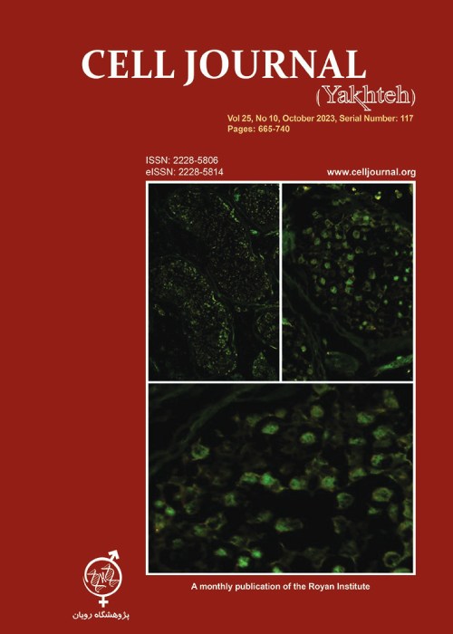
ResultsSperm parameters, chromatin and DNA integrity were significantly lower, and sperm lipid peroxidation significantly increased in varicocele-induced rats in comparison with control rats. During spermatogenesis in rat testis, 5-mC and 5-hmC epigenetic marks, and TET1-3 mRNA and proteins were expressed. In contrast to the 5-mC fluorescent signal which was presented in all testicular cells, the 5-hmC fluorescent signal was presented exclusively in spermatogonia and a few spermatids. In varicocele-induced rats, the 5-mC signal decreased in all cells within the tubules, whereas a strong signal of 5-hmC was detected in seminiferous tubules compared to the control group. As well, the levels of TET2 mRNA and protein expression were significantly upregulated in varicocele-induced rats in comparison with the control group. Also, our results showed that the varicocele-induced animals exhibited strong fluorescent signals of TET1-3 in testicular cells, whereas weak fluorescent signals were identified in the seminiferous tubules of the control animals.
ConclusionConsequently, we showed TET2 upregulation and the 5-hmC gain at testicular levels are associated with varicocele and sperm quality decline, and therefore they can be exploited as potential biomarkers of spermatogenesis.
Keywords: DNA Methylation, Male Infertility, Sperm, Varicocele, 5-Methylcytosine -
Pages 717-726Objective
Vaccinium arctostaphylos has traditionally been employed in Iranian folk medicine to treat diabetes. However, the precise molecular mechanisms underlying its antidiabetic properties remain incompletely understood. The current experiment intended to explore the modulatory effects of V. arctostaphylos fruit ethanolic extract (VAE) on biochemical and molecular events in the livers of diabetic rats.
Materials and MethodsIn this experimental study, male Wistar rats were randomly assigned to four groups: normal control, normal rats with VAE treatment, diabetic control, and diabetic rats with VAE treatment. Following 42 days of treatment, the impact of VAE on diabetes-induced rats was assessed by measuring various serum biochemical parameters, including insulin, free fatty acids (FFA), tumor necrosis factor-α (TNF-α), reactive oxygen species (ROS), and adiponectin levels. The activities of hepatic carbohydrate metabolic enzymes and glycogen content were determined. Additionally, expression levels of selected genes implicated in carbohydrate/lipid metabolism and miR-27b expression were evaluated. H&E-stained liver sections were prepared for light microscopy examination.
ResultsTreatment with VAE elevated levels of insulin and adiponectin that reduced levels of FFA, ROS, and TNF-α in the serum of diabetic rats. VAE-treated rats exhibited increased activities of hepatic glucokinase (GK), glucose-6-phosphate dehydrogenase (G6PD), and glycogen concentrations, in conjunction with decreased activities of glucose-6-phosphatase (G6Pase) and fructose-1,6-bisphosphatase (FBPase). Furthermore, VAE significantly upregulated the transcription levels of hepatic insulin receptor substrate 1 (Irs1) and glucose transporter 2 (Glut2), while considerably downregulated the expression of peroxisome proliferator-activated receptor gamma (Pparg) and sterol regulatory element-binding protein 1c (Srebp1c). VAE remarkably enhanced the expression of miR27-b in the hepatic tissues of diabetic rats. Abnormal histological signs were dramatically normalized in diabetic rats receiving VAE compared to those in the diabetic control group.
ConclusionOur findings underscore the hypoglycemic and hypolipidemic activities of V. arctostaphylos and assist in better comprehension of its antidiabetic properties.
Keywords: Diabetic Rat, Liver, miR27-b, Vaccinium arctostaphylos -
Pages 727-737Objective
Varicocele is a common cause of male infertility, affecting a substantial proportion of infertile men. Recent studies have employed transcriptomic analysis to identify candidate genes that may be implicated in the pathogenesis of this condition. Accordingly, this study sought to leverage rat gene expression profiling, along with protein-protein interaction networks, to identify key regulatory genes, related pathways, and potentially effective drugs for the treatment of varicocele.
Materials and MethodsIn this in-silico study, differentially expressed genes (DEGs) from the testicular tissue of 3 rats were screened using the edgeR package in R software and the results were compared to 3 rats in the control group. Data was obtained from GSE139447. Setting a -11 and P<0.05 as cutoff points for statistical significance, up and down-regulated genes were identified. Based on Cytoscape plugins, protein-protein interaction (PPI) networks were drawn, and hub genes were highlighted. ShinyGO was used for pathway enrichment. Finally, effective drugs were identified from the drug database.
ResultsAmong the 1277 DEGs in this study, 677 genes were up-regulated while 600 genes were down-regulated in rats with varicocele compared to the control group. Using protein-protein interaction networks, we identified the top five up-regulated genes and the top five down-regulated genes. Enrichment analysis showed that the up-regulated genes were associated with the cell division cycle pathway, while the down-regulated genes were linked to the ribosome pathway. Notably, our findings suggested that dexamethasone may be a promising therapeutic option for individuals with varicocele.
ConclusionThe current investigation indicates that in varicocele the cell division cycle pathway is up-regulated while the ribosome pathway is down-regulated compared to controls. Based on these findings, dexamethasone could be considered a future candidate drug for the treatment of individuals with varicocele.
Keywords: Cell Division Cycle, RNA-SEQ, Varicocele -
Pages 738-740
"Theory of Forms" implies that a genuine version of creatures exists beyond the shapes in this world. Stem cell technology has adopted developmental cues to mimic real life. However, the functionality of the lab-made cells is far from primary ones. Perhaps it is time to switch from analytical to systematic perspective in stem cell science. This may be the way to define new horizons based on the systematic perspective and convergence of science in stem cell biology, bridging the current gap between the shadows of real knowledge in current research and reality in future.
Keywords: Theory of Forms, Stem Cell Science, Systems Biology


