فهرست مطالب

Iranian Endodontic Journal
Volume:19 Issue: 1, Winter 2024
- تاریخ انتشار: 1402/11/02
- تعداد عناوین: 10
-
-
Page 1
-
Pages 2-12
Invasive cervical root resorption (ICRR) is a dental pathology, marked by unexpected destruction originating in the cervical region of the tooth. This comprehensive literature review provides a holistic view into the pathogenesis, clinical manifestation, and precise management of ICRR, aiming to guide endodontists and enhance patient care and treatment outcomes. The review delves into the potential etiology of ICRR, covering contributing factors such as trauma, orthodontic treatment, and other pertinent conditions. It outlines the clinical and radiographic indicators, underscoring the crucial role of early detection and precise diagnosis in effectively managing and halting ICRR progression. The exploration of treatment approaches is thorough, ranging from non-surgical methods like vital pulp therapy or root canal treatment to surgical interventions. This review accentuates the essential role of interdisciplinary collaboration among diverse dental specialties in enhancing ICRR management. It highlights the importance of a consolidated strategy in enhancing treatment outcomes and preserving tooth structure and function. Moreover, it investigates prevention methods, risk evaluation, and identifies prospective research pathways to address the existing knowledge gaps.
Keywords: Endodontics, Invasive Cervical Root Resorption, Interdisciplinary Approach, Pathogenesis, Pulpotomy, Radiographic Examination, Treatment Strategies -
Pages 13-21Introduction
This non-randomized clinical trial investigated the outcomes of full pulpotomy in adult molars with irreversible pulpitis, comparing those with calcified and non-calcified pulp chambers over 6 and 12 months.
Materials and MethodsA total of 101 adult permanent molars with irreversible pulpitis, in individuals over 12 years old, were categorized based on pulp chamber calcification observed in radiographic images by two endodontists. Subsequently, full pulpotomy procedures were performed, achieving hemostasis, and applying a 2 mm layer of calcium-enriched mixture (CEM) cement as a pulp covering agent. After 48 hours, the setting of the CEM cement was verified, followed by the application of a layer of resin-modified glass-ionomer. The tooth was then restored using amalgam. Clinical and radiographic evaluations were conducted at 6-month and 1-year follow-ups by blinded endodontists. Success rates were compared using Fisher's exact test and logistic regression tests with a significance level of 0.05.
ResultsAmong the 97 patients with 6-month and 1-year follow-ups, all achieved clinical success. Radiographic success rates were 99% at 6 months and 96.9% at 1 year, regardless of pulp calcification. In the 6-month follow-up, success rates were 98.07% for non-calcified pulp chambers and 100% for calcified pulp chambers. At the 1-year follow-up, success rates were 96.1% and 97.8%, respectively. Statistical analysis showed no significant difference in radiographic success rate between the two groups at both follow-ups (P>0.05).
ConclusionsFull pulpotomy using CEM cement is a successful treatment for adult permanent teeth with calcified and non-calcified pulp chambers presenting signs and symptoms of irreversible pulpitis up to a 1-year follow-up. This study provides compelling evidence that vital pulp therapy can be effectively employed in the pulpotomy of calcified teeth, at least in the short term.
Keywords: Calcified Teeth, Calcium-Enriched Mixture, CEM Cement, Full Pulpotomy, Deep Caries, Irreversible Pulpitis, Vital Pulp Therapy -
Pages 22-27Introduction
The aim of this study was to assess the effectiveness of filling removal material from the apical third of curved mesial root canals of mandibular molars. Reciprocating instrumentation followed by additional rotary instrumentation with instruments made of alloys with different heat treatments was evaluated.
Materials and MethodsThirty-six mesial roots of mandibular molars were divided into two groups: Group Class IV consisted of 16 roots with two independent canals, and Group Class II consisted of 20 roots with two canals that merged into one at their apical level. Each of these two groups were further divided into two subgroups, according to the additional rotary instrument used after the reciprocating instrumentation: Group RH and Group RM for Hyflex and Mtwo, respectively. After each procedural step, the roots were scanned by micro-tomography. After each step of filling removal, the Wilcoxon matched pair test and the Mann-Whitney test were used for the evaluation between groups. The significance level adopted was 5%.
ResultsSignificant differences were observed between groups with different Class II and Class IV anatomies, regarding filling removal after Reciproc (P<0.05). After the use of an additional rotary instrumentation, no differences were observed between the two groups (P>0.05).
ConclusionsIn the apical third of mesial roots of mandibular molars with Class II anatomy, an additional rotary instrumentation was shown to be necessary for improving the removal of filling material after using the single -file reciprocating instrumentation technique.
Keywords: Curved Root Canals, Micro-computed Tomography, Root Canal Retreatment, RotaryInstrument -
Pages 28-34Introduction
This study investigates the influence of root length in mandibular molars with irreversible pulpitis on the success of supplemental intraligamentary injection following an inferior alveolar nerve (IAN) block. Various factors, including anatomical location, tooth type, and anesthetic solution, may affect supplemental anesthesia success.
Materials and MethodsA total of 251 patients diagnosed with irreversible pulpitis in mandibular first or second molars underwent buccal infiltration anesthesia (4% articaine with 1:100,000 epinephrine) after IAN block injection (3% prilocaine and 0.03 IU/mL of felypressin). Fifty patients experiencing pain during accesscavity preparation received supplemental intraligamentary injection (0.3 mL of 2% lidocaine with 1:80,000 epinephrine) at each mesial and distal line angle. The root length of treated teeth was recorded using an apex locator. Data analysis involved independent t-tests, Chi-square tests, and logistic regression.
ResultsSuccessful supplemental intraligamentary injection was observed in 21 (42%) out of 50 patients. No significant correlation was found between the mean length of mesiobuccal (P=0.61), mesiolingual (P=0.34), or distal (P=0.60) canals of mandibular molars and the injection's success. Logistic regression analysis, however, revealed a significant impact of mesiolingual canal length on the success rate [OR 0.09 (0.01-0.79) , P=0.030].
ConclusionThe root length of mandibular first and second molars does not significantly affect the success of supplemental intraligamentary injection.
Keywords: Intraligamentary Injection, Irreversible Pulpitis, Mandibular Molar, Root Length -
Pages 35-38Introduction
Microbial agents play a crucial role in periapical lesions and despite mechanical preparation, presence of persistent bacteria in root canal system is a challenge. Photodynamic therapy offers a debridement method, utilizing photosensitizers such as Curcumin, Indocyanine Green (ICG), and Methylene Blue (MB). This study aimed to assess and compare the penetration depth of these photosensitizers on the lateral surface of the root canal.
Materials and MethodsThe crown of 30 single-rooted teeth were separated by a diamond disc. The canals were prepared using a rotary systemand were rinsed with 10 mL of 2.5% NaOCl. In order to remove the smear layer debris, 17% EDTA was placed in the root canal for 1 min, then rinsed with NaOCl and saline. The teeth were sterilized by autoclave and randomly assigned to three groups thatfilled with curcumin, ICG, or MB. Subsequently, they were incubated for 10 min and dried up by paper. Longitudinal sections were cut, and penetration depth of the photosensitizers in coronal, middle, and apical sections were measured using a stereomicroscope.
ResultsCurcumin demonstrated a higher average penetration depth (3000 μm) than MB, and MB showed higher penetration depth than ICG. Significantly different penetration depths were observed in pairwise comparisons among all three groups (P<0.005).
ConclusionCurcumin with its superior average penetration depth, emerges as a promising choice for effective root canal disinfection in endodontic treatments. Consideration of these findings may enhance the selection of photosensitizers in clinical applications.
Keywords: Curcumin, Indocyanine Green, Methylene Blue, Photodynamic Therapy, Photosensitizers -
Pages 39-45Introduction
Mechanical root canal preparations and irrigation solutions are essential for reducing microbial counts in the root canal system. However, these methods do not completely eliminate microorganisms. Intracanal medicaments are used to further decrease microbial counts. This study aims to assess the cytotoxicity of various intracanal medicaments.
Materials and methodsIn this in vitro study, murine fibroblast cell lines (L929) were cultured in a controlled environment. The MTT assay was employed to evaluate the cytotoxicity of different medicament combinations, including calcium hydroxide and triamcinolone (D1), niosomal doxycycline and triamcinolone (D2), calcium hydroxide (D3), and a combination of doxycycline and triamcinolone (D4). Statistical analysis was performed using ANOVA and Dunnett’s test.
ResultsThe results indicated that D1 and D2 had lower cytotoxicity, while D4 exhibited the highest cytotoxicity. D1 was found to be non-cytotoxic up to a concentration of 500 µg/mL over a period of 72 hours. D2 and D3 showed similar effects up to concentrations of 250 µg/mL and 100 µg/mL, respectively, for 72 hours. In contrast, D4 exhibited cytotoxicity at concentrations above 75 µg/mL at 72 hours.
ConclusionThis study suggests that encapsulating doxycycline in niosomal structures (D2) reduces cytotoxicity in murine fibroblast cell lines (L929) for at least 24 and 48 hours. These findings offer promising implications for the development of endodontic medicaments with improved biocompatibility.
Keywords: Calcium hydroxide, corticosteroid, cytotoxicity, niosomal doxycycline -
Pages 46-49
This case report highlights a rare complication of root canal treatment involving the inadvertent extrusion of sodium hypochlorite solution, resulting in a sodium hypochlorite-induced facial hematoma. A 44-year-old female patient presented significant right hemifacial swelling and ecchymosis following root canal therapy. Computed tomography imaging confirmed a hematoma involving the facial region without active signs of bleeding. Sodium hypochlorite, a potent cytotoxic agent commonly used in root canal procedures, was identified as the causative agent. Treatment consisted of prednisone, antibiotics, and NSAIDs, resulting in gradual improvement over a month. The cytotoxic properties of sodium hypochlorite, its variable concentrations, and risk factors associated with facial hematomas are discussed. It is essential to emphasize the rarity of such hematomas and highlight the need for precise technique, vigilant monitoring, and interdisciplinary collaboration to mitigate risks and prioritize patient safety.
Keywords: Case Report, Facial Hematoma, Sodium Hypochlorite, Root Canal Treatment -
Pages 50-55
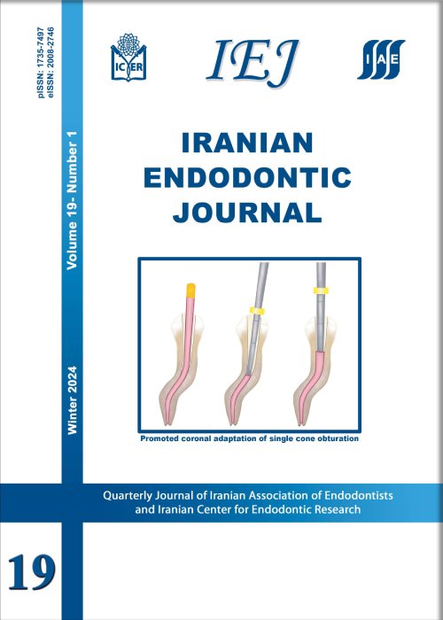
The single-cone technique, also known as the hydraulic condensation technique, is widely employed in endodontics. However, the aforementioned method is presented with certain limitations; specifically concerning the coronal seal and the adaptation of the coronal third of a master gutta-percha (GP) with a round cross-section to the coronal dentinal walls of root canals with semi-round or oval cross- sections. Through two case reports, the current article introduces the coronal vertical condensation (CVC) technique; aiming to enhance GP adaptation to canal walls in similar scenarios. In fact, the coronal vertical condensation technique amalgamates the different aspectsof warm vertical condensation and single-cone techniques. In CVC , following the placement ofthe master GP cone, an electrical heat carrier is inserted immediately a few millimeters apical from the canal orifice to remove the coronal portion of the master GP cone. Subsequently, a hand plugger is used to condense GP in the vertical dimension, and the coronal space is backfilled using melted GP. The implementation of CVC technique has demonstrated an improved coronal adaptation of GP with canal walls. The stated technique seems beneficial; especially in the obturation of severely curved canals or root canals with a final preparation shape of variable taper.
Keywords: Root Canal Filling Materials, Root Canal Obturation, Root Canal Preparation, Root Canal Therapy -
Pages 56-60
Invasive cervical root resorption (ICRR) is a rare and clinically complex condition marked by the progressive loss of dental hard tissues below the junctional epithelium. This case report outlines the management of a 32-year -old female patient presenting with ICRR class 3 affecting a maxillary incisor. Despite the absence of symptoms, the expansivenature of the defect warranted conservative surgical intervention. The procedure involved the surgical removal of inflamed tissues, followed by an ultraconservative modified pulpotomy utilizing calcium-enriched mixture (CEM) cement through a surgical window. The selected intervention is substantiated by its potential benefits, such as minimal removal of tooth structure and the inherent biocompatibility and sealing capabilities of CEM cement. A one-year follow-up revealed arrested resorption, re-establishment of periodontal attachment, and successful esthetic restoration, affirming the efficacy of vital pulp therapy in surgically addressing advanced ICRR. Accurate diagnosis, strategic treatment planning, and a patient-centered approach proved critical in achieving favorable outcomes.
Keywords: Calcium-enriched Mixture, CEM Cement, Endodontics, Invasive Cervical Root Resorption, Pulpotomy, Vital Pulp TherapyReceived

