فهرست مطالب

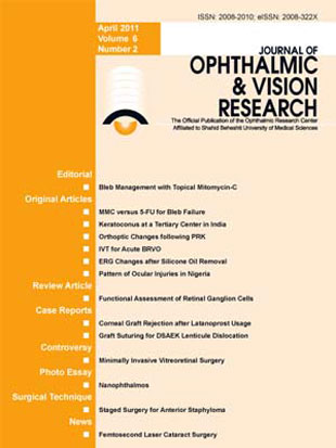
Journal of Ophthalmic and Vision Research
Volume:6 Issue: 2, Apr-Jun 2011
- تاریخ انتشار: 1390/03/05
- تعداد عناوین: 14
-
-
Page 78PurposeTo compare the efficacy and safety of topical mitomycin C (MMC) drops with that of subconjunctival 5 fluorouracil (5 FU) injections for management of early bleb failure after trabeculectomy or combined phacoemulsification and trabeculectomy with posterior chamber intraocular lens implantation (PT+PCIOL).MethodsIn a randomized comparative study, 37 eyes of 37 patients with impending early bleb failure received MMC 0.02% eye drops for 2 or 4 weeks (19 eyes) or subconjunctival 5 FU injections, 5 mg per dose (18 eyes). Complete success was defined as 5 < IOP? 18 mmHg without medications.ResultsBaseline characteristics were comparable between the study groups. However, there were more cases of combined PT+PCIOL in the MMC group [11 (57.9%) eyes versus 3 (16.7%) eyes, P = 0.017]. Mean preoperative IOP was 20.5±8.85 mmHg in the MMC group and 25.82±11.35 mmHg in the 5 FU group (P = 0.129), which was decreased to 13.2±6.1 and 10.6±4.8 mmHg respectively after 12 months (P = 0.159). There was no significant difference between the study groups in terms of bleb extent (P = 0.170), height (P = 0.178) or vascularity (P = 0.366). At the end of the study, complete success was achieved in 13 eyes (68.4%) in the MMC group and 14 eyes (77.8%) in the 5 FU group (P = 0.714). The survival of success at 8 months (median follow-up) was 89.5% and 86.5% in the MMC and 5 FU groups respectively; the number of glaucoma medications (P = 0.707) and best-corrected visual acuity (P = 0.550) were also comparable. Complication rates were similar in the study groups (P = 0.140).ConclusionTopical MMC 0.02% has comparable safety and efficacy to subconjunctival 5 FU injections for management of early bleb failure. Topical MMC 0.02% drops are more convenient and can be initiated first, while 5 FU injections may be reserved for eyes with an insufficient response to topical MMC.
-
Page 87PurposeTo evaluate the presentation and characteristics of patients with keratoconus at a tertiary eye care center in Mumbai, India.MethodsThis single center, non-comparative, retrospective cohort analysis was performed on patients with keratoconus who presented to the Clear Vision Eye Center clinic from April 2007 to March 2009. Data was collected to characterize correlations among visual acuity, corneal biomicroscopic findings, and refractive and topographic findings in keratoconus.ResultsRecords of 274 patients including 189 male and 85 female subjects with mean age of 20.1±3.5 (range, 13 to 29) years at the time of diagnosis were assessed. There was history of skin allergy in 73 (26.6%), symptomatic ocular allergy in 67 (24.45%) and asthma in 31 (11.31%) patients. The most frequent corneal sign was Fleischer's ring which was observed in 81% of cases. Corneal topography revealed mean simK (simulated keratometry) of 53.3±6.1 (range, 41.2 to 69.0) diopters. Corneal topography analysis with the Cone Location Magnitude Index disclosed the presence of inferior cones in 93% of patients.ConclusionThis group of patients had younger age at presentation and more severe keratoconus as compared to western populations; contact lenses were used only in a minority of patients.
-
Page 92PurposeTo report orthoptic changes after photorefractive keratectomy (PRK).MethodsThis interventional case series included 297 eyes of 150 patients scheduled for PRK. Complete ophthalmologic evaluations focusing on orthoptic examinations were performed before and 3 months after PRK.ResultsBefore PRK, 2 (1.3%) patients had esotropia which remained unchanged; 3 (2%) patients had far exotropia which improved after the procedure. Of 12 cases (8%) with initial exotropia at near, 3 (2%) cases became orthophoric, however 6 patients (4%) developed new near exotropia. A significant reduction in convergence and divergence amplitudes (P < 0.001) and a significant increase in near point of convergence (NPC) (P < 0.006) were noticed after PRK. A reduction of 10 PD or more in convergence amplitude and 5 PD or more in divergence amplitude occurred in 10 and 5 patients, respectively. Four patients had initial NPC > 10 cm which remained unchanged after surgery. Out of 9 (6%) patients with baseline stereopsis > 60 seconds of arc, 2 (1.33%) showed an improvement in stereopsis following PRK. No patient developed diplopia postoperatively.ConclusionPreexisting strabismus may improve or remain unchanged after PRK, and new deviations can develop following the procedure. A decrease in fusional amplitudes, an increase in NPC, and an improvement in stereopsis may also occur after PRK. Preoperative evaluation of orthoptic status for detection of baseline abnormalities and identification of susceptible patients seem advisable
-
Page 101PurposeTo evaluate the therapeutic effect of intravitreal triamcinolone (IVT) injection for recent branch retinal vein occlusion (BRVO).MethodsIn a randomized controlled clinical trial, 30 phakic eyes with recent (less than 10 week's duration) BRVO were assigned to two groups. The treatment group (16 eyes) received 4 mg IVT and the control group (14 eyes) received subconjunctival sham injections. Changes in visual acuity (VA) were the main outcome measure.ResultsVA and central macular thickness (CMT) changes were not significantly different between the study groups at any time point. Within group analysis showed significant VA improvement from baseline in the IVT group up to three months (P < 0.05); the amount of this change was 0.53 ± 0.46, 0.37 ± 0.50, 0.46 ± 0.50, and 0.29 ± 0.45 logMAR at 1, 2, 3, and 4 months, respectively. Corresponding VA improvements in the control group were 0.20 ± 0.37, 0.11 ± 0.46, 0.25 ± 0.58, and 0.05 ± 0.50 logMAR (all P values > 0.05). Significant reduction in CMT was noticed only in the treatment group (172 ± 202 microns, P = 0.029) and at 4 months. Ocular hypertension occurred in 4 (25%) and 2 (14.3%) eyes in the IVT and control groups, respectively.ConclusionA single IVT injection had a non-significant beneficial effect on VA and CMT in acute BRVO as compared to the natural history of the condition. The 3-month deferred treatment protocol advocated by the Branch Vein Occlusion Study Group may be a safer option than IVT injection considering its potential side effects.
-
Page 109PurposeTo evaluate electroretinogram (ERG) changes after silicone oil removal.MethodsScotopic and photopic ERGs, and best-corrected visual acuity (BCVA) were checked before and shortly after silicone oil removal in eyes that had previously undergone vitrectomy and silicone oil injection for complex retinal detachment. Pre- and postoperative ERG a- and b-wave amplitudes were compared.ResultsTwenty-eight eyes of 28 patients including 20 male and 8 female subjects with mean age of 39.3 ± 0.06 (range, 12 to 85) years were studied. Mean interval from primary vitreoretinal surgery to silicone oil removal was 21.04 ± 0.52 (range, 7 to 39) months. Mean duration from silicone oil removal to second ERG was 13.04 ± 1.75 (range, 10 to 16) days. Before silicone oil removal, mean a-wave amplitudes in maximal combined response, rod response and cone response ERGs were 27.4 ± 19.9, 7.2 ± 4.5 and 5.5 ± 3.4 microvolts, respectively. These values increased to 48.8 ± 31.9, 15.1 ± 14.4 and 17.4 ± 22.2 microvolts, respectively after silicone oil removal (P < 0.001). Mean b-wave amplitudes in the same order, were 69.41 ± 51, 41.2 ± 30.4 and 25.1 ± 33.9 microvolts before silicone oil removal, increasing to 165.6 ± 102.5, 81.7 ± 53.7 and 44.7 ± 34.1 microvolts respectively, after silicone oil removal (P < 0.001). Mean BCVA significantly improved from 1.10 ± 0.34 at baseline to 1.02 ± 0.33 logMAR after silicone oil removal (P < 0.001).ConclusionThe amplitudes of ERG a- and b-waves under scotopic and photopic conditions increased significantly shortly after silicone oil removal. An increase in BCVA was also observed. These changes may be explained by the insulating effect of silicone oil on the retina.
-
Page 114PurposeTo determine the pattern of ocular injuries in patients presenting to the eye clinic and the accident and emergency department of Federal Medical Center, Owo, Ondo State, Nigeria.MethodsThis prospective study was conducted between January and December 2009. Federal Medical Center, Owo is the only tertiary hospital in Ondo State, Nigeria. The eye center located at this medical center was the only eye care facility in the community at the time of this study. All patients were interviewed with the aid of an interviewer-administered questionnaire and underwent a detailed ocular examination.ResultsOf 132 patients included in the study, most (84.1%) sustained blunt eye injury while (12.1%) had penetrating eye injury. A considerable proportion of patients (37.9%) presented within 24 hours of injury. Vegetative materials were the most common (42.4%) offending agent, a minority of patients (22%) was admitted and none of the patients had used eye protection at the time of injury.ConclusionIn the current series, blunt eye injury was the most common type of ocular trauma. The community should be educated and informed about the importance of preventive measures including protective eye devices during high risk activities. Patients should be encouraged to present early following ocular injury.
-
Page 119The information generated by cone photoreceptors in the retina is compressed and transferred to higher processing centers through three distinct types of ganglion cells known as magno, parvo and konio cells. These ganglion cells, which travel from the retina to the lateral geniculate nucleus (LGN) and then to the primary visual cortex, have different structural and functional characteristics, and are organized in distinct layers in the LGN and the primary visual cortex. Magno cells are large, have thick axons and usually collect input from many retinal cells. Parvo cells are smaller, with fine axons and less myelin than mango cells. Konio cells are diverse small cells with wide fields of input consisting of different cells types. The three cellular pathways also differ in function. Magno cells respond rapidly to changing stimuli, while parvo cells need time to respond. The distinct patterns of structure and function in these cells have provided an opportunity for clinical assessment of their function. Functional assessment of these cells is currently used in the field of ophthalmology where frequency-doubling technology perimetry selectively assesses the function of magno cells. Evidence has accrued that the three pathways show characteristic patterns of malfunctions in multiple sclerosis, schizophrenia, Parkinson's and Alzheimer's diseases, and several other disorders. The combination of behavioral assessment with other techniques, such as event related potentials and functional magnetic resonance imaging, seems to bear promising future clinical applications
-
Page 127PurposeTo report endothelial corneal graft rejection after administration of topical latanoprost eye drops. CASE REPORT: Two eyes of two patients with a history of multiple intraocular procedures prior to penetrating keratoplasty developed endothelial graft rejection one month after administration of topical latanoprost. Cystoid macular edema developed simultaneously in one patient.ConclusionLatanoprost may trigger endothelial graft rejection in susceptible eyes.
-
Graft Suturing for Lenticule Dislocation after Descemet Stripping Automated Endothelial KeratoplastyPage 131PurposeTo report the mid-term outcomes of graft suturing in a patient with lenticule dislocation after Descemet stripping automated endothelial keratoplasty (DSAEK). CASE REPORT: A 78-year old woman was found to have graft dislocation involving the nasal half of the cornea after uneventful DSAEK. Graft repositioning, refilling the anterior chamber with air, and placement of four full-thickness 10/0 nylon sutures over the detached area were performed two weeks after the initial surgery. The sutures were removed 6 weeks later. Serial specular microscopy and anterior segment optical coherence tomography were performed. At 18 months, there was good lenticule apposition and a clear graft.ConclusionAnchoring sutures seem to be effective for management of graft detachment following DSAEK.
-
Page 145
-
Page 147Herein we describe a staged surgical technique consisting of penetrating sclerokeratoplasty (PSKP) followed by penetrating keratoplasty (PKP) and present its clinical course and complications over two years of follow-up. A 23-year-old man presented with cosmetically unacceptable protrusion of the globe corresponding to the cornea and sclera. PSKP was performed transplanting a full-thickness beveled 13 mm corneoscleral tectonic graft. Hypotony developed subsequently and was successfully managed medically, however corneal graft failure occurred. After 15 months, a 7.5 mm PKP was performed for optical reasons, which subsequently remained clear with a healthy epithelium. In this particular case, cosmetic, tectonic, therapeutic, and optical requirements were met. PSKP is a surgical procedure which entails a high rate of complications but may be the only alternative when the main goal of intervention is restoration of the globe in complicated cases such as our patient.
-
Page 151

