فهرست مطالب

Cell Journal (Yakhteh)
Volume:13 Issue: 1, Spring 2011
- تاریخ انتشار: 1390/04/01
- تعداد عناوین: 10
-
-
Page 1ObjectiveMelatonin, the pineal gland hormone as a direct or indirect antioxidant and free radical scavenger, is involved in the process of both aging and age-related diseases. This study investigates the effects of melatonin on the histology of testicular seminiferous tubules in aged mice.Materials And MethodsTwenty male, white mice, aged 16 months, that weighed 20-23 gr were equally divided into control and experimental groups. The experimental group was intraperitoneally injected with a daily single dose of 10 mg/kg melatonin for 14 days. The control group received only saline. Six days after the last injection, all mice were sacrificed and the testes were excised and processed for light microscope observation. In the morphometric study, we evaluated testicular seminiferous tubule parameters such as height of germinal epithelium, seminiferous tubule diameter, thickness of interstitial connective tissue and spermatogenesis index (SI). SPSS software and student's t-test analyzed all parameters to assess the significance of changes between control and experimental groups.ResultsMelatonin-treated mice had seminiferous tubules with a wide lumen lined by low height germinal epithelium. The interstitial connective tissue thickened significantly in the experimental group (p<0.05), tubular diameter and germinal epithelium height decreased significantly (p<0.01), and the SI reduced compared to the control group (p<0.001).ConclusionThe results of this study showed the disadvantages of melatonin on seminiferous tubules of aged mice testes.
-
Page 5ObjectiveToxic fumes generated during the soldering process contain various contaminants released at sufficient rates to cause both short- and long-term health problems. Studies have shown that these fumes change the quality and quantity of semen fluid in exposed workers. The aim of the present study was to determine the potentially toxic effects of solder fumes on spermatogenesis in seminiferous tubules of rats as an experimental model, with conditioned media in an exposed chamber.Materials And MethodsA total number of 48 male Sprague Dawley adult rats were randomly divided into experimental (n=30) and control (n=18) groups. Based on exposure time, each group was further subdivided into two, four and six subgroups. Rats in the experimental groups were exposed to solder fumes in an exposure chamber for one hour/day. The concentrations of fumes [formaldehyde, stanum (Sn) and lead (Pb)] were measured by a standard method via atomic absorption and spectrophotometry. According to a timetable, under deep anesthesia, the rats of both experimental and control subgroups were killed. After fixation of testes, specimens were weighed and routinely processed. Paraffin sections were stained by hematoxylin and eosin. Spermiogenesis index was calculated and data analyzed by Mann Whitney NPAR test.ResultsAnalysis of air samples in the exposure chamber showed the following fume concentrations: 0.193 mg/m3 for formaldehyde, 0.35 mg/m3 for Sn and 3 mg/m3 for Pb. Although there was no significant difference in testes weight between control and experimental subgroups, there was only a significant difference in spermiogenesis index between the six week experimental and control subgroups (p<0.02).ConclusionThe results of this study showed that solder fumes can change the spermiogenesis index in experimental groups in a time dependent manner.
-
Page 11ObjectiveOsteoblasts arise from multipotent mesenchymal stem cells (MSCs) present in the bone marrow stroma and undergo further differentiation to osteocytes or bone cells. Many factors such as proteins present in the Wnt signaling pathway affect osteoblast differentiation. ROR2 is an orphan tyrosine kinase receptor that acts as a co-receptor in the non-canonical Wnt signaling pathway. However, ROR2 has been shown to be regulated by both canonical and non-canonical Wnt signaling pathways. ROR2 expression increases during differentiation of MSCs to osteoblasts and then decreases as cells differentiate to osteocytes. On the other hand, research has shown that ROR2 changes MSC fate towards osteoblasts by inducing osteogenic transcription factor OSTERIX. Here we speculated whether ROR2 gene expression regulation during osteoblastogenesis is epigenetically determined.Materials And MethodsMSCs from bone marrow were isolated, expanded and characterized in vitro according to standard procedures. ROR2 promoter methylation status was determined using methylation specific PCR in a multipotent state and during differentiation to osteoblasts.ResultsWe determined that the demethylation process in ROR2 promoter occurs during the differentiation process. The process of demethylation begins at day 8 and continues until 21 days of differentiation.ConclusionThis result is in concordance with previous works on the role of ROR2 on osteoblast differentiation, which have shown an upregulation of ROR2 expression during this process.
-
Page 19ObjectiveRetinoids are recognized as important regulators of cell differention and tissue function. Previous studies, performed both in vivo and in vitro, indicate that retinoids influence several reproductive events. In this study, we investigated the effect of all-trans retinoic acid (t-RA) on maturation and fertilization rate of immature oocytes (germinal vesicle).Materials And MethodsGerminal vesicle (GV) oocytes were recovered from 4-6 week old female mice 48 hours after injection of 10 IU pregnant mare serum gonadotropin (PMSG). Collected oocytes were divided into seven groups: control, sham and five experimental groups. t-RA at concentrations of 1, 2, 4, 6, 8 µM were added to oocyte maturation medium in the experimental groups. The maturation rate was recorded after 24 hours of culture in a humidified atmosphere of 5% CO2 at 37°C. Fertilization and developmental rates of matured oocytes were recorded after in vitro fertilization (IVF) and 24 hour culture.ResultsThe rate of oocytes that developed to the metaphase ІІ stage of maturation significantly increased with 2 and 4 µM t-RA compared to the control and sham groups (p<0.05). In addition, the number of fertilized oocytes was significantly higher in 4 μΜ retinoic acid compared to the control (p<0.05), but the difference between the number of fertilized oocytes which developed to the 2-cell stage was not significant between the two groups.ConclusionThe results show that t-RA enhanced mouse oocyte maturation in vitro and improved fertilization and development rates in a dose dependent manner.
-
Page 25ObjectiveThe aim of the present study was to investigate the neuroprotective effects of Melissa officinalis, a major antioxidant plant, against neuron toxicity in hippocampal primary culture induced by 3,4-methylenedioxymethamphetamine (MDMA) or ecstasy, one of the most abused drugs, which causes neurotoxicity.Materials And Methods3-(4,5-dimethyl-2 thiazoyl)-2,5-diphenyl-tetrazolium bromide (MTT) assay was used to assess mitochondrial activity, reflecting cell survival. Caspase-3 activity assay and Hoechst / propiedium iodide (PI) staining were done to show apoptotic cell death.ResultsA high dose of ecstasy caused profound mitochondrial dysfunction, around 40% less than the control value, and increased apoptotic neuronal death to around 35% more than the control value in hippocampal neuronal culture. Co-treatment with Melissa officinalis significantly reversed these damages to around 15% and 20% respectively of the MDMA alone group, and provided protection against MDMA-induced mitochondrial dysfunction and apoptosis in neurons.ConclusionMelissa officinalis has revealed neuroprotective effects against apoptosis induced by MDMA in the primary neurons of hippocampal culture, which could be due to its free radical scavenging properties and monoamine oxidase (MAO) inhibitory effects.
-
Page 31ObjectiveDiabetic neuropathy is the most common complication of diabetes mellitus affecting the nervous system. In this study, we investigated the in vivo effects of combined administration of 4-methylcatechol (4-MC) and progesterone (P) as a potential therapeutic tool for sciatic nerve function improvement and its role in histomorphological alterations in diabetic neuropathy in rats.Materials And MethodsMale adult rats were divided into 3 groups: sham operated control (CO), untreated diabetic (DM) and diabetic treated with progesterone and 4-methylcatechol (DMP4MC) groups. Diabetes was induced by a single dose injection of 55 mg/kg streptozotocin (STZ). Four weeks after the STZ administration, the DMP4MC group was treated with P and 4-MC for 6 weeks. Then, following anesthesia, the animal's sciatic nerves were removed and processed for light and transmission electron microscopy (TEM) as well as histological evaluation.ResultsDiabetic rats showed a statistically significant reduction in motor nerve conduction velocity (MNCV), nerve blood flow (NBF), mean myelinated fiber (MF) diameters and myelin sheath thickness of the sciatic nerve after 10 weeks. In the sciatic nerve of the untreated diabetic group, endoneurial edema and increased number of myelinated fibers with myelin abnormalities such as infolding into the axoplasm, irregularity of fibers and alteration in myelin compaction were also observed. Treatment of diabetic rats with a combination of P and 4-MC significantly increased MNCV and NBF and prevented endoneurial edema and all myelin abnormalities.ConclusionOur findings indicated that co-administration of P and 4-MC may prevent sciatic nerve dysfunction and histomorphological alterations in experimental diabetic neuropathy.
-
Page 39ObjectiveAlzheimer’s disease is the most common type of neurodegenerative disorder. It has been suggested that oxidative stress can be one of the pathological mechanisms of this disease. Carnosic acid (CA) is an effective antioxidant substance and recent studies have shown that its electrophilic compounds play a role in reversing oxidative stress. Thus we tried to find out whether CA administration protects hippocampal neurons, preventing neurodegeneration in rats.Materials And MethodsAnimals were divided into four groups: Sham-operated (sham), CA-pretreated sham-operated (sham+CA), untreated lesion (lesion) and CA-pretreated lesion (lesion+CA). Animals in all groups received vehicle or vehicle plus CA (CA: 10mg/kg) intra-peritoneally one hour before surgery, again the same solution injected 3-4 hours after surgery (CA: 3 mg/kg) and repeated each afternoon for 12 days. A lesion was made by bilateral intra-hippocampal injection of 4 μl of beta amyloid protein (1.5 nmol/μl) or vehicle in each side. 14 days after surgery, the brains were extracted for histochemical studies. Data was expressed as mean ± SEM and analyzed using SPSS statistical software.ResultsResults showed that pretreatment with carnosic acid can reduce cellular death in the cornu ammonis 1 (CA1) region of the hippocampus in the lesion+CA group, as compared with the lesion group.ConclusionCarnosic acid may be useful in protecting against beta amyloid-induced neurodegeneration in the hippocampus.
-
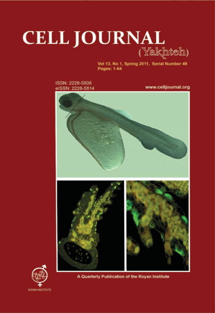
Page 45ObjectiveOn a global scale, stratospheric ozone depletion has caused an increase in UV-B radiation reaching the earth's surface. Ultraviolet radiation has long been suspected to be harmful to aquatic organisms.Materials And MethodsIn order to study ionocyte localization (by Na+/K+-ATPase immunolocalization) and the effects of UV radiation on the ionocytes of skin and gills, the alevins of Salmo trutta caspius were exposed to different doses of UV radiation [unit low doses (ULD) of: 60 µw/cm2 UVC; 100 µw/cm2 UVB and 40 µw/cm2 UVA and unit high doses (UHD) of: 90 µw/cm2 UVC; 130 µw/cm2 UVB and 50 µw/cm2 UVA] using two adjustable F8T5 UV-B, 302 nm lamps (Japan) for 15 minutes once a day in laboratory conditions. Alevins not subjected to UV exposure served as a control group.ResultsIn both UV exposure groups, all the alevins died on the ninth day. No mortality was observed in the control group. The Na+/K+-ATPase immunolocalization study indicated that ionocytes were located, in lessening order, on the yolk sac, trunk, gills, opercula and rarely on the head skin. Immunohistochemical results showed significant reduction in the number of ionocytes on the yolk sac, with lesser reduction on the trunk in both UV exposure groups. In contrast, the number of immunofluorescence cells on the gill was significantly elevated. Our results also showed that the size of ionocytes was reduced on the trunk and yolk sac in the UV exposure groups, but not significantly. Deformation and destruction of ionocytes on the yolk sac and trunk were observed with scanning electron microscope (SEM) in the UV exposure groups.ConclusionOur results showed that ionocytes were located mainly on the yolk sac, in lesser amounts on the trunk, gills and opercula, and rarely also on the head skin of alevins. UV radiation caused deformation and reduction in the number and size of ionocytes on the trunk and yolk sac. As the skin cells of trout alevins possess essential functions for respiration, osmoregulation, excretion and defense during this stage of life, the observed damage may have contributed to their suddenly mortality in the UV exposure condition.
-
Page 55ObjectiveTo determine the frequency of DYT1 mutation in Iranian patients affected with primary dystonia.Materials And MethodsIn this study, we investigated 60 patients with primary dystonia who referred to the Tehran Medical Genetics Laboratory (TMGL) to determine the deletional mutation of 904-906 del GAG in the DYT1 gene. DNA extracted from patients’ peripheral blood was subjected to PCR-sequencing for exon 5 of the DYT1 gene. The collection of samples was based on random sampling.ResultsThe deletional mutation of 904-906 del GAG in the DYT1 gene (15099 to 15101 based on reference sequence: NG_008049.1) was identified in 11 patients (18.33%). The average age of affected patients with this mutation was 13.64 ± 7.4 years.ConclusionIt can be concluded that the DYT1 deletional mutation of 904-906 del GAG has a high frequency in Iranian patients in comparison with other non-Jewish populations. Therefore, this particular mutation may be the main representative of pathogenic DYT1 gene for a large proportion of Iranian patients with primary dystonia.
-
Color PhotographsPage 59


