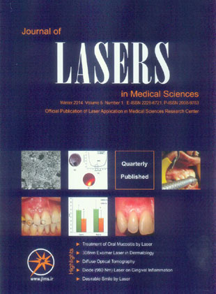
فهرست مطالب

Journal of Lasers in Medical Sciences
Volume:5 Issue: 1, Winter 2014
- تاریخ انتشار: 1392/10/09
- تعداد عناوین: 8
-
-
Pages 1-7IntroductionOral mucositis is considered a severe complication in cancer patients receiving radiotherapy or chemotherapy for head and neck cancer. The aim of this review study was to assess the effect of low level laser therapy for prevention and management of oral mucositis in cancer patients.MethodsThe electronic databases searched included Pubmed, ISI Web of Knowledge and Google scholar with keywords as “oral mucositis”, “low level laser therapy” from 2000 to 2013.ResultsThe results of most studies showed that photobiomodulation (PBM) reduced the severity of mucositis. Also, it can delay the appearance of severe mucositis.ConclusionLow level laser therapy is a safe approach for management and prevention of oral mucositis.Keywords: low level laser therapy, mucositis, prevention, management
-
Pages 8-12308nm xenon-chloride excimer laser, a novel mode of phototherapy, is an ultraviolet B radiation system consisting of a noble gas and halide. The aim of this systematic review was to investigate the literature and summarize all the experiments, clinical trials and case reports on 308-nm excimer laser in dermatological disorders. 308-nm excimer laser has currently a verified efficacy in treating skin conditions such as vitiligo, psoriasis, atopic dermatitis, alopecia areata, allergic rhinitis, folliculitis, granuloma annulare, lichen planus, mycosis fungoides, palmoplantar pustulosis, pityriasis alba, CD30+ lymphoproliferative disorder, leukoderma, prurigo nodularis, localized scleroderma and genital lichen sclerosus. Although the 308-nm excimer laser appears to act as a promising treatment modality in dermatology, further large-scale studies should be undertaken in order to fully affirm its safety profile considering the potential risk, however minimal, of malignancy, it may impose.Keywords: excimer, laser, dermatology
-
Pages 13-18IntroductionIn this study, we intend to use diffuse optical Tomography (DOT) as a noninvasive, safe and low cost technique that can be considered as a functional imaging method and mention the importance of image reconstruction in accuracy and procession of image. One of the most important and fastest methods in image reconstruction is the boundary element method (BEM). This method is introduced and employed in our works.MethodGenerally, to image a biological tissue we must obtain its optical properties. In order to reach this goal we benefit from diffusion equation because tissue is highly scattering medium. Diffusion equation is solved by boundary element equation (BEM) in our research. First, we assume a double layer phantom with different scattering and absorption coefficients to simulate and verify precession and accuracy of image reconstruction by BEM. Light absorption can be affected by volume fraction of blood in skin. For a specific skin species the volume fraction is calculated and then the results are compared with the reconstructed values obtained by BEM. Since the depth of tissue is important in light absorption a two layer phantom with known values is made and the depths of layers are reconstructed by BEM then they are compared with the expected values. A homogenous phantom with known scattering and absorption coefficients was made and then these coefficients were reconstructed by BEM. Finally, an inhomogeneous phantom (phantom with defect) whose defect was in a known position was made and the absorption and scattering coefficients were reconstructed and compared with real values.ResultsComparison between real or simulated values and reconstructed values of scattering and absorption coefficients, volume fraction of blood and thickness of phantom layers by BEM shows maximum errors of 24%, 7% and 35%, respectively.ConclusionComparison between BEM data and real or simulated values shows an acceptable agreement. Consequently, we can rely on BEM as a beneficial method in diffuse optical tomography image reconstruction.Keywords: optical tomography, phantom, laser
-
Pages 19-26IntroductionInfecting microorganisms of the root canals are difficult to eliminate during endodontic treatment. In this study the effect of root canal disinfection with photodynamic therapy (PDT) at different time intervals in comparison to 2.5% sodium hypochlorite (NaOCl) irrigation and passive ultrasonic irrigation (PUI) in extracted teeth colonized with Enterococcus faecalis and Candida albicans was tested to assess which treatment reaches the best disinfection rate.MethodsOne hundred and fifty-six extracted single-rooted teeth were collected, sterilized, and incubated with Enterococcus faecalis (ATCC 29212) and Candida albicans (ATCC 60193). The two groups were further divided into 6 groups depending on the treatment mode; HELBO®Endo Blue photosensitizer dye application followed by HELBO laser irradiation, with the output power 100 mW and emission of 660 nm, for a 1, 3 and 5 minutes, irrigation with 2.5% NaOCl, 10 second PUI with 2.5% NaOCl and control group. Flow cytometry and scanning electron microscopic (SEM) analysis were used to determine the effectiveness of the different disinfecting methods.ResultsThe different disinfecting methods had a significantly different effect on the percent of dead cells (p<0.001). A statistical significance of dead cells between organisms (p<0.001) was observed. Interaction between the disinfecting method and both of organisms had shown the statistical significance (p=0.045). Percent of dead cells in treatment groups were significantly higher compared to control group for both organisms (p<0.001).ConclusionsPUI still remains the most effective method for disinfection of infected root canalsin endodontics compared to hand instrumentation for both microorganisms. SEM analysis only confirmed the results. Other results ex vivo suggested that prolonging the time from 1 to 5 minutes of PDT increased the number of killed microorganisms significantly, therefore longer times of photodynamic therapy were recommended. Irrigation with 2.5% NaOCl showed similar results to 5 min irradiation.Keywords: enterococcus faecalis, Candida albicans, infection, root canal
-
Pages 27-31IntroductionPeriodontitis is an inflammatory disease, for which, scaling and root planning (SRP) is the common approach for non-surgical control of inflammation. Using lasers is another approach in the first phase of periodontal treatment for control of inflammation. Diode laser has some beneficial effects such as acceleration of wound healing, promotion of angiogenesis and augmentation of growth factor release. Thus the aim of this study is the evaluation of diode laser (980 nm) effect on gingival inflammation when it is used between the first and second phase of periodontal treatment, in comparison with common treatment (SRP) modality alone.MethodsIn this study, 21 patients with moderate to severe chronic periodontitis were selected and divided in to control group (SRP) and test group (SRP + laser). Two months after the last scaling and laser radiation, indexes including gingival level (GL), bleeding on probing (BOP) and modified gingival index (MGI) were recorded and compared with baseline.ResultsTwo months after the beginning of the study, all indices improved in both groups. The indices were not different between two groups except for BOP which was lower in laser group.ConclusionBased on overall improvement in parameters such as superiority of laser application in some indices, lack of thermal damage and gingival recession with the specific settings used in this study, the application of laser as an adjunctive treatment together with common methods is preferable.Keywords: periodontitis, diode Laser, gingival
-
Pages 32-38IntroductionThe aim of this study was to evaluate the morphological changes and quantitatively assess the roughness of dentin after the ablation with a Ytterbium-Doped Potassium Yttrium Tungstate (YB: KYW) thin-disk femtosecond pulsed laser of different fluences, scanning speeds and scanning distances.MethodTwelve extracted human premolars were sectioned into crowns and roots along the cementum-enamel junction, and then the crowns were cut longitudinally into sheets about 1.5 mm thick with a cutting machine. The dentin samples were fixed on a stage at focus plane. The laser beam was irradiated onto the samples through a galvanometric scanning system, so rectangular movement could be achieved. After ablation, the samples were examined with a scanning electron microscope and laser three-dimensional profile measurement microscope for morphology and roughness study.With increasing laser fluence, dentin samples exhibited more melting and resolidification of dentin as well as debris-like structure and occluded parts of dentinal tubules.ResultsWhen at the scanning speed of 2400mm/s and scanning distance of 24μm, the surface roughness of dentin ablated with femtosecond pulsed laser decreased significantly and varied between values of dentin surface roughness grinded with two kinds of diamond burs with different grits. When at the scanning speed of 1200mm/s and scanning distance of 12μm, the surface roughness decreased slightly, and the surface roughness of dentin ablated with femtosecond pulsed laser was almost equal to that grinded with a low grit diamond bur.ConclusionThis study showed that increased laser influence may lead to more collateral damage and lower dentin surface roughness, while scanning speed and scanning distance were also negatively correlated with surface roughness. Adequate parameters should be chosen to achieve therapeutic benefits, and different parameters can result in diverse ablation results.Keywords: laser, dentin, morphology
-
Pages 39-46IntroductionTo study the effects of Polarized Polychromatic Noncoherent Light (Bioptron) therapy on patients with carpal tunnel syndrome (CTS).MethodsThis study was designed as a randomized clinical trial. Forty four patients with mild or moderate CTS (confirmed by clinical and electrodiagnostic studies) were assigned randomly into two groups (intervention and control goups). At the beginning of the study, both groups received wrist splinting for 8 weeks. Bioptron light was applied for the intervention group (eight sessions, for 3/weeks). Bioptron was applied perpendicularly to the wrist from a 10 centimeters distance. Pain severity and electrodiagnostic measurements were compared from before to 8 weeks after initiating each treatment.ResultsEight weeks after starting the treatments, the mean of pain severity based on Visual Analogue Scale (VAS) scores decreased significantly in both groups. Median Sensory Nerve Action Potential (SNAP) latency decreased significantly in both groups. However, other electrophysiological findings (median Compound Motor Action Potential (CMAP) latency and amplitude, also SNAP amplitude) did not change after the therapy in both groups. There was no meaningful difference between two groups regarding the changes in the pain severity.ConclusionBioptron with the above mentioned parameters led to therapeutic effects equal to splinting alone in patients with carpal tunnel syndrome. However, applying Bioptron with different therapeutic protocols and light parameters other than used in this study, perhaps longer duration of therapy and long term assessment may reveal different results favoring Bioptron therapy.Keywords: syndrome, carpal tunnel, noncoherent light, electrodiagnostic study
-
Pages 47-50IntroductionProviding desirable smile is one of the main concerns in cosmetic dentistry. Hyperpigmentation is one of the esthetic concerns especially in gummy smile patients. Lasers with different wavelength are used for oral surgery including Carbon Dioxide Laser (CO2), Neodymium-Doped Yttrium Aluminium Garnet (Nd:YAG), Erbium family and diode laser. In this case, all esthetic procedures including gingival depigmentation, caries detection and removal were done by laser technology in one session.Case study: A 40- year-old male with a chief complaint of black gingiva in upper jaw was referred. The right side of maxillary was anesthetized and depigmentation was done by Erbium, Chromium doped Yttrium Scandium Gallium Garnet (Er-Cr: YSGG) laser. Due to scores obtained from Diagnodent which indicated caries in dentin, the cavities were prepared by Er-Cr:YSGG laser. The cavities were restored by composite resin. The patient was advised to keep oral hygiene instructions and use mouthwash.ResultsThe patient reported no pain after surgery and did not use any systemic antibiotic. After 4 weeks, complete healing was observed.ConclusionConsidering acceptable clinical outcome, Er-Cr: YSGG laser can be considered as an effective method for combination of soft and hard tissue treatment.Keywords: gingiva, laser, YSGG Laser

