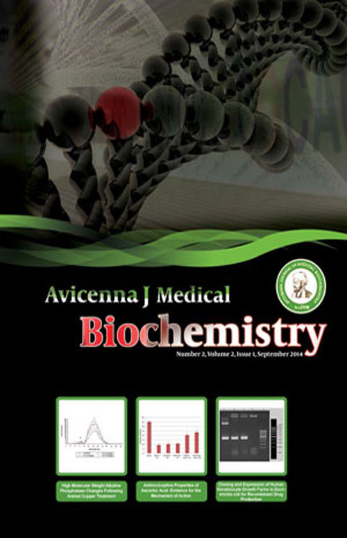
فهرست مطالب

Avicenna Journal of Medical Biochemistry
Volume:2 Issue: 1, Sep 2014
- تاریخ انتشار: 1394/04/30
- تعداد عناوین: 8
-
-
Page 2BackgroundMalathion is an insecticide of the grouping of organophosphate pesticides (OPs), which shows strong insecticidal effects. In addition, vitamin E reacting to cell membrane site may prevent OP-induced oxidative injury.ObjectivesThe aim of this study was to examine the protective function of vitamin E on toxicity of malathion, by measuring the activities of liver and liver mitochondrial superoxide dismutase (SOD), catalase (CAT),lipid peroxidation (LPO),and glutathione peroxidase (GPx) in rats.Materials And MethodsThe mitochondrial viability was determined in liver. Effective doses of malathion(200 mg/kg/day) and vitamin E (alpha-tocopherylacetate [AT]; 15 mg/kg/day) were administered alone or in combination for 14 days. At the end of the experiment, the liver tissue and liver mitochondria of the animals were harvested and examined.ResultsIn liver tissue, the activity of LPO and CAT was higher in the malathion group in comparison to controls. AT reduced malathion-induced LPO, SOD, CAT, and GPx in rat liver. Coadministration of AT with malathion improved LPO, SOD, and CAT levels in liver as well as CAT and GPx in liver mitochondria. Malathion-induced mitochondria toxicity was recovered by AT.ConclusionsIn conclusion, AT measurement can be beneficial for the safety or recovery of malathion-induced toxic injury in liver tissue and liver mitochondria.Keywords: Malathion, Mitochondria, Liver, Oxidative Stress, Rats, Vitamin E
-
Page 3BackgroundAscorbic acid is amongst important water-soluble vitamins and when used orally in high-doses it has been observed to relieve pain and reduce opioid use in patients. However no controlled trial has compared the antinociceptive effects of ascorbic acid with other analgesic groups on animal models, and investigated the involved mechanisms.ObjectivesIn the present study, the antinociceptive effect of vitamin C on male mice was investigated and compared with morphine and diclofenac. Also, possible mechanisms were assayed.Materials And MethodsMale albino mice were used in this study. Antinociception was measured using the writhing test, tail flick and formalin tests. Ascorbic acid was used in three doses (30, 150 and 300 mg/kg, IP) and compared with the antinociceptive effects of 10 mg/kg of morphine as an opioid analgesic agent and 5-10 mg/kg of diclofenac as a nonsteroidal anti-inflammatory drug (NSAID) analgesic agent.The antinociceptive effect of ascorbic acid (300 mg/kg) was compared before and after treatment with naloxone (4 mg/kg), ondansetron (0.5 mg/kg), atropine (5 mg/kg) and metoclopramide (1 mg/kg) in the writhing test.ResultsVitamin C caused dose-dependent antinociceptive effects in acetic acid writhing test (P < 0.05). It had no significant effect in the tail flick test. Meanwhile, vitamin C in high doses reduced pain in the second phase of the formalin test (P < 0.05). Morphine had higher nociceptive effects in comparison to ascorbic acid in the writhing test (P < 0.05). In the second phase of the formalin test the antinociceptive effects of vitamin C (300 mg/kg) was not significantly different with morphine at dose of 10 mg/kg. There was not significant difference between vitamin C (300 mg/kg) and diclofenac (10 mg/kg) in the second phase of the formalin test. Metoclopramide and ondansetrone reduced the antinociceptive effects of vitamin C.ConclusionsThe results obtained from the acetic acid induced writhing test and second phase of the formalin test indicate that vitamin C possess antinociceptive activity especially on inflammatory pain.Ondansetrone and metoclopramide reduced the effects of ascorbic acid, which may be because ascorbic acid produced antinociception through mechanisms that may be involved in dopaminergic and serotoninergic systems.Keywords: Ascorbic Acid, Nociception, Diclofenac, Morphine, Dopaminergic, Cholinergic Agents
-
Page 4BackgroundAlthough trace amounts of copper (Cu) are necessary to maintain proper body functions, the excess amount can contribute to the development of hepatic dysfunction.ObjectivesThis study aimed to investigate the relationship between copper treatment and changes in the serum concentration of high molecular weight alkaline phosphatase (HMW-ALP).Materials And MethodsMale Wistar rats were injected intraperitoneally (IP) with copper (Cu) as copper chloride (CuCl2. 4H2O) 4, 2 and 1 mg/kg for 10, 30 and 60 days respectively. Animals were killed at indicated time and blood samples were collected, and sera was separated and used for alkaline phosphatase activity determinations and also for isoenzymes gel filtration chromatography and Sephacryl S-300 was used.ResultsObtained data showed that with increasing administration of copper, the ALP activity was elevated significantly. In comparison with the control group the elevations were between 20%-56% using gel filtration chromatography. It was found that the elevation of serum ALP was mostly due to HMW-ALP.ConclusionsThe elevation of HMW-ALP activity in Cu treated animal suggests the occurrence of biliary disease. This may be used as a biomarker for the diagnosis of copper toxicity.Keywords: Alkaline Phosphatase, Copper, Liver
-
Page 5BackgroundKeratinocyte growth factor (KGF) is a member of fibroblast growth factor (FGF) family which induces proliferation and differentiation in a wide variety of epithelial tissues. KGF plays an important role in protection, repair of various types of epithelial cells, and re-epithelialization of wounds. Therefore, in patients with hematologic malignancies receiving high doses of chemotherapy and radiotherapy, treatment with KGF decreases the incidence and duration of severe oral mucositis.ObjectivesThe aim of this study was to express the recombinant form of human keratinocyte growth factor in Escherichia coli.Materials And MethodsKGF gene was amplified by PCR and cloned into the expression vector pET28a(+). The recombinant vectors were transformed into E. coli BL21(DE3) as expression host and expression of the desired protein was induced by IPTG. The expression was evaluated at RNA and protein levels by reverse transcriptase PCR (RT-PCR) and SDS-PAGE analyses, respectively and the expressed protein was confirmed through western blotting.ResultsCloning was confirmed by PCR and restriction digestion. RT-PCR and SDS-PAGE represented expression of KGF in E. coli. The optimized expression was achieved 16 hours after induction with 0.3 mM IPTG at 37°C in luria broth (LB) containing kanamycin. The 18 kDa protein was confirmed by western blotting, using anti-His antibodies.ConclusionsThe result of the present study indicated that E. coli expression system was suitable for overexpression of recombinant human KGF and the expressed protein can be considered as a homemade product.Keywords: Cloning, Recombinant Protein, Keratinocyte Growth Factor
-
Page 6BackgroundThe malonyl-CoA decarboxylase (MCD, EC.4.1.1.9) enzyme regulates malonyl-CoA levels. The effect of aerial parts extracts of Urtica dioica (UD) on MCD is poorly understood.ObjectivesThe present experiment was undertaken to evaluate the effect of UD aerial parts extracts on MCD level.Materials And MethodsIn this experimental study, two groups of rats were used: normal and hyperglycemic group. Then UD aerial parts extracts (5 mg /500 µL) administrated to the hyperglycemic group of rats and finally, the MCD and insulin levels were measured in both groups.ResultsInterestingly, we observed that the UD aerial parts extracts powder caused a significant (P < 0.05) increase in insulin level during the experiment, from the base level of 0.36 ± 0.07 μg/L to the peak value of 0.52 ± 0.15 μg/L. Also, it caused a significant (P < 0.05) decrease in MCD level, from the base level of 29.68 ±1.29 pg/mL to the bottom value of 22.12 ± 2.41 pg/mL.ConclusionsThe results of the present study indicate that UD aerial part extracts would decrease MCD level in hyperglycemic rats.Keywords: Urtica dioica_Malonyl Coenzyme A Decarboxylase_Streptozotocin
-
Page 7BackgroundEdible salt is the most commonly used food additive worldwide. Therefore, any contamination of table salt could be a health hazard.ObjectivesThe present study aimed to determine the levels of heavy metals in table and bakery refined salts.Materials And MethodsEighty-one table refined salt samples and the same number of bakery refined salt samples were purchased from retail market in the province of Hamadan, Iran. The levels of lead (Pb), cadmium (Cd), mercury (Hg), copper (Cu), and iron (Fe) were determined using atomic absorption spectroscopy method.ResultsThe levels (mean ± SD, μg/g) of Pb, Cd, Hg, Cu, Fe in table refined salt samples were 0.852 ± 0.277, 0.229 ± 0.012, 0.054 ± 0.040, 1.25 ± 0.245 and 0.689 ± 1.58, respectively. The results for the same metals in bakery refined salt samples were as follows (mean ± SD, μg/g): 22 ± 0.320 for Pb, 0.240 ± 0.018 for Cd, 0.058 ± 0.007 for Hg, 1.89 ± 0.218 for Cu, and 8.75 ± 2.10 for Fe. Heavy metal concentrations were generally higher in bakery refined salt.ConclusionsThe results obtained in the present study were compared with the literature and legal limits. All values for these metals in the table and bakery refined salts were lower than the permitted consumption level defined by Codex (2 µg/g of Pb, 0.5 µg/g of Cd, 0.1 µg/g of Hg, and 2 µg/g of Cu).Keywords: mium, Lead, Heavy Metals, Mercury, Sodium Chloride
-
Page 8BackgroundLipids play an important role in the functional activity of sperm cells.ObjectivesThe main goal of this study was to assess the correlation between the levels of cholesterol, phospholipids and triacylglycerols found in serum, with the lipid levels of semen in infertile men.Patients andMethodsCholesterol, phospholipids and triacylglycerols in sperm cells, seminal plasma and serum were assayed in 60 infertile men.ResultsThere were no significant relationships between the concentration of sperm and seminal plasma cholesterol with serum cholesterol (r = 0.003, P = 0.9 and r = 0.055, P = 0.67, respectively), between the concentration of sperm and seminal plasma triglycerides with serum triglycerides (r = 0.16, P = 0.2 and r = - 0.039, P = 0.77, respectively), or between the concentration of sperm and seminal plasma phospholipids with serum phospholipids (r = 0.18, P = 0.16 and r = 0.053, P = 0.69, respectively).ConclusionsThese results suggest that serum cholesterol, phospholipids and triacylglycerols have no effect on the levels of cholesterol, phospholipids and triacylglycerols of spermatozoa and seminal plasma. Our findings suggest that sperm lipid content is regulated locally within the male reproductive tract.Keywords: Cholesterol, Lipids, Phospholipid, Serum, Semen Analysis, Spermatozoa

