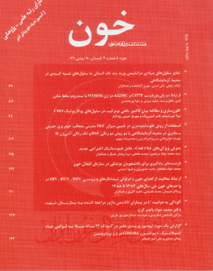
فهرست مطالب

فصلنامه خون
سال هشتم شماره 2 (پیاپی 31، تابستان 1390)
- تاریخ انتشار: 1390/04/04
- تعداد عناوین: 9
-
-
صفحه 79سابقه و هدف سلول های بنیادی مزانشیمی جدار ورید بند ناف انسانی، به عنوان سلول های چند توان، اخیرا جداسازی شده و پتانسیل تمایزی آن ها به سلول های مختلفی نظیر سلول های عصبی و قلبی بررسی شده است. در این مطالعه، توانایی سلول های بنیادی مزانشیمی ورید بند ناف در تمایز به سلول های کبدی با تمرکز بر پروفایل بیان ژن های اختصاصی کبد ارزیابی شد. مواد و روش ها در یک مطالعه تجربی، سلول های بنیادی مزانشیمی جدار ورید بند ناف انسان پس از جداسازی به مدت یک ماه با استفاده از یک روش دو مرحله ای تحت تیمار با فاکتورهای القایی نظیر فاکتور رشد کبدی و انکوستاتین M قرار گرفتند. سپس بیان ژن های اختصاصی سلول های کبدی از طریق روش RT-PCR و ایمونوسیتوشیمی مورد بررسی قرار گرفت. یافته ها پس از تیمار سلول ها با فاکتور های القایی، تغییر شکل سلول ها از حالت فیبروبلاستی و دو قطبی به فرم اپیتلیال و چند وجهی مشاهده گردید. بیان یا افزایش بیان ژن های اختصاصی سلول های کبدی نظیر CK8، CK18، HNF3β، c-Met، AFP، TTR، G6P، Transthyretin و ALB در طول دوره القایی تایید شد. هم چنین واکنش سلول ها به آنتی بادی بر علیه آنتی ژن سیتوکراتین 18 در سلول های تمایز یافته مثبت بود. نتیجه گیری با توجه به داده های کسب شده، می توان نتیجه گرفت که سلول های بنیادی مزانشیمی مشتق از جدار ورید بند ناف، توانایی تمایز به سلول های شبه کبدی را دارا می باشند.
کلیدواژگان: وریدهای بند ناف، سلول های بنیادی مزانشیمی، فاکتور رشد کبدی، انکوستاتین M -
صفحه 88سابقه و هدف یکی از فاکتورهای مطرح در ایجاد ترومبوفیلی در زنان مبتلا به سقط مکرر، پلی مورفیسم های A1298C و C677T در ژن MTHFR است. هدف از این تحقیق، بررسی ارتباط این دو پلی مورفیسم با سندروم سقط مکرر به عنوان یکی از عوامل خطر ژنتیکی برای این سندروم بود.
مواد و روش ها در یک مطالعه مورد شاهدی از میان مراجعین بیمارستان بقیه اله و مرکز ناباروری ابن سینا، 30 زن با سابقه سقط مکرر خود به خود با علت نامشخص به عنوان گروه بیمار و 10 زن بدون سابقه سقط مکرر و دارای حداقل دو باروری موفق، به عنوان گروه شاهد انتخاب شدند. دو پلی مورفیسم ژن MTHFR با واکنش زنجیره ای پلی مراز و هضم آنزیمی محصولات PCR با آنزیم های اندونوکلئاز محدودالاثر (PCR-RFLP) بررسی شدند. نتایج به دست آمده از تعیین ژنوتیپ هر پلی مورفیسم با نرم افزار 16 SPSS، دو آزمون χ2 و همبستگی اسپیرمن تجزیه و تحلیل شدند.
یافته ها بین دو پلی مورفیسم C677T و A1298C ارتباط متقابل وجود داشت. 17 نفر از گروه مورد(6/56%) و 5 نفر از گروه شاهد(50%)، برای پلی مورفیسم C677T هتروزیگوت بودند. فراوانی آلل موتانت T در زنان دچار سقط، بیشتر از زنان گروه شاهد بود(4/28% در زنان دچار سقط مکرر و 25% در زنان گروه شاهد، 05/0 p<). فراوانی پلی مورفیسم A1298C در میان زنان دچار سقط مکرر و زنان گروه شاهد به ترتیب 3/63% و 50% بود.
نتیجه گیری نتایج به دست آمده در این پژوهش نشان داد که هیچ کدام از دو پلی مورفیسم MTHFR، نمی توانند توجیه کننده علت سقط مکرر در زنان مورد بررسی محسوب گردند.
کلیدواژگان: متیلن تتراهیدروفولات ردوکتاز، ترومبوفیلی، سقط خود به خود، پلی مورفیسم (ژنتیک) -
صفحه 96سابقه و هدف فاکتور بافتی(TF)، اصلی ترین آغازکننده آبشار انعقادی است. برای بررسی مسیر خارجی انعقاد، اندازه گیری زمان پروترومبین بهترین روش است که در این روش با توجه به TF به کار گرفته شده، نوساناتی در نتیجه آزمایش دیده می شود. در این مطالعه با هدف دستیابی به نتایجی با تکرارپذیری بیشتر، از TF نوترکیب استفاده شده است.
مواد و روش ها مطالعه انجام شده از نوع تجربی بود. cDNA فاکتور بافتی در پلاسمید pcDNA3.1 کلون و به سلول های CHO منتقل گردید. پروتئین های سلول های CHO ترانسفکت شده استخراج و پس از جداسازی با روش SDS-PAGE با وسترن بلات آنالیز شدند. سلول های CHO که TF را بیان می کردند در حضور کلروکلسیم به پلاسمای سیتراته افزوده شدند و زمان لخته شدن پلاسما ثبت گردید. فعال سازی فاکتور VII در پلاسما با روش الایزا مورد بررسی قرار گرفت.
یافته ها در آنالیز پروتئین های جدا شده از سلول های ترانسفکت شده با روش SDS-PAGE، باندی حدود 40 کیلودالتون مشاهده شد که با آنتی بادی مونوکلونال TF در آنالیز وسترن بلات مورد تایید قرار گرفت. زمان انعقاد پلاسما در مقایسه با سلول های CHO ترانسفکت نشده، از 3 دقیقه به 3 ± 21 ثانیه کاهش یافت. هم چنین اضافه کردن غشای این سلول ها به پلاسما در حضور کلرور کلسیم، موجب فعال سازی فاکتور VII به میزان ng/ml 35 ±810 شد.
نتیجه گیری TF نوترکیب بیان شده در سلول های CHO، قادر به لخته کردن پلاسمای سیتراته و فعال سازی فاکتور VII بود.
کلیدواژگان: سلول های CHO، فاکتور VII، زمان پروترومبین -
صفحه 104سابقه و هدف فلوسایتومتری روشی مناسب، سریع، دقیق و قابل اعتماد جهت ارزیابی میزان خونریزی های جنینی مادری (FMH) می باشد. هدف از این مطالعه، بررسی میزان FMH با استفاده از شاخص آنتی ژنیک RhD وهموگلوبین جنینی(HbF) به روش فلوسایتومتری است.
مواد و روش ها در یک مطالعه تجربی، 34 نمونه خون بند ناف نوزاد Rh مثبت با خون فرد بالغ RhD منفی به صورت رقیق سازی سریالی در 6 رقت(125/0، 25/0، 5/0، 1، 5 و 10 درصد) که بیانگر 9 / 99 درصد احتمال FMH بود، شبیه سازی شد و به روش فلوسایتومتری دو رنگی HbF و کربونیک انهیدراز (CA) وتک رنگی RhD مورد مطالعه قرار گرفت. از آزمون های آماری T test، آنالیز واریانس و رگرسیون جهت تحلیل نتایج استفاده شد.
یافته ها همبستگی قابل قبولی بین نتایج HbF و RhD به روش فلوسایتومتری مشاهده شد(897/0 r=). میزان خونریزی محاسبه شده توسط هر دو پارامتر HbF و RhD در مقایسه با مقادیر خونریزی مورد انتظار، نشان داد که آنالیز دو رنگی HbF و CA ازحساسیت بالاتری نسبت به آنالیز تک رنگی RhD در تعیین مقادیر اندک خونریزی برخوردار می باشد. نتایج روش RhD با وجود همبستگی بالا(984/0 r=)، در تعیین مقادیر پایین FMH در مقایسه با میزان خونریزی به دست آمده از روش HbF/CA، افزایش کاذبی را نشان داد ولی توانایی آن درتعیین مقادیر بالاتر خونریزی در مقایسه با HbF/CA قابل توجه بود.
نتیجه گیری استفاده از مونوکلونال آنتی بادی های اختصاصی در روش فلوسایتومتری، امکان ارزیابی دقیق میزان خونریزی و به تبع آن تعیین دقیق دوز ایمونوگلبولین Rh جهت محافظت مادران از آلو ایمونیزاسیون بر علیه آنتی ژن D را ایجاد می کند.
کلیدواژگان: خونریزی، انتقال خون جنینی، مادری، فلوسایتومتری -
صفحه 115سابقه و هدف خونریزی کنترل نشده، عامل مرگ و میر در صدمات نظامی و در حوادث غیر نظامی است. توقف غیر فشاری خونریزی از جمله اقداماتی است که امکان زنده ماندن افراد مجروح را افزایش می دهد. هدف از این مطالعه، بررسی کارآیی Cool Clot در کنترل خونریزی های شریانی و کاهش زمان تشکیل لخته بود.
مواد و روش ها در یک مطالعه تجربی، سه قلاده سگ هیبریدی انتخاب گردیده و پونکسیون شریانی به وسیله سوزن و یا آرتریوتومی در شریان های فمورال حیوان انجام شد. در شریان های کنترل، تنها از دستور عمل 4 دقیقه فشار بر روی موضع با گاز استریل و در سمت مقابل از عامل هموستاتیک Cool Clot استفاده شد. برای اندازه گیری زمان تشکیل لخته، از 10 دانشجوی داوطلب، نمونه گیری خون صورت گرفت. جهت تحلیل نتایج از آزمون های ANOVA و دانت و نرم افزار 15 SPSS استفاده شد.
یافته ها در مدل حیوانی سگ، مدت زمان توقف خونریزی در شریان تیمار شده با روش های معمول(بدون عامل هموستاتیک) بیش از سه برابر شریان تیمار شده با عامل هموستاتیک Cool Clot بود. در فاز بعدی مطالعه و در نمونه های خون انسانی، میانگین زمان تشکیل لخته در نمونه های خون کنترل و نمونه های خون تیمار شده با بنتونیت، زئولیت و عامل انعقادی Cool Clot به ترتیب معادل 1/44 ± 4/253، 0/50 ± 5/149، 5/74 ± 3/162 و 6/114 ± 4/143 ثانیه بود(05/0 p<).
نتیجه گیری نتایج به دست آمده نشان دهنده تاثیر عامل انعقادی Cool Clot در کاهش دادن زمان لازم برای توقف خونریزی در مدل حیوانی سگ و کاهش زمان تشکیل لخته در نمونه های خون انسانی است.
کلیدواژگان: خونریزی، هموستاز، بنتونیت، زئولیت -
صفحه 122سابقه و هدف نگرانی از عوارض ناشی از انتقال خون سبب گردیده تا به افزایش دانش در حوزه طب انتقال خون به عنوان راه کاری برای بهبود کیفیت مراقبت از بیمار و کاهش هزینه های سلامت توجه شود. این مطالعه قصد دارد با نشان دادن فرصت های یادگیری در سازمان انتقال خون برای دانشجویان پزشکی، این مراکز را به عنوان بخشی از عرصه آموزشی طب انتقال خون معرفی نماید.
مواد و روش ها در یک مطالعه نیمه تجربی، تعداد 14 دانشجوی پزشکی در یک دوره 6 روزه در سازمان انتقال خون اصفهان، فرصت های یادگیری طب انتقال خون را تجربه کردند. در پایان دوره، دانش دانشجویان طی آزمونی سنجیده شد و پس از شش ماه، ماندگاری اطلاعات آن ها اندازه گیری گردید. نمرات دانشجویان با استفاده از آزمون ویلکاکسون و استفاده از نرم افزار 18 SPSS مقایسه شد.
یافته ها میانگین نمرات دانشجویان در آزمون اول 2 ± 7/15(از 20) بود و در آزمون شش ماه بعد، میانگین نمرات به 2 ± 8/12 از بیست تقلیل یافت(001/0 p<). در رابطه با نظر سنجی انجام شده، 7/85% شرکت کنندگان این دوره را برای دانشجویان پزشکی دوره بالینی توصیه کرده و 4/71% آن ها در پایان دوره، خود را در مورد انتخاب فرآورده های خونی توانمند می دانستند.
نتیجه گیری آموزش طب انتقال خون باید به صورت همکاری گروه های آموزشی انجام گیرد و آموزش محیط های بالینی کافی نمی باشد. سازمان انتقال خون به عنوان مرکز تهیه و توزیع فرآورده های خونی می تواند فرصت های یادگیری ویژه ای برای دانشجویان پزشکی فراهم آورد.
کلیدواژگان: انتقال خون، آموزش پزشکی، یادگیری -
صفحه 130سابقه و هدف در سال های اخیر در مراکز انتقال خون برای انتخاب اهداکننده اقداماتی دقیق تر، چند مرحله ای با کنترل شدید صورت گرفته است. این مطالعه با هدف ارزیابی این شرایط، میزان بروز آلودگی در واحدهای خون اهدایی را در ارتباط با معافیت های اهداکنندگان با علت رفتارهای پرخطر مورد بررسی قرار داد.
مواد و روش ها در یک مطالعه توصیفی تحلیلی، 542705 واحد خون اهدا شده از مرداد 1383 تا پایان سال 1388 در انتقال خون اصفهان با روش سرشماری مورد بررسی قرار گرفت. درصد فراوانی معافیت های ناشی از خطر انتقال بیماری های ویروسی هپاتیت و ایدز و هم چنین درصد فراوانی نشانگرهای HBs Ag، HCVAb HIV Ab، با روش های تاییدی در واحدهای خون اهدایی محاسبه و ارتباط آن ها با استفاده از برنامه نرم افزاری نگاره و 17 SPSS تجزیه و تحلیل شدند.
یافته ها درصد فراوانی نشانگرها در مراجعه کنندگان به انتقال خون از 83 تا 88 به ترتیب54/0، 45/0، 34/0، 25/0، 22/0 و 22/0 درصد بود. درصد فراوانی معافیت های مرتبط با خطر انتقال بیماری های ویروسی در کل مراجعه کنندگان نیز طی همین سال ها به ترتیب 69/3، 71/4، 29/5، 19/5، 93/3 و 04/4 درصد بود. درصد فراوانی معافیت ها رابطه ضعیف و معکوسی با درصد فراوانی نشانگرهای ویروسی هپاتیت B (78/0= p، 148/0- (r= و هپاتیت C (75/0= p، 165/0- (r= داشت.
نتیجه گیری اقدامات سازمان انتقال خون در سال های اخیر با کاهش نشانگرهای ویروسی و افزایش سلامت خون همراه بوده است ولی به علت احتمال مخفی کاری معدودی از اهداکنندگان، آموزش و انجام آزمایش رایگان و دادن اطلاعات پس از اهدا ممکن است کمک بیشتری به سلامت خون کند.
کلیدواژگان: اهداکنندگان خون، آنتی ژن های سطحی هپاتیت B، ایران -
صفحه 137سابقه و هدف هر چند غربالگری خون باعث کاهش بروز عفونت HCV شده است، این مشکل هنوز از علل مهم مرگ و میر و از کارافتادگی در بیماران تالاسمی می باشد.
مواد و روش ها طی یک مطالعه توصیفی در 206 بیمار تالاسمی مراجعه کننده به درمانگاه تالاسمی بیمارستان کرج، از فروردین 1388 لغایت فروردین 1389 به صورت غربالگری، Anti-HCV اندازه گیری شد. برای موارد مثبت، آزمایش RIBA-II انجام شد. اطلاعات توسط نرم افزار 18 SPSS و آزمون های کای دو و t تجزیه و تحلیل شدند.
یافته ها 31 بیمار(15 درصد) دارای آنتی بادی هپاتیت C بودند(11 مذکر و 20 مؤنث) که از این تعداد، در 29 نفر آزمایش RIBA نیز مثبت بود(11 مذکر و 18 مؤنث). یک بیمار تزریق خون بعد از سال 1375 (شروع غربالگری فرآورده های خونی از نظر HCV) داشت.
نتیجه گیری با توجه به شیوع بالای آلودگی به ویروس هپاتیت C در بیماران تالاسمی ماژور نسبت به جمعیت عمومی و نیز خطر ابتلای بیش از 80 درصد افراد آلوده به بیماری های مزمن کبدی و فیبروز، با اقدامات پیشگیرانه از جمله بررسی اهداکنندگان خون از نظر آلودگی ویروسی و پیگیری های مکرر بیماران، می توان قدم مهمی در شناسایی و درمان زودرس این بیماری و کنترل انتشار آن برداشت.
کلیدواژگان: هپاتیت C، تالاسمی، آنتی بادی های هپاتیت C -
صفحه 143سابقه و هدف ترومبوز یکی از عوارض غیر شایع در بیماران مبتلا به لوکمی حاد لنفوبلاستیک حین کموتراپی است که دارای شیوع متغیری می باشد.
مورد بیمار پسر بچه ای 12 ساله بود که در نیمه اول سال 1388 با تشخیص لوکمی حاد لنفوبلاستیک از نوع Pre B، تحت کموتراپی با دستور عمل BFM گروه پر خطر با پاسخ سریع قرار گرفت. در مرحله دوم درمان بر اساس دستور عمل فوق، بیمار دچار سردرد شد. بعد از بررسی های تشخیصی، ترومبوز وریدی مغز مطرح و اثبات گردید. به دنبال بررسی علل مستعد کننده، موتاسیون هتروزیگوت در ژن پروترومبین به شکل G20210A به اثبات رسید.
کلیدواژگان: لوکمی، ترمبوز وریدی، موتاسیون، پروترومبین
-
Page 79Background and ObjectivesUmbilical vein mesenchymal stem cells (UVMSC) which are multipotent precursors have been recently isolated. They are capable of differentiating into various cell types including neural and cardiac cells. In this study, the differentiation ability of human UVMSC into hepatocyte cells has investigated with concentration on hepatic genes expression profile. Materials and MethodsThe UVMSC were isolated and cultured in differentiation medium containing hepatocyte growth factor (HGF) and oncostatin M (OSM) using two step protocol. Differentiation of UVMSCs into cells expressing liver-specific genes was investigated by RT-PCR and immunocytochemistry.ResultsThe cells showed the remarkable transition from bipolar fibroblast-like morphology to epithelial and polygonal shapes. The temporal gene expression pattern of hepatocyte-specific genes such as CK8, CK18, HNF3β, c-Met, AFP, TTR, G6P, Transthyretin, and ALB were detected during differentiation. The immunoflourescent analysis also showed that the differentiated cells were stained positively for CK-18 protein. ConclusionsFindings indicate that UVMSC has a potential for differentiation into the hepatic like cells.
-
Page 88Background and ObjectivesTwo factors known to cause thrombophilia in women with unexplained recurrent spontaneous abortion (RSA) are MTHFR polymorphisms including C677T and A1298C. This study aimed to determine the association between RSA and two polymorphisms of MTHFR in Iranian patients.Materials and MethodsIn this case-control study, 30 patients with the background of two or more consecutive unexplained abortions and 10 women with at least two live births without a miscarriage who referred to Baqiyatallah Hospital and Avicenna Infertility Clinic were analyzed for MTHFR C677T and A1298C polymorphisms by the PCR-RFLP method. Results achieved from estimating the genotype of each polymorphism were analyzed by the SPSS16. The Spearman method was also used to evaluate the correlation between the two polymorphisms.ResultsData has shown a significant correlation between MTHFR C677T and MTHFR A1298C polymorphisms. Seventeen women (56.6%) with recurrent spontaneous abortions and 5 women (50%) among the controls were heterozygote for MTHFR C677T polymorphism. T allele frequency in the patient group was more than the control group (28.4% for patients and 25% for controls). Frequency of the MTHFR A1298C polymorphism was 63.3% in patients and 50% in controls. For A1298C polymorphism, 43.3% of patients and 20% of controls were heterozygote. Furthermore, 20% of patients and 30% of controls were homozygote for this polymorphism.ConclusionsThe prevalence of MTHFR C677T and MTHFR A1298C polymorphisms were slightly higher in RSA patients compared to controls. These findings failed to support the relationship between thrombophilia polymorphisms and the increasing risk of RSA in evaluated Iranian women.
-
Page 96Background and ObjectivesTissue factor (TF), a 45-kDa transmembrane glycoprotein, is the major cellular initiator of the coagulation cascade. Prothrombin Time (PT) is the test that evaluates extrinsic pathway of coagulation. Thromboplastin used in this test is mostly prepared of rabbit brain. Thus it causes variation in the PT results. There is an important advantage of using recombinant TF in PT test to get more reproducible results. Materials and MethodsTF mRNA was isolated from human lung fibroblast cells. After preparation of cDNA it has cloned in pcDNA3 plasmid. CHO cells were transfected with recombinant plasmid. Transfected cells were grown in presence of Geneticin. Total proteins were extracted from CHO cells and separated with SDS/PAGE electrophoresis and analyzed by western blotting technique. CHO cells expressing TF were added to citrated plasma in presence of Cacl2 and clotting time was measured. In addition, factor VII activation by recombinant TF in the plasma was assessed by ELISA method. ResultsExtracted proteins from tarnsfected CHO cells separated on SDS/PAGE showed an approximately 40-kDa band that verified with a monoclonal antibody against TF on western blot analysis. Adding transfected cells (106cell/ml) to the citrated plasma in presence of Cacl2 could decrease clotting time from 3 minutes to 21 ± 3 seconds compared with untransfected CHO cells. The level of factor VIIa in citrated plasma was 810 ± 35 ng/ml in presence of transfected cells’ membrane.ConclusionsRecombinant TF expressed in CHO cells was able to clot citrated plasma and to activate factor VII.
-
Page 104Background and ObjectivesThe quantification of fetal cells in the maternal circulation is an important goal to determine the amount of anti-D for prevention of active immunization of a D-negative mother giving birth to a D-positive baby. The aim of this study was to evaluate two flowcytometric staining technics for determination of fetal erythrocytes in maternal blood and to recognize the best.Materials and MethodsIn this experimental study, 34 adult D-negative blood samples were spiked with six serial dilutions of D-positive cord blood (0.125, 0.25, 0.5, 1, 5 and 10%) which was representative of 99.9% of the clinical fetomaternal hemorrhage (FMH); they were stained for flow cytometric analysis. The Fetal Cell Count Kit was used for HbF dual staining and monoclonal anti-D for RhD single staining.ResultsA comparison between RhD and HbF percentages and FMH volume in spiking samples was performed. A significant correlation between two different parameters in flowcytometric percentages was observed (anti-HbF versus anti-D, r= 0.897, p<0.05). FMH volume was calculated and a significant correlation between expected and observed FMHs in RhD and HbF was obtained (p<0.05, r=0.984, r=0.874). The anti-HbF flowcytometric dual staining allowed better distinction between fetal RBCs (HbF+,CA-), F cells (HbF+,CA+), and adult RBCs (HbF-,CA+). Although Rh-D showed better correlation, but higher values with the RhD in suspensions lower than 10% were noted.ConclusionsData showed that anti-HbF labeling is significantly much more accurate than RhD labeling. Quantification of FMH using these two techniques described allows precise dosage of RhD immunoglobulin for protection against anti-D allo-immunization.
-
Page 115Background and ObjectivesUncontrolled bleeding is the first leading cause of preventable death in the battlefield and the second cause of mortality in civil accidents. Incompressible hemorrhage control is among the interventions that drastically increase the survival rate of the wounded individuals. Materials and MethodsThree healthy hybrid dogs were selected and after induction of anesthesia with Ether, either arterial puncture by a needle or arteriotomy was performed on both groin regions of the dogs. For control arteries, only a 4 min pressure using a sterile gauze was performed, while for the femoral arteries of the opposite side, Cool Clot hemostatic agent was topically used before the pressure. To assess the Coagulation Time (CT), blood samples were collected from 10 volunteer students. CT was measured by a chronometer according to standard Lee -White method.ResultsIn dog animal model, the Bleeding Time (BT) in control arteries (no hemostatic agent) was more than 15 minutes, while in case of the opposite sides which were treated with Cool Clot hemostatic agent, the BT was only up to 5 minutes. In the human phase of the study, the mean coagulation time in control blood samples was 253.4 ± 4.1 sec, while for blood samples treated with bentonite, zeolite and Cool Clot the mean times were 149.5 ± 50.0, 162.3 ± 74.5 and 143.4 ± 114.6 sec, respectively.ConclusionsThe results obtained in this study indicate the significant efficacy of Cool Clot in shortening the BT in dog model or CT in human blood samples.
-
Page 122Background and ObjectivesThe concerns about blood transfusion have given prominence to the role knowledge improvement can play as a strategy to improve health care quality and reduce health cost. This study is aimed at defining the learning opportunities available in blood centers for medical students. Materials and MethodsIn this quasi-experimental research study, fourteen medical students participated in a five day course designed to introduce learning opportunities in blood centers. At the end of the course, participants’ knowledge was measured through a test; they were also retested after six months. Students’ marks were then compared by means of Wilcoxon “T” test. SPSS18 was used for data analysis.ResultsThe mean of students’ marks in the first exam was 15.7 +2 (out of 20) but in the second exam it declined to 12.8+2 (p< 0.001). However, 85.7% of participants recommended this course for other medical students and 71.4% believed they were competent enough in selecting appropriate blood products. ConclusionsTransfusion medicine should be educated through a multi-disciplinary manner and current clinical fields seem inadequate for this purpose. Iranian Blood Transfusion Organization can be considered as an effective center to provide learning opportunities for medical students.
-
Page 130Background and ObjectivesIn recent years we have been faced with more precise, multi-stage, and highly stringent blood donor screening criteria. The present study aimed at evaluating these criteria through comparing the rate of infection in blood donations with deferral rate for high-risk behaviors.Materials and MethodsThis descriptive analytic study was carried out in Esfahan Blood Transfusion Organization (EBTO) with census method through 2004 to 2009. Annual deferral rate due to high-risk behaviors and major viral markers were analyzed separately by using Pearson correlation. The data were obtained by Negareh software. ResultsThe viral markers frequencies were 54, 45, 34, 25, 22 and 22 percent in volunteers that referred to EBTO from 2004 to 2009. Overall deferral rates due to high-risk behaviors were 3.69, 4.71, 5.29, 5.19, 3.93, and 4.04 percent, respectively. It had a slightly negative correlation with HBV (r=0.148; p=0.78) and HCV (r=0.165; p=0.75) markers.ConclusionsData showed that recent procedures of Iranian Blood Transfusion Organization declined viral markers considerably. However, slightly negative correlation of deferral rate due to TTDs with hepatitis B and C markers makes it necessary for donor selection criteria to be reviewed. More appropriate counselling procedures and post-donation counselling can help improve blood safety.
-
Page 137Background and Objectives Although recent screening policies have markedly decreased the incidence of HCV infection in transfusion dependent thalassemia paitients, this problem is still one of the most important causes of mortality and morbidity in thalassemic patients.Materials and Methods A retrospective cross-sectional study was conducted on 206 thalassemia patients referred to the Shahid Bahonar Hospital during 2009-2010. Serum specimens were tested with anti –HCV assay and RIBA –II technique to asses HCV infection. Statistical analysis was done with SPSS18 (χ2 and T-test). Results The overall prevalence rate of anti-HCV was 15%)11 male, 20 female). Moreover 29 anti-HCV positive patients were RIBA –II positive(11 male, 18 female). All HCV affected patients but one had received their first transfusion before 1996 when HCV screening had became mandatory. Conclusions HCV infection rate compared with the general population is higher in multi transfused thalassemia patients. Out of HCV infected thalassemias, 80% are at risk of developing chronic liver diseases and fibrosis; thus preventive measures such as blood donor screening for anti-HCV and stringent infection control procedures are crucial steps to be implemented for HCV prevention.
-
Page 143Background and ObjectivesCerebral vein thrombosis is a relatively rare but important complication during treatment of Acute Lymphoblastic Leukemia(ALL) in children.CaseA 12 year old boy with pre-B ALL during the consolidation phase of treatment with BFM protocol was admitted with severe headache; imaging study showed cerebral venous thrombosis in left sigmoid and lateral sinuses. Diagnostic evaluation revealed heterozygot mutation in G20210A prothombin gene. ConclusionsSome hereditary hypercoagulability states such as prothrombin G20210A heterogeneity, play an important role in childhood thrombotic events during treatment of hematologic malignancy with medications such as L-asparginase. So venous thrombosis prophylaxis in this situation should be considered.


